Original Article
Suppressive role of microRNA-29 in
hepatocellular carcinoma via targeting IGF2BP1
Jianyi Yang, Xuejun Gong, Jing Yang, Linghua Ouyang, Rou Xiao, Xing You, Yanlan Ouyang
Department of General Surgery, Xiangya Hospital of Central South University, Changsha, Hunan, PR China Received December 25, 2016; Accepted March 14, 2017; Epub March 1, 2018; Published March 15, 2018
Abstract: Hepatocellular carcinoma (HCC) is the most common primary liver cancer, ranking as the second leading cause of male cancer death worldwide. MicroRNA-29 (miR-29) has been demonstrated to act as a tumor suppres-sor in HCC. However, the regulatory mechanism of miR-29 underlying HCC growth and metastasis still remains obscure. In the present study, we showed that the expression of miR-29 was significantly reduced in HCC tissues and cell lines, and low miR-29 expression was associated with disease progression and shorter survival time of HCC patients. In vitro experiments showed that restoration of miR-29 expression caused a significant reduction in HCC cell proliferation, migration and invasion. Insulin like growth factor 2 mRNA binding protein 1 (IGF2BP1) was identified as a novel target gene of miR-29. The expression of IGF2BP1 was significantly increased in HCC tissues and cell lines. Moreover, IGF2BP1 was negatively regulated by miR-29 at the post-transcriptional levels in HCC cells. Furthermore, overexpression of IGF2BP1 attenuated the suppressive effects of miR-29 on the proliferation, migration, and invasion of HCC cells. According to these above findings, our study suggests that miR-29 may play a suppressive role in HCC growth and metastasis through directly targeting IGF2BP1. Therefore, miR-29 may be used as a potential candidate for the treatment of HCC.
Keywords: Hepatocellular carcinoma, microRNA, insulin like growth factor 2 mRNA binding protein 1, IGF2BP1, tumor suppressor
Introduction
As one of the most common malignant can- cers worldwide, hepatocellular carcinoma (HC- C), with a rapidly increased incidence, brings about a large amount of deaths every year [1, 2]. In recent years, the treatment outcomes for advanced HCC have been not improved, despite great efforts have been paid on surgical rese- ction combined with radiotherapy and/or che-motherapy [2]. Understanding the molecular mechanism underlying HCC growth and metas-tasis may be benefit for the development of novel therapeutic strategies for this disease. MicroRNAs (miRs) are a class of 18-25 nucleo-tides in length non-coding RNAs. It has been well established that miRNAs can directly bind to 3’ untranslated regions (3’UTRs) of their tar-get mRNAs, and lead to mRNA degradation or translation inhibition, and thus act as impor- tant regulators for gene expression [3, 4]. In re- cent years, miRs have been demonstrated to
be involved in many physiological and patho-logical processes, such as development, differ-entiation, angiogenesis, as well as tumorigene-sis [5-12]. Recently, many miRs have been re- ported to play promoting or suppressive roles in liver cancers including HCC [13, 14]. For ins- tance, MiR-935 promotes liver cancer cell pro-liferation and migration by targeting SOX7 [15]. MiR-133b inhibits hepatocellular carcinoma cell progression by targeting Sirt1 [16].
bit HCC growth in vitro and in vivo, and induce cell apoptosis, and downregulation of miR-29 was significantly associated with worse dis-ease-free survival of HCC patients [20]. Lin et al. reported that miR-29 could inhibit HCC me- tastasis through targeting TET1 [21]. However, the molecular mechanism of miR-29 underly- ing HCC growth has not been fully uncovered. Therefore, our study aimed to investigate the regulatory mechanism of miR-29 underlying HCC growth and metastasis.
Materials and methods
Tissue collection
All experiments in our study were approved by the Ethics Committee of Xiangya Hospital, Central South University. A total of 64 HCC tis-sues as well as their paired adjacent tistis-sues were collected from our department at Xiangya Hospital from October 2010 to June 2012. The written informed consents have been obtained from these HCC patients. Tissues were
immedi-(Thermo Fisher), according to the manufac-ture’s instruction. After 48 h for transfection, the expression analysis was performed using real-time PCR or western blot.
Real-time PCR assay
[image:2.612.91.353.96.398.2]Total RNA of tissues and cells was extracted using Trizol Reagent (Thermo Fisher), accord- ing to the manufacturer’s instruction. After that, RNA was converted into cDNA using the Reverse Transcription Kit (Thermo Fisher). For mRNA expression detection, Standard SYBR-Green RT-PCR Kit (Takara) was used to con- duct real-time PCR using on ABI 7500 ther- mocycler (Thermo Fisher), according to the ma- nufacturer’s instruction. For miR expression detection, PrimeScript® miRNA RT-PCR Kit (Takara, Tokyo, Japan) was used to conduct real-time PCR using on ABI 7500 thermocycler (Thermo Fisher), according to the manufac-ture’s instruction. U6 or GAPDH were used as an internal reference for miR or mRNA expres-sion, respectively. The reaction condition was
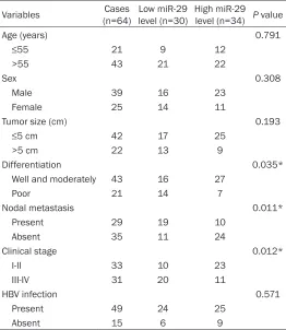
Table 1. Association between miR-29 expression and clinico-pathological characteristics in hepatocellular carcinoma Variables (n=64)Cases level (n=30)Low miR-29 High miR-29 level (n=34) P value
Age (years) 0.791
≤55 21 9 12
>55 43 21 22
Sex 0.308
Male 39 16 23
Female 25 14 11
Tumor size (cm) 0.193
≤5 cm 42 17 25
>5 cm 22 13 9
Differentiation 0.035*
Well and moderately 43 16 27
Poor 21 14 7
Nodal metastasis 0.011*
Present 29 19 10
Absent 35 11 24
Clinical stage 0.012*
I-II 33 10 23
III-IV 31 20 11
HBV infection 0.571
Present 49 24 25
Absent 15 6 9
*Means the difference has statistical significance.
ately snap-frozen in liquid nitrogen after surgical removal, and stored at -80°C. The clinical characteris-tics were summarized in Table 1.
Cell culture and transfection
95°C for 3 min, followed by 40 cycles of 95°C for 30 sec and 60°C for 30 sec. The express- ion analysis was conducted using the 2-ΔΔCt
method.
Western blot assay
Cells were lysed in RIPA buffer, and the lysates were then centrifuged at 12,000×g for 30 min at 4°C. BCA protein assay kit (Beyotime Bio- technology, Shanghai, China) was used to de- termine the protein concentration, according to the manufacturer’s instruction. The protein (60 μg) was then separated in 10% SDS-PAGE gel, which was then transferred onto a polyvi-nylidene fluoride (PVDF) membrane (Millipore, Billerica, MA, USA). The membrane was then blocked in 5% non-fat milk (Mengniu, Beijing, China) overnight at 4°C. After that, the mem-brane was incubated with rabbit anti-human IGF2BP1 and GAPDH antibodies(Abcam, Cam- bridge, MA, USA) at room temperature for 3 h. Then, the membrane was incubated with the mouse anti-rabbit secondary antibody (Abcam) at room temperature for 1 h. The immunoblots on the membrane were visualized using an en- hanced chemiluminescence (ECL) kit (Thermo Fisher).
MTT assay
MTT assay was used to examine cell prolifera-tion. HepG2 cells (50000 cells per well) in 100 μL of serum-free DMEM containing 0.5 g/L MTT (Sigma, USA) were seeded in a 96-well plate, and then incubated at 37°C for 0 h, 24 h, 48 h and 72 h. After that, the medium was removed, and 50 μL of DMSO (Sigma) was added. After incubated at 37°C for 10 min, the A570 of cells was measured using a spectrophotometer (UV-3600, Shimadzu, Kyo- to, Japan).
Transwell assay
Transwell assay was used to examine cell invasion using transwell chambers (BD, USA). HepG2 cell suspension (5×10⁵ cells/ml) was
prepared in DMEM, 300 μl of which was added into the upper chamber. After that, 500 μl of DMEM added with 10% FBS was added into the lower chamber. After incubated in 37°C for 24 h, a cotton-tipped swab was used to wipe out those HepG2 cells not through the pores. After that, the filter was fixed in 90% alcohol, and
stained by crystal violet (Sigma). Cells through the pores were counted and photographed under an inverted microscope (IX71, Olympus, Tokyo, Japan).
Wound healing assay
HepG2 cells were cultured to full confluence in 24-well plates. A plastic scriber was used to create wounds (about 1 mm width). After that, HepG2 cells were washed with DPBS (Thermo Fisher), and then cultured in DMEM added with 10% FBS 37°C for 48 h. After that, HepG2 cells were photographed under an inverted micro-scope (IX71, Olympus, Tokyo, Japan).
Luciferase reporter assay
The putative target genes of miR-29 were pre-dicted using Targetscan (www.targetscan.org/) and miRanda (www.microrna.org), according to the manufacture’s instruction. The wild type (WT) of IGF2BP1 3’-UTR were constructed using PCR. The mutant type (MT) of IGF2BP1 3’-UTR was generated using the Quick-Change Site-Directed Mutagenesis Kit (Stratagene, La Jolla, CA, USA), according to the manufacturer’s in- struction. The WT or MT IGF2BP1 3’-UTR was inserted into the psiCHECK vector (Promega, Madison, WI, USA), generating WT-IGF2BP1-3’UTR or MT-IGF2BP1-WT-IGF2BP1-3’UTR plasmid, respec-tively. HepG2 cells a 24-well plate were co-transfected with 100 ng of WT-IGF2BP1-3’UTR or MT-IGF2BP1-3’UTR plasmid, and 50 nM of miR-29 mimics or miR-NC, respectively, using Lipofectamine 2000. After transfection for 48 h, the renilla and firefly luciferase activities were detected using the Dual-luciferase Re- porter Assay System (Promega), according to the manufacturer’s instruction. After that, Re- nilla luciferase activity was normalized to firefly luciferase activity.
Statistical analysis
characteris-tics in HCC. P value less than 0.05 was consid-ered as statistically significant.
Results
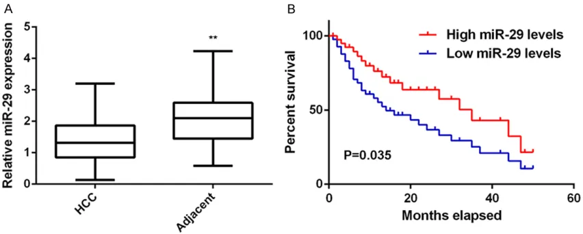
MiR-29 is downregulated in HCC, associated with HCC progression and poor prognosis
The regulatory mechanism of miR-29 underly-ing HCC progression still remains largely un- known. Here we firstly conducted real-time PCR to examine the miR-29 expression in HCC tis-sues and adjacent non-tumor tistis-sues. The miR-29 levels were significantly decreased in HCC tissues compared to adjacent non-tumor tis-sues (Figure 1A). The clinical significance of miR-29 expression in HCC was then investi- gated. Our data indicated that low miR-29 ex- pression was significantly associated with poor differentiation, node metastasis, and clinical stage in HCC (Table 1). In addition, those HCC patients with low miR-29 levels showed shor- ter survival time, when compared with those with high miR-29 expression (Figure 1B). Accor- dingly, our data demonstrate that the reduced expression of miR-29 associated with HCC pro-gression and poor prognosis.
Restoration of miR-29 decreases the prolifera-tion, migration and invasion of HepG2 cells
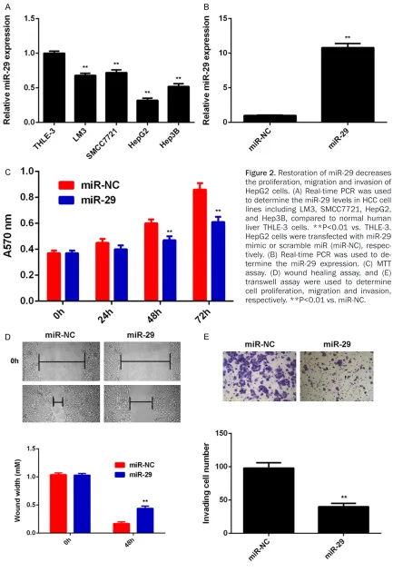
To further confirm these above findings, we detected the expression of miR-29 in several common HCC cell lines including LM3, HepG2, Hep3B, and SMCC7721. The normal liver
THLE-3 cells were used as the control group. As indi-cated in Figure 2A, miR-29 was also significant-ly downregulated in HCC cell lines compared with THLE-3 cells.
As HepG2 cells showed the most significant decrease in miR-29 expression, we used this cell line in the following experiments in vitro. To restore the expression levels of miR-29 in HepG2 cells, miR-29 mimic were used to per-form cell transfection. Our data indicated that transfection with miR-29 mimic caused a sig-nificant increase in the expression of miR-29 (Figure 2B). MTT assay, wound healing assay and transwell assay were then conducted to determine the cell proliferation, migration and invasion, respectively. As indicated in Figure 2C-E, restoration of miR-29 expression signifi-cantly reduced the proliferation, migration and invasion of HepG2 cells. Therefore, miR-29 may play a suppressive role in HCC growth and me- tastasis.
IGF2BP1, upregulated in HCC, is identified as a novel target gene of miR-29
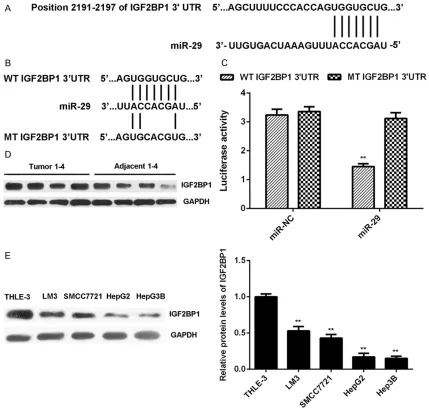
As miRs function through regulating the expres-sion of their target genes, the putative target genes of miR-29 were predicted. As shown in
[image:4.612.93.519.75.246.2]Figure 3A, IGF2BP1 was a putative target ge- ne of miR-29. To confirm this targeting rela- tionship, the luciferase vectors containing WT or MT of IGF2BP1 3’-UTR were constructed, re- spectively (Figure 3B). Luciferase reporter as-
Figure 2. Restoration of miR-29 decreases the proliferation, migration and invasion of HepG2 cells. (A) Real-time PCR was used to determine the miR-29 levels in HCC cell lines including LM3, SMCC7721, HepG2, and Hep3B, compared to normal human liver THLE-3 cells. **P<0.01 vs. THLE-3. HepG2 cells were transfected with miR-29 mimic or scramble miR (miR-NC), respec-tively. (B) Real-time PCR was used to de-termine the miR-29 expression. (C) MTT assay. (D) wound healing assay, and (E) transwell assay were used to determine cell proliferation, migration and invasion, respectively. **P<0.01 vs. miR-NC.
say data further showed that the luciferase
elimi-nated when transfection with MT-IGF2BP1-3’UTR plasmid (Figure 3C). As no previous study has revealed their targeting relationship, the present study for the first time reports that IGF2BP1 is a novel target gene of miR-29. As miRs negatively regulate the expression of their targets at the post-transcriptional levels, we then examined the expression of IGF2BP1 in HCC tissues and cell lines. The protein levels of IGF2BP1 were significantly higher in HCC tis-sues compared to adjacent non-tumor tistis-sues (Figure 3D). As indicated in Figure 3E, the IGF-
2BP1 protein levels were also increased in HCC cells compared with THLE-3 cells. These find-ings suggest that the reduced expression of miR-29 may contribute to the upregulation of IGF2BP1 in HCC tissues and cell lines.
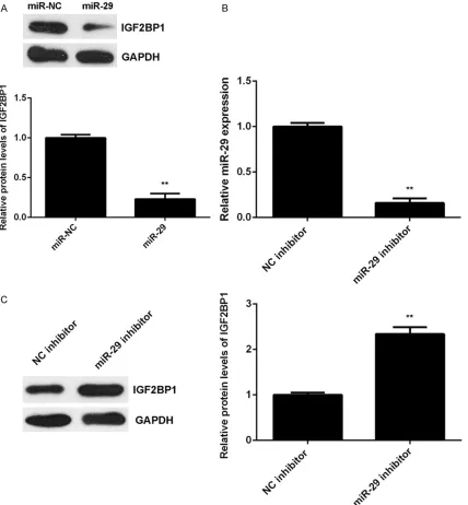
IGF2BP1 is negatively regulated by miR-29 at the post-transcriptional levels in HepG2 cells
[image:6.612.91.520.73.487.2]After that, we examined the effects of miR-29 expression on the protein expression of IGF- 2BP1 in HepG2 cells. As shown in Figure 4A, overexpression of miR-29 led to a significant
reduction of IGF2BP1 protein expression in HepG2 cells. To further confirm these findings, HepG2 cells were transfected with miR-29 in- hibitor or NC inhibitor, respectively. After trans-fection, the miR-29 levels were significantly de- creased in miR-29 inhibitor group compared with NC inhibitor group (Figure 4B). We then found that knockdown of miR-29 significantly enhanced the protein expression of IGF2BP1
(Figure 4C). Therefore, miR-29 negatively re- gulates the protein expression of IGF2BP1 in HepG2 cells.
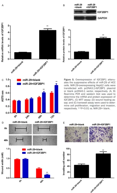
Overexpression of IGF2BP1 attenuates the suppressive effects of miR-29 of HCC cells
Based on the above findings, we speculated that IGF2BP1 might be involved in
[image:7.612.95.523.74.536.2]diated malignant phenotypes of HepG2 cells. To clarify this speculation, miR-29-overexpre- ssing HepG2 cells were transfected with pc- DNA3.1-IGF2BP1 plasmid or blank pcDNA3.1 vector, respectively. As indicated in Figure 5A,
5B, the IGF2BP1 levels were significantly incre- ased in the miR-29+IGF2BP1 group compared with miR-29+ blank group. MTT assay, wound healing assay and transwell assay were then conducted to determine the cell proliferation, migration and invasion, respectively. Our data showed that the proliferation, migration and invasion of HepG2 cells were significantly in- creased in miR-29+IGF2BP1 group compared with miR-29+ blank group, indicating that over-expression of IGF2BP1 attenuates the sup- pressive effects of miR-29 of the malignant phenotypes of HCC cells. These findings sug-gest that miR-29 may inhibit HCC growth and metastasis via targeting IGF2BP1.
Discussion
The present study investigated the underlying regulatory mechanism of miR-29 in HCC, and showed that miR-29 was significantly down- regulated in HCC tissues and cell lines, which was significantly associated with disease pro-gression as well as shorter survival time of HCC patients. In vitro experiments data indicated that restoration of miR-29 expression caused a significant reduction in HCC HepG2 cell pro- liferation, migration and invasion. IGF2BP1, sig-nificantly upregulated in HCC, was identified as a novel target of miR-29, and its protein ex- pression was negatively regulated by miR-29 in HepG2 cells. Moreover, overexpression of IGF- 2BP1 attenuated the suppressive effects of miR-29 on the malignant phenotypes of HepG2 cells.
MiR-29 has been demonstrated to play a sup-pressive role in different cancers types, such as breast cancer [22], acute megakaryoblastic leukemia [23], ovarian cancer [24], and gastric cancer [25]. Recently, miR-29 was found to play a suppressive role in HCC. For instance, Xiong et al. found that miR-29 was downregulated in HCC, which was significantly associated with poor prognosis of HCC patients. Moreover, they showed that miR-29 could sensitize HCC cells to apoptosis triggered by either serum starva-tion and hypoxia or chemotherapeutic drugs, and inhibit HCC cell growth in vitro and in vivo [20]. In the present study, we also found that
miR-29 was downregulated in HCC tissues and cell lines, and low expression of miR-29 was associated with the poor differentiation, me- tastasis, and advanced clinical stage in HCC. We further showed that restoration of miR-29 significantly decreased HepG2 cell prolifera-tion, migration and invasion.
Moreover, several targets of miR-29 have been identified in HCC, including TET1, Bcl-2 and Mcl-1 [20, 21]. We speculated that other tar-gets of miR-29 may also play important roles in HCC. In this study, bioinformatics analysis indicated that IGF2BP1 was a putative target gene of miR-29. We then conducted luciferase reporter gene assay, and confirmed their tar- get relationship. IGF2BP1 is a member of the IGF2BP family, and contains four K homology domains and two RNA recognition motifs [26]. IGF2BP1 can bind to and stabilize the mRNAs of some genes, such as c-MYC, MKI67, IGF2, and beta-actin, through which it participates in regulating cell survival and proliferation [26-28]. Recently, IGF2BP1 was reported to pro-mote tumor cell proliferation, migration and invasion in several cancers including HCC [27-29]. Knockdown of IGF2BP1 could significantly decrease HCC growth in vivo, suggesting that IGF2BP1 may become a promising molecular target for HCC treatment [28]. Moreover, sev-eral miRs have been reported to directly tar- get HCC, and thus function as tumor suppres-sors. For instance, miR-1275 has suppressive effects on HCC tumor growth through target- ing IGF2BPs and IGF1R [30]. MiR-625 could inhibit HCC cell migration and invasion by tar-geting IGF2BP1 [31]. In this study, we found that IGF2BP1 was significantly upregulated in HCC tissues and cell lines, and IGF2BP1 was negatively regulated by miR-29 at the post- transcriptional level in HepG2 cells. Moreover, we showed that overexpression of IGF2BP1 attenuated the suppressive effects of miR-29 on the proliferation, migration and invasion of HepG2 cells, which further suggests that the inhibitory effects of miR-29 on HCC is probably through inhibition of IGF2BP1.
Disclosure of conflict of interest
None.
Address correspondence to: Yanlan Ouyang, De- partment of General Surgery, Xiangya Hospital of Central South University, 87 Xiangya Road, Chang- sha 410008, Hunan, PR China. Tel: +86-731-897- 53607; Fax: +86-731-89753607; E-mail: xyouyang-yanlan@qq.com
References
[1] Jemal A, Bray F, Center MM, Ferlay J, Ward E and Forman D. Global cancer statistics. CA Cancer J Clin 2011; 61: 69-90.
[2] Siegel RL, Miller KD and Jemal A. Cancer sta-tistics, 2015. CA Cancer J Clin 2015; 65: 5-29. [3] Zhu K, He Y, Xia C, Yan J, Hou J, Kong D, Yang Y
and Zheng G. MicroRNA-15a inhibits prolifera-tion and induces apoptosis in CNE1 nasopha-ryngeal carcinoma cells. Oncol Res 2016; 24: 145-151.
[4] Ambros V. The functions of animal microRNAs. Nature 2004; 431: 350-355.
[5] Zhou W, Zou B, Liu L, Cui K, Gao J, Yuan S and Cong N. MicroRNA-98 acts as a tumor sup-pressor in hepatocellular carcinoma via target-ing SALL4. Oncotarget 2016; 7: 74059-74073. [6] Luo T, Yan Y, He Q, Ma X and Wang W. miR-328-5p inhibits MDA-MB-231 breast cancer cell proliferation by targeting RAGE. Oncol Rep 2018; [Epub ahead of print].
[7] Xiao F, Li Y, Wan Y and Xue M. MircroRNA-139 sensitizes ovarian cancer cell to cisplatin-based chemotherapy through regulation of ATP7A/B. Cancer Chemother Pharmacol 2018; [Epub ahead of print].
[8] Xu R, Zhu X, Chen F, Huang C, Ai K, Wu H, Zhang L, Zhao X. LncRNA XIST/miR-200c regu-lates the stemness properties and tumourige-nicity of human bladder cancer stem cell-like cells. Cancer Cell Int 2018; 18: 41.
[9] Zhang Z, Li X, Xiao Q and Wang Z. MiR-574-5p mediates the cell cycle and apoptosis in thy-roid cancer cells via Wnt/beta-catenin signal-ing by represssignal-ing the expression of Quaksignal-ing proteins. Oncol Lett 2018; 15: 5841-5848. [10] Ma Z. Downregulation of SETD8 by miR-382 is
involved in glioma progression. Pathol Res Pract 2018; 214: 356-360.
[11] Sun J, Zheng G, Gu Z and Guo Z. MiR-137 in-hibits proliferation and angiogenesis of hu- man glioblastoma cells by targeting EZH2. J Neurooncol 2015; 122: 481-489.
[12] Song H, Zhang Y, Liu N, Wan C, Zhang D, Zhao S, Kong Y and Yuan L. miR-92b regulates glio-ma cells proliferation, migration, invasion, and apoptosis via PTEN/Akt signaling pathway. J Physiol Biochem 2016; 72: 201-211.
[13] Wan D, Shen S, Fu S, Shen C, Wu J, Wang S, Xie W, Chen B, Liya A, Guo Y, Zheng D, Zhi Q and Peng B. miR-203 suppresses the proliferation and metastasis of hepatocellular carcinoma by targeting oncogene ADAM9 and oncogenic long non-coding RNA HULC. Anticancer Agents Med Chem 2016; 16: 414-23.
[14] Sheng Y, Li J, Zou C, Wang S, Cao Y, Zhang J, Huang A and Tang H. Downregulation of miR-101-3p by hepatitis B virus promotes prolifera-tion and migraprolifera-tion of hepatocellular carcinoma cells by targeting Rab5a. Arch Virol 2014; 159: 2397-410.
[15] Liu X, Li J, Yu Z, Sun R and Kan Q. MiR-935 promotes liver cancer cell proliferation and migration by targeting SOX7. Oncol Res 2017; 25: 427-435.
[16] Tian Z, Jiang H, Liu Y, Huang Y, Xiong X, Wu H and Dai X. MicroRNA-133b inhibits hepatocel-lular carcinoma cell progression by targeting Sirt1. Exp Cell Res 2016; 343: 135-147. [17] Trehoux S, Lahdaoui F, Delpu Y, Renaud F,
Leteurtre E, Torrisani J, Jonckheere N and Van Seuningen I. Micro-RNAs 29a and miR-330-5p function as tumor suppressors by tar-geting the MUC1 mucin in pancreatic cancer cells. Biochim Biophys Acta 2015; 1853: 2392-2403.
[18] Muniyappa MK, Dowling P, Henry M, Meleady P, Doolan P, Gammell P, Clynes M and Barron N. MiRNA-29a regulates the expression of numerous proteins and reduces the invasive-ness and proliferation of human carcinoma cell lines. Eur J Cancer 2009; 45: 3104-3118. [19] Cui Y, Su WY, Xing J, Wang YC, Wang P, Chen
XY, Shen ZY, Cao H, Lu YY and Fang JY. MiR-29a inhibits cell proliferation and induces cell cycle arrest through the downregulation of p42.3 in human gastric cancer. PLoS One 2011; 6: e25872.
[20] Xiong Y, Fang JH, Yun JP, Yang J, Zhang Y, Jia WH and Zhuang SM. Effects of microRNA-29 on apoptosis, tumorigenicity, and prognosis of hepatocellular carcinoma. Hepatology 2010; 51: 836-845.
[21] Lin LL, Wang W, Hu Z, Wang LW, Chang J and Qian H. Negative feedback of miR-29 family TET1 involves in hepatocellular cancer. Med Oncol 2014; 31: 291.
[22] Wu Z, Huang X, Zou Q and Guo Y. The inhibitory role of Mir-29 in growth of breast cancer cells. J Exp Clin Cancer Res 2013; 32: 98.
[23] Sharifi M and Salehi R. Blockage of miR-92a-3p with locked nucleic acid induces apoptosis and prevents cell proliferation in human acute megakaryoblastic leukemia. Cancer Gene Ther 2016; 23: 29-35.
ovari-an covari-ancer cells. Int J Covari-ancer 2014; 134: 542-551.
[25] Chen L, Xiao H, Wang ZH, Huang Y, Liu ZP, Ren H and Song H. miR-29a suppresses growth and invasion of gastric cancer cells in vitro by targeting VEGF-A. BMB Rep 2014; 47: 39-44. [26] Bell JL, Wachter K, Muhleck B, Pazaitis N,
Kohn M, Lederer M and Huttelmaier S. Insulin-like growth factor 2 mRNA-binding proteins (IGF2BPs): post-transcriptional drivers of can-cer progression? Cell Mol Life Sci 2013; 70: 2657-2675.
[27] Luo Y, Sun R, Zhang J, Sun T, Liu X and Yang B. miR-506 inhibits the proliferation and inva-sion by targeting IGF2BP1 in glioblastoma. Am J Transl Res 2015; 7: 2007-2014.
[28] Gutschner T, Hammerle M, Pazaitis N, Bley N, Fiskin E, Uckelmann H, Heim A, Grobeta M, Hofmann N, Geffers R, Skawran B, Longerich T, Breuhahn K, Schirmacher P, Muhleck B, Hut- telmaier S and Diederichs S. Insulin-like grow- th factor 2 mRNA-binding protein 1 (IGF2BP1) is an important protumorigenic factor in he- patocellular carcinoma. Hepatology 2014; 59: 1900-1911.
[29] Qu Y, Pan S, Kang M, Dong R and Zhao J. MicroRNA-150 functions as a tumor suppres-sor in osteosarcoma by targeting IGF2BP1. Tumour Biol 2016; 37: 5275-5284.
[30] Fawzy IO, Hamza MT, Hosny KA, Esmat G, El Tayebi HM and Abdelaziz AI. miR-1275: a single microRNA that targets the three IGF2-mRNA-binding proteins hindering tumor growth in hepatocellular carcinoma. FEBS Lett 2015; 589: 2257-2265.