Review Article
A meta-analysis: could we predict the malignancy of
solid pseudopapillary neoplasm?
He Song1, Ming Dong1, Huimin Xiao2, Jianping Zhou1, Weiwei Sheng1, Banghua Zhong1, Wei Gao1
1Department of Gastrointestinal Surgery, The First Hospital of China Medical University, Shenyang 110001, China; 2Department of General Surgery, The People’s Hospital of China Medical University, Shenyang 110016, China
Received May 12, 2017; Accepted July 26, 2017; Epub September 1, 2017; Published September 15, 2017
Abstract: Objective: To evaluate the risk factors influencing the malignant potential of solid pseudopapillary neo-plasm (SPN). Methods: This meta-analysis used MEDLINE (PubMed), EMBASE and web of science including 14 cohort studies reporting the risk factors influencing the malignant potential after the initial operation on SPN up to March 2017. Review Manager Software 5.2 was used for meta-analysis. Results: 14 studies with a total of 763 patients were included in our meta-analysis. In all the variables, age and tumor size were significantly correlated with malignancy. Conclusion: Malignant SPNs tended to be larger in diameter and younger in age than benign type. In particularly, larger tumor size may be a crucial factor for decision of aggressive resection.
Keywords: Solid pseudopapillary neoplasm, pancreas, malignancy
Introduction
Solid pseudopapillary neoplasm (SPN) is a ra- re pancreatic tumor predominantly affecting young women with low malignant potential [1]. It usually has a favorable prognosis, with just over 95% of patients reported as disease free after surgical resection and with less than 2% mortality [2]. In the year of 2010, the World Health Organization (WHO) classified SPN as a low-grade malignant neoplasm. Approximately 10% to 15% cases of SPN are malignant, which could require the good survival with the resec-tion [3], however due to higher risk of recur-rence and mortality, aggressive surgical app- roach is warranted especially in the malignant cases such as local invasion, metastasis [4-7]. There has been a dramatic increase of report-ed SPN in the world over the past few decades [2]. Unfortunately the detection of risk factors of malignancy is of utmost importance, but remains unclear.
Materials and methods
Search strategy
Studies evaluating the factors influencing the malignancy of SPN were retrieved from the
March 2017. We used the following free-text search terms in “All fields”: “solid pseudopapil-lary neoplasm” and “risk factors” and “malig-nancy”. There was no language restriction and no methodological filters. A recursive search of the reference of selected studies, review arti-cles and guidelines were performed manually to identify the rest of potential relative articles.
Study inclusion/exclusion criteria
Risk factors to predict the malignancy of solid pseudopapillary neoplasm
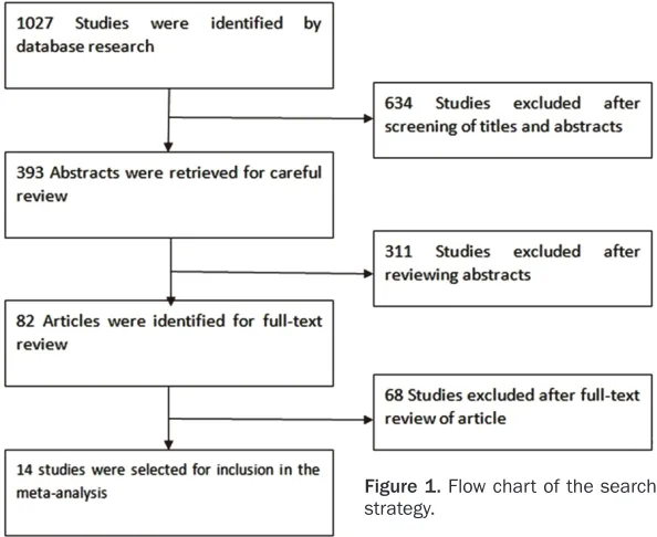
sufficient to calculate them). Fourth, to avoid overlapping data that may result from duplica-tions, only the articles with the largest sample size were included. Editorials, review articles or case series without association between risk factors and malignancy, duplicate publications and case reports were excluded. As is shown in the Figure 1.
Data extraction and management
All data were extracted onto a standardized form. The primary data extracted from each
individual outcomes were integrated with the meta-analysis software Review Manager Software 5.2 (Cochrane Collaborative, Oxford, United Kingdom).
Results
[image:2.612.90.387.73.316.2]There were 14 eligible studies with the compa-rision of clinicopathlogical paremeter which were published from 2007 to 2015 (Table 1). One of these studies were from America, one from Africa, one from Australia, and eleven from Asia (six from Korea, five from China, one Figure 1. Flow chart of the search
strategy.
Table 1. Characteristics of studies concluded
Referrance Country Year Malignant/Total
Cai [8] China 2011 17:33
Chuang [9] Korea 2009 12:30
GOH [10] Singapore 2007 9:16
Hokim [11] Korea 2011 5:30
Hwang (child) [12] Korea 2014 9:45
Jean [13] U.S. 2010 9:45
Kang [3] Korea 2006 11:33
Kim [5] Korea 2014 17:106
Lee (adult and child) [14] Korea 2008 10:62
Nakeeb [15] Eygpt 2013 6:24
Yang [16] China 2009 2:26
Yu [4] China 2015 16:97
Yucai [17] China 2014 35:116
Tang [18] China 2015 24:100
article included the first authorship, country of ori-gin, year of publication (Ta- ble 1).
Assessment of risk of bias in included studies
Risk of bias across studies may be present, particular-ly with regard to publication bias. As the topic involves surgical procedures and outcomes, it is very likely that smaller-sample stud-ies or those with negative outcomes may not be pub-lished in the literature. A funnel plot was created to assess publication bias.
Statistical analysis
If a specific factor was reported in at least three studies and supported by compara-ble methodologies, the odds ratios (OR) and 95% confidence intervals (CIs) were calculated to estimate the association between binary factors and malignancy of SPN. When mean values and SDs for a certain risk factor were provided, we calculated the mean differences (MDs) between patients with malignant SPN and with benign SPN. Depending on the presence or absence of significant het-erogeneity, meta-analysis was conducted using the random-effects model or the fixed-effect model. Statistical heteroge-neity of treatment effects between stud-ies was formally tested with Cochran’s Chi-squared statistics and with signifi-cance set at P<0.10. The I2 statistic was
[image:2.612.92.327.351.550.2]from Singapore). There were 763 patients in total and 101 patients of malignant SPN. There were 9 potential risk factors for malignant SPN identified for analysis.
Gender
With the statistical method of Mantel-Haenszel in the fixed-effect model, it showed no statisti-cally significant difference in malignancy in the patients with male vs. female (OR = 1.09, 95%
CI: 0.77-1.56) without heterogeneity (I2 = 0%, P
= 0.78) (Figure 2).
Age
With the statistical method of Inverse Variance in the fixed-effect model, it showed the statisti-cally significant difference in malignancy in the patients about age (MD = -4.02, 95% CI: -5.98 to -2.07) without much heterogeneity (I2 = 36%,
[image:3.612.98.520.72.269.2] [image:3.612.101.521.307.397.2]P = 0.20) (Figure 3). Figure 2. Forest plot for risk factors for malignancy of SPN categorized by male vs. female.
Figure 3. Forest plot for risk factors for malignancy of SPN categorized by age.
[image:3.612.103.519.435.560.2]Risk factors to predict the malignancy of solid pseudopapillary neoplasm
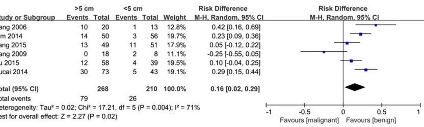
Tumor size
There were 2 meta-analyses to calculate the OR and MD respectively according to the dichotomous data type and continuous date type in the studies evaluated. In the dichoto-mous data type, tumor size were classified as two groups size >5 cm vs. size <5 cm. Analysis of the pooled data showed the risk factors of malignancy was significantly higher among the
patients in the size >5 cm group (OR = 2.82, 95% CI: 1.13-7.01) with heterogeneity (I2 =
61%, P = 0.03) using the statistical method of Mantel-Haenszel in the random-effect model (Figure 4).
[image:4.612.94.521.75.160.2] [image:4.612.95.520.201.353.2]In the continuous data type, the meta-analysis showed no statistically significant mean differ-ence in malignancy in the patients about tumor diameter (MD = 1.58, 95% CI: -0.29 to 3.44) Figure 5. Forest plot for risk factors for malignancy of SPN categorized by tumor size in the continuous date type.
Figure 6. Forest plot for risk factors for malignancy of SPN categorized by symptom.
[image:4.612.101.519.391.559.2]with heterogeneity (I2 = 61%, P = 0.05) using
the statistical method of Inverse Variance in the random-effect model (Figure 5). In the sen-sitivity analysis, removal of Lee study, there was significant mean difference (MD = 2.67, 95% CI: 1.57 to 3.76) without heterogeneity (I2
= 0%, P = 0.47) (figure not shown).
Symptom
A meta-analysis showed no statistically signifi-cant difference in malignancy in the patients with present vs. absent (OR = 1.41, 95% CI: 0.86-2.30) without heterogeneity (I2 = 1%, P =
0.42) using the statistical method of Mantel-Haenszel in the fixed-effect model (Figure 6).
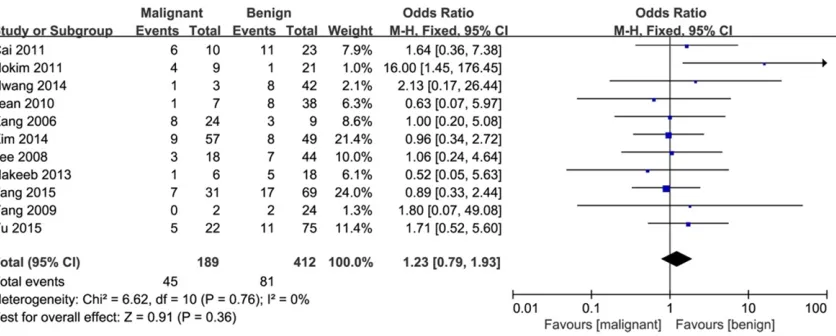
Calcification
A meta-analysis showed no statistically signifi-cant difference in malignancy in the patients with present vs. absent (OR = 1.23, 95% CI:
0.79-1.93) without heterogeneity using the sta-tistical method of Mantel-Haenszel in the fixed-effect model (I2 = 0%, P = 0.76) (Figure 7).
Tumor location
The tumor location was defined as head+neck vs. body+tail. Comparisons of patients with head+neck vs. body+tail, there was no statisti-cally significant difference between different tumor location (OR = 0.88, 95% CI: 0.60-1.28) without heterogeneity (I2 = 4%, P = 0.41) using
the statistical method of Mantel-Haenszel in the fixed-effect model (Figure 8).
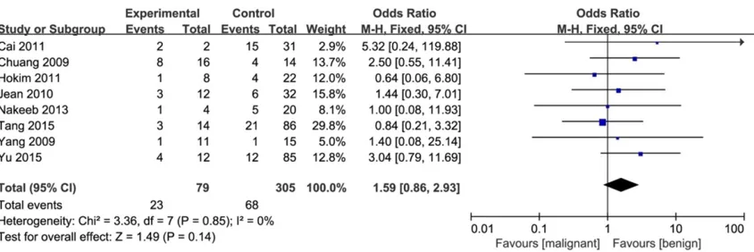
Tumor nature
[image:5.612.93.520.72.250.2]Tumor nature was defined as predominantly solid vs. predominantly cystic or mixed. The meta-analysis showed no statistically signifi-cant difference in malignancy in the patients about tumor nature (OR = 1.59, 95% CI: 0.86-Figure 8. Forest plot for risk factors for malignancy of SPN categorized by tumor location.
[image:5.612.98.520.288.429.2]Risk factors to predict the malignancy of solid pseudopapillary neoplasm
2.93) without heterogeneity (I2 = 0%, P = 0.85)
using the statistical method of Mantel-Haens- zel in the fixed-effect model (Figure 9).
Tumor marker CA199
The tumor marker CA199 was defined as ele-vated vs. normal. Comparisons of patients with elevated vs. normal, there was no statistically significant difference between different tumor marker (OR = 1.65, 95% CI: 0.47-5.80) without heterogeneity (I2 = 0%, P = 0.88) using the
sta-tistical method of Mantel-Haenszel in the fixed-effect model (Figure 10).
and calculated the pooled ORs of the rest of studies. No significant differences were obser- ved between the corresponding results and the robust overall results (data not shown), except removal of Lee study leading to signifi-cant result in the continuous data type of the tumor diameter.
Publication bias
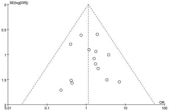
[image:6.612.92.523.74.168.2] [image:6.612.96.521.209.306.2]No obvious asymmetry was observed in the funnel plot of the meta-analysis evaluating the risk factors of malignancy in SPN (Figure 12). Figure 10. Forest plot for risk factors for malignancy of SPN categorized by tumor marker.
Figure 11. Forest plot for risk factors for malignancy of SPN categorized by tumor hemorrhage or necrosis.
Figure 12. Funnel plot for risk factors for malignancy of SPN is symmetry.
Tumor hemorrhage or ne-crosis
A meta-analysis showed no statistically significant dif-ference in malignancy in the patients with present vs. absent (OR = 0.65, 95% CI: 0.31-1.34) without het-erogeneity (I2 = 0%, P =
0.99) using the statistical method of Mantel-Haens- zel in the fixed-effect mo- del (Figure 11).
Sensitivity analyses
[image:6.612.91.391.346.542.2]Discussion
In the recent days, a relatively large case series of single center [19-25] or the multicenter [26] reporting SPN have tried to enrich the unsuffi-cient clinical data in the current literature, but the results were limited to descriptive clinical analysis and failed to enclose the relation between clinicopathologic feature and poten-tial malignancy. Many factors may affect malig-nancy of SPN, yet there still existed some con-troversy so far. The prevalence of malignancy in SPN patients should not be negligible, there-fore malignancy-related variables involved in SPN patients’ outcome are crucial. This study was the first meta-analysis of the literature on exploring the factors for SPN patients with malignancy. Included in our analysis were 14 unique studies from 2007 to 2017 with 763 patients. Data shows that age and tumor size were associated with a significantly increased risk of malignancy.
For the gender, the report of 34 cases [27] from Brazil regarded SPN were more aggressive in male patients. Men had a two-times higher inci-dence of metastases and a three-times higher death rate [28]. However, the similar tendency was not confirmative in our meta-analysis. There were no significant sex differences in his-topathologic or immunohistochemical features of SPN [29, 30]. So maybe the report [27] was limited to the small sample case series to draw the conclusion.
For the age, although SPN had different clinical features in adults and children, children were not likely to appear malignant potential [14]. However, in our meta-analysis the younger age (in adults) could be related to the malignant potential. Only four studies were included, the- re would be more specific clinical data report-ing age.
For the tumor size, there were several studies [3, 31] indicating that larger tumor size was related to malignancy, which is consistent with our meta-analysis. Moreover, tumor size is the significant clinical feature associated with met-astatic disease and decreased disease-free survival [32]. Therefore, we suggest a more pre-cise and aggressive resection of the SPN for larger tumors (>5 cm). Tumor (<5 cm) can be managed in conservative surgery such as enu-leation or distal pancreatectomy preserving spleen. Especially, tumor locating in the head
of pancreas more than 5 cm was likely proceed-ed with duodenum-preserving pancreatic head resection rather than enucleation [33]. In the sensitivity analysis, removal of Lee study led to significant result in the continuous data type of the tumor diameter. It was probable that Lee study covered the combination of data among both children and adults, children’s clinical da- ta will interfere with the overall results. In com-parison with the adult population, children with SPN generally have a better prognosis and a different clinical feature [34].
Clinical presentation of the SPN is usually non-specific. At the time of diagnosis, SPN may appear with a significantly enlarged size leading to abdominal pain or distention [35]. However, presence of symptom is not related to malig-nancy in our ananlysis.
Tumor location, tumor marker and presence of hemorrhage or necorsis were not associated with malignancy in our study, which has the similar results as the Tang [18].
There were some studies showing calcification [36] and tumor nature [12] were related to the malignancy which is not consistent with our meta-analysis.
Few studies reported other important risk fac-tors, such as tumor capsule [9], Ki-67 [4] and peripancreatic lymphadenopathy [18], a meta-analysis of these factors could not be per-formed due to unsufficient data.
The malignancy incidence of SPN varies from 3.6 to 56% [37]. FDG-PET may help distinguish the malignant SPN from benign type, however no statistical analysis could be performed because the PET is not the routine examination [37, 38]. Maybe the accumulation of FDG in the imaging would be the new orientation for pre-dictive factors of malignancy.
Risk factors to predict the malignancy of solid pseudopapillary neoplasm
In conclusion, malignant SPNs tended to be larger in diameter and younger in age than benign type. In particularly, larger tumor size may be a crucial factor for decision of aggres-sive resection.
Acknowledgements
This work was supported by Chinese National Science Foundation (NO. 81672835) to Ming Dong.
Disclosure of conflict of interest
None.
Address correspondence to: Dr. Ming Dong, Depart- ment of Gastrointestinal Surgery, First Hospital of China Medical University, Shenyang 110001, China. Tel: +86-24-83282886; E-mail: dongming@cmu. edu.cn
References
[1] Papavramidis T, Papavramidis S. Papavramid-is, solid pseudopapillary tumors of the pancre-as: review of 718 patients reported in English literature. J Am Coll Surg 2005; 200: 965-72. [2] Law JK, Ahmed A, Singh VK, Akshintala VS,
Ol-son MT, Raman SP, Ali SZ, Fishman EK, Kamel I, Canto MI, Dal Molin M, Moran RA, Khashab MA, Ahuja N, Goggins M, Hruban RH, Wolfgang CL, Lennon AM. A systematic review of solid-pseudopapillary neoplasms: are these rare le-sions? Pancreas 2014; 43: 331-7.
[3] Kang CM, Kim KS, Choi JS, Kim H, Lee WJ, Kim BR. Solid pseudopapillary tumor of the pan-creas suggesting malignant potential. Pancre-as 2006; 32: 276-80.
[4] Yu P, Cheng X, Du Y, Yang L, Xu Z, Yin W, Zhong Z, Wang X, Xu H, Hu C. Solid pseudopapillary neoplasms of the pancreas: a 19-year multi-center experience in China. J Gastrointest Surg 2015; 19: 1433-40.
[5] Kim MJ, Choi DW, Choi SH, Heo JS, Sung JY. Surgical treatment of solid pseudopapillary neoplasms of the pancreas and risk factors for malignancy. Br J Surg 2014; 101: 1266-71. [6] Wang WB, Zhang TP, Sun MQ, Peng Z, Chen G,
Zhao YP. Solid pseudopapillary tumor of the pancreas with liver metastasis: clinical fea-tures and management. Eur J Surg Oncol 2014; 40: 1572-7.
[7] Park JK, Cho EJ, Ryu JK, Kim YT, Yoon YB. Natu-ral history and malignant risk factors of solid pseudopapillary tumors of the pancreas. Post-grad Med 2013; 125: 92-9.
[8] Cai H, Zhou M, Hu Y, He H, Chen J, Tian W, Deng Y. Solid-pseudopapillary neoplasms of the pancreas: clinical and pathological fea-tures of 33 cases. Surg Today 2013; 43: 148-54.
[9] Chung YE, Kim MJ, Choi JY, Lim JS, Hong HS, Kim YC, Cho HJ, Kim KA, Choi SY. Differentia-tion of benign and malignant solid pseudopap-illary neoplasms of the pancreas. J Comput Assist Tomogr 2009; 33: 689-94.
[10] Goh BK, Tan YM, Cheow PC, Chung AY, Chow PK, Wong WK, Ooi LL. Solid pseudopapillary neoplasms of the pancreas: an updated expe-rience. J Surg Oncol 2007; 95: 640-4.
[11] Kim HH, Yun SK, Kim JC, Park EK, Seoung JS, Hur YH, Koh YS, Cho CK, Shin SS, Kweon SS, Kim HS, Kim HJ. Clinical features and surgical outcome of solid pseudopapillary tumor of the pancreas: 30 consecutive clinical cases. Hepa-togastroenterology 2011; 58: 1002-8. [12] Hwang J, Kim DY, Kim SC, Namgoong JM, Hong
SM. Solid-pseudopapillary neoplasm of the pancreas in children: can we predict malignan-cy? J Pediatr Surg 2014; 49: 1730-3.
[13] Butte JM, Brennan MF, Gönen M, Tang LH, D’Angelica MI, Fong Y, Dematteo RP, Jarnagin WR, Allen PJ. Solid pseudopapillary tumors of the pancreas. Clinical features, surgical out-comes, and long-term survival in 45 consecu-tive patients from a single center. J Gastroin-test Surg 2011; 15: 350-7.
[14] Lee SE, Jang JY, Hwang DW, Park KW, Kim SW. Clinical features and outcome of solid pseudo-papillary neoplasm: differences between adults and children. Arch Surg 2008; 143: 1218-21.
[15] El Nakeeb A, Abdel Wahab M, Elkashef WF, Azer M, Kandil T. Solid pseudopapillary tumour of the pancreas: Incidence, prognosis and out-come of surgery (single center experience). Int J Surg 2013; 11: 447-57.
[16] Yang F, Jin C, Long J, Yu XJ, Xu J, Di Y, Li J, Fu de L, Ni QX. Solid pseudopapillary tumor of the pancreas: a case series of 26 consecutive pa-tients. Am J Surg 2009; 198: 210-5.
[17] Cai Y, Ran X, Xie S, Wang X, Peng B, Mai G, Liu X. Surgical management and long-term follow-up of solid pseudopapillary tumor of pancreas: a large series from a single institution. J Gas-trointest Surg 2014; 18: 935-40.
[18] Tang X, Zhang J, Che X, Chen Y, Wang C. Peri-pancreatic lymphadenopathy on preoperative radiologic images predicts malignancy in pan-creatic solid pseudopapillary neoplasm. Int J Clin Exp Med 2015; 8: 16315-21.
immuno-histochemical study of 33 cases from a single institution in Southern India. Indian J Pathol Microbiol 2015; 58: 163-9.
[20] Ren Z, Zhang P, Zhang X, Liu B. Solid pseudo-papillary neoplasms of the pancreas: clinico-pathologic features and surgical treatment of 19 cases. Int J Clin Exp Pathol 2014; 7: 6889-97.
[21] Guerrache Y, Soyer P, Dohan A, Faraoun SA, Laurent V, Tasu JP, Aubé C, Cazejust J, Boudiaf M, Hoeffel C. Solid-pseudopapillary tumor of the pancreas: MR imaging findings in 21 pa-tients. Clin Imaging 2014; 38: 475-82. [22] Yagci A, Yakan S, Coskun A, Erkan N, Yıldırım
M, Yalcın E, Postacı H. Diagnosis and treat-ment of solid pseudopapillary tumor of the pancreas: experience of one single institution from Turkey. World J Surg Oncol 2013; 11: 308.
[23] Chen YL, Huang ZQ, Dong JH, Zhang WZ, Huang XQ, Wang YB, Chen MY, Feng J, Liu ZW, Wan T, Leng JJ, Chen JY. [Surgical manage-ment and outcome of solid-pseudopapillary tumor of pancreas: a series of 58 cases]. Zhonghua Wai Ke Za Zhi 2012; 50: 615-7. [24] Yu P, Cheng X, Guo J, Wang X, Zhang Y. Solid
pseudopapillary tumor of the pancreas: clini-cal analysis of 11 cases. Hepatogastroenterol-ogy 2011; 58: 192-7.
[25] Patil TB, Shrikhande SV, Kanhere HA, Saoji RR, Ramadwar MR, Shukla PJ. Solid pseudopapil-lary neoplasm of the pancreas: a single institu-tion experience of 14 cases. HPB (Oxford) 2006; 8: 148-50.
[26] Kang CM, Choi SH, Kim SC, Lee WJ, Choi DW, Kim SW; Korean Pancreatic Surgery Club. Pre-dicting recurrence of pancreatic solid pseudo-papillary tumors after surgical resection: a multicenter analysis in Korea. Ann Surg 2014; 260: 348-55.
[27] Machado MC, Machado MA, Bacchella T, Juke-mura J, Almeida JL, Cunha JE. Solid pseudo-papillary neoplasm of the pancreas: distinct patterns of onset, diagnosis, and prognosis for male versus female patients. Surgery 2008; 143: 29-34.
[28] Lin MY and Stabile BE. Solid pseudopapillary neoplasm of the pancreas: a rare and atypi-cally aggressive disease among male patients. Am Surg 2010; 76: 1075-8.
[29] Hirabayashi K, Kurokawa S, Maruno A, Yama-da M, Kawaguchi Y, Nakagohri T, Mine T, Sugi-yama T, Tajiri T, Nakamura N. Sex differences in immunohistochemical expression and capil-lary density in pancreatic solid pseudopapil-lary neoplasm. Ann Diagn Pathol 2015; 19: 45-9.
[30] Tien YW, Ser KH, Hu RH, Lee CY, Jeng YM, Lee PH. Solid pseudopapillary neoplasms of the pancreas: is there a pathologic basis for the observed gender differences in incidence? Surgery 2005; 137: 591-6.
[31] Butte JM, Brennan MF, Gönen M, Tang LH, D’Angelica MI, Fong Y, Dematteo RP, Jarnagin WR, Allen PJ. Solid pseudopapillary tumors of the pancreas. Clinical features, surgical out-comes, and long-term survival in 45 consecu-tive patients from a single center. J Gastroin-test Surg 2011; 15: 350-7.
[32] Lubezky N, Papoulas M, Lessing Y, Gitstein G, Brazowski E, Nachmany I, Lahat G, Goykhman Y, Ben-Yehuda A, Nakache R, Klausner JM. Solid pseudopapillary neoplasm of the pan-creas: management and long-term outcome. Eur J Surg Oncol 2017; 43: 1056-1060. [33] Naar L, Spanomichou DA, Mastoraki A,
Smyrni-otis V, Arkadopoulos N. Solid pseudopapillary neoplasms of the pancreas: a surgical and ge-netic enigma. World J Surg 2017; 41: 1871-1881.
[34] Escobar MA, Bond BJ, Schopp J. Solid pseudo-papillary tumour (Frantz’s tumour) of the pan-creas in childhood. BMJ Case Rep 2014; 2014. [35] Huang Y, Feng JF. Clinicopathologic character-istics and surgical treatment of solid pseudo-papillary tumor of the pancreas. Hippokratia 2013; 17: 68-72.
[36] Kim HH, Yun SK, Kim JC, Park EK, Seoung JS, Hur YH, Koh YS, Cho CK, Shin SS, Kweon SS, Kim HS, Kim HJ. Clinical features and surgical outcome of solid pseudopapillary tumor of the pancreas: 30 consecutive clinical cases. Hepa-togastroenterology 2011; 58: 1002-8.
[37] Morikawa T, Onogawa T, Maeda S, Takadate T, Shirasaki K, Yoshida H, Ishida K, Motoi F, Nai-toh T, Rikiyama T, Katayose Y, Egawa S, Unno M. Solid pseudopapillary neoplasms of the pancreas: an 18-year experience at a single Japanese Institution. Surg Today 2013; 43: 26-32.