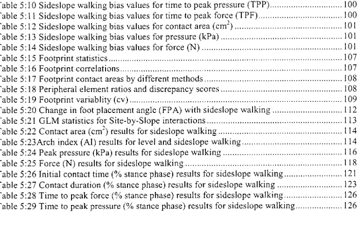
Pressure distribution beneath the foot in sideslope walking
226
0
0
Full text
Figure
+2
Related documents