Original Article
Gallbladder neuroendocrine carcinoma: report of 10
cases and comparision of clinicopathologic features
with gallbladder adenocarcinoma
Chen Chen1, Lin Wang1, Xi Liu2, Guanjun Zhang2, Yaling Zhao3, Zhimin Geng1
Departments of 1Hepatobiliary Surgery, 2Pathology, The First Affiliated Hospital of Xi’an Jiaotong University, Xi’an
710061, China; 3Department of Epidemiology and Biostatistics, School of Public Health, Xi’an Jiaotong University,
Health Science Center, Xi’an 710061, China
Received May 5, 2015; Accepted June 23, 2015; Epub July 1, 2015; Published July 15, 2015
Abstract: Few cases of neuroendocrine carcinoma (NEC) of the gallbladder (GB-NEC) have been reported. Data ob-tained from the 10 patients with GB-NEC treated in our hospital between January 2008 and December 2012 were retrospectively analyzed and compared with those of 377 patients with gallbladder adenocarcinoma. GB-NEC ac-counted for 2.2% of all gallbladder cancers. The patients (8 females and 2 males) were 59.0 ± 10.0 years old. Four patients presented mixed adenocarcinoma, while six had pure NEC. Immunohistochemical examinations showed a positive rate of 100% for CgA, NSE, and CK; the positive rates for Syn, EMA, and CD56 were 88.9, 87.5, and 75%, respectively. TNM grades II, IVA, and IVB were found in 1, 2, and 7 patients, respectively. GB-NEC patients showed
significantly higher N2 lymphatic metastasis rates than gallbladder adenocarcinoma patients (70.0 vs. 34.0%; P
< 0.05). Two patients were treated with radical resection and the remaining 8 with palliative operation. The 1-, 2-, and 3-year survival rates were 20, 10, and 0%, respectively (median survival time, 3.0 m); the 1-, 2-, 3-, and 5-year
survival rates for all gallbladder adenocarcinoma patients were 38.0, 31.0, 30.1, and 28.4%, respectively (median survival time, 6.0 m), the difference was statistically significant (P = 0.038). The results demonstrate that GB-NEC was mainly found in aged females and shows high malignancy. Its prognosis is poorer than that of gallbladder ad-enocarcinoma, and surgical resection combined with TACE, radiotherapy, and chemotherapy could increase patient survival.
Keywords: Gallbladder carcinoma, neuroendocrine carcinoma, gallbladder adenocarcinoma, clinical feature, sur-gery
Introduction
Neuroendocrine carcinomas (NECs), also known as APUD (Amine Precursor Uptake Decarboxylation) tumors, originate from dis-seminated neuroendocrine cells. NECs account for less than 1% of all malignant tumors. Most NECs are found in gastrointestinal (66%) and respiratory (31%) tracts [1]. In the gastrointesti-nal tract, most NECs are found in the rectum, jejuno-ileum, and pancreas [2], and NEC of the gallbladder (GB-NEC) is very rare [3]. Modlin et al reported that NECs of the extrahepatic duct and gallbladder only account for 0.2-2% and 0.2% of all gastrointestinal tract NECs [4, 5]. As GB-NEC cases are very rare, only very few
Patients and methods
Methods
Data were retrospectively analyzed, from 10 patients with GB-NEC and 377 patients with gallbladder adenocarcinoma that had been treated between January 2008 and December 2012 at the Department of Hepatobiliary Sur- gery, First Affiliated Hospital of Xi’an Jiaotong University. General characteristics, clinical pre-sentations, imaging data, laboratory examina-tion results, pathological findings, surgical pro-cedures, and survival were compared between the patients with GB-NEC and those with gall-bladder adenocarcinomas.
Imaging and laboratory examinations
Imaging examinations included abdominal ultrasound and enhanced CT scanning. Morning fasting venous blood was collected from all patients; after centrifugation, serum was col-lected before surgery and the levels of tumor biomarkers including CA-125, CA-199, carcino-embryonic antigen (CEA), and neuron-specific enolase (NSE) were measured within 6 hours using a chemiluminescence kit (E170, Roche, Switzerland). CA-125 > 35.00 U/ml, CA-199 > 37.00 U/ml, CEA > 3.40 ng/ml, or NSE > 15.20 ng/ml was considered positive.
Surgical procedures
According to surgical findings (including inva-sive range of tumor, lymphatic metastasis, and distant metastasis), radical resection, exten-sive radical resection, and palliative operation
were performed, respectively, for patients with TNM grade II, III, and IV tumors. For radical resection, cholecystectomy, wedge resection of the liver, and dissection of lymph nodes at the hepatoduodenal ligament were performed. Pathological examination
The extent of disease was determined using the TNM classification of extrahepatic bile duct tumors according to the American Joint Committee on Cancer/International Union Against Cancer guidelines (AJCC/UICC), 7th edi-tion [6]. Immunohistochemical examinaedi-tion of NSE, chromogranin A (CgA), synaptophysin (Syn), cytokeratin (CK), epithelial membrane antigen (EMA), and CD56 were performed by the immunohistochemical streptavidin-perosi-dase (SP) method. GB-NEC was diagnosed based on the WHO classification published in 2010 [7].
Follow-up of the patients
All patients were followed up by telephone, mail, or outpatient visits. Patient data were analyzed to the last follow-up before June 1, 2014. Patient survival was calculated from the time of surgery to the time of death or most recent follow-up.
Statistical analysis
[image:2.612.92.524.72.227.2]Table 1. General characteristics, stage, and surgical procedures for the 10 patients with GB-NEC
ID Sex Age T N M TNM stage Surgical procedure Margin
1 F 65 3 2 0 IVB Radical resection R1
2 M 68 4 2 0 IVB Palliative resection of gallbladder cancer, T-tube drainage R2
3 F 59 3 2 0 IVB Palliative resection of gallbladder cancer, liver radiofrequency ablation R2
4 F 71 4 1 0 IVA Cholecystectomy, U-tube drainage R2
5 F 63 3 2 1 IVB Cholecystectomy, T-tube drainage R2
6 M 53 4 2 1 IVB Palliative resection of gallbladder cancer, left lobectomy, partial gastrectomy, gastroenterostomy, common bile duct exploration, and T-tube drainage R2 7 F 58 4 2 0 IVB Palliative resection of gallbladder cancer, radiofrequency ablation of metastases, and T-tube drainage R2
8 F 49 2 0 0 II Radical resection, resection of liver metastases, and TACE R0
9 F 67 4 1 0 IVA Cholecystectomy, Y-tube drainage R2
using the χ2 test. Survival curves were
generat-ed using the Kaplan-Meier method and com-pared using the log-rank test. A value of P < 0.05 was considered statistically significant.
Results
General characteristics and clinical presenta-tions
Among the 10 included patients with GB-NEC, 8 (80.0%) were females and 2 (20.0%) males. The mean patient age was 59.0 ± 10.0 (ranging from 40 to 71) years. The clinical presentations of the patients were similar to common gall-bladder adenocarcinoma. The main presenta-tions were: epigastric pain (5/10, 50.0%), jaun-dice (2/10, 20.0%), epigastric mass (2/10, 20.0%), emaciation (2/10, 20.0%), poor appe-tite (1/10, 10.0%), and weakness (1/10, 10.0%). In addition, 8 (80%), 3 (30%), and 1 (10%) patients also had cholecystolithiasis, hypertension, and type 2 diabetes, respect- ively.
Imaging data
Ultrasound examination was performed for all 10 patients, and CT scanning was performed for 8 of them. Both ultrasound data and CT scans revealed space occupying lesions in the gallbladder; the imaging results were similar to those obtained with common gallbladder carci-noma patients (Figure 1).
Tumor biomarkers
Serum levels of tumor biomarkers were mea-sured for all 10 patients. The results showed
positive rates of 57.1% (4/7), 25.0% (2/8), and 12.5% (1/8) for CEA, CA-19-9, and CA-125, respectively. In addition, serum NSE levels were measured for 2 of the 10 patients, and both samples were positive.
Treatments
Surgical treatments were performed for all 10 patients, and the surgical procedures are listed in Table 1. Radical resection was carried out in 2 patients (R1 for 1 patient, and R0 for the other); the remaining 8 patients received pallia-tive surgery.
Postoperative radiotherapy and chemotherapy were performed in 3 patients (Table 2). 3-D radiotherapy with 10 MV-X-ray and DT4000 cGy/20f was performed for Patient 3. Liver metastasis was found for Patient 8 at 6-month after radical resection (Figure 1B), and resec-tion of the liver metastases was performed twice followed by 3 cycles of transhepatic arte-rial chemoembolization (TACE). Eight cycles of chemotherapy were performed for Patient 10 at 11-month after palliative surgery (VP-16 150 mg/d1-3; CDDP 50 mg/d1-3) and radiotherapy with 10 MV-X-ray and 3D-CRTDT 50 Gy/25f was performed.
Pathological results
[image:4.612.96.523.84.242.2]Pathological examination showed that the tumors in 4 patients (40.0%) were combined with adenocarcinoma with different differentia-tion characteristics (2 poorly differentiated, 1 with moderate differentiation, and 1 undiffer-entiated), while the remaining 6 patients (60.0%) had pure NEC. The NECs in all 10
Table 2. Pathology and adjuvant therapy of the 10 patients with NEC of the gallbladder
ID position Tumor size (cm) MorphologyTumor Combination with adenocarcinoma Differentiation of adenocarcinoma tumor thrombiIntravascular Chemo-therapy therapyRadio- Survival (d) 1 Fundus 3 × 1.5 × 1.3 Infiltrative Yes Undifferentiated No No No 170 2 Fundus 3.5 × 2.8 × 1.8 Infiltrative No NA Yes No No 86
3 Body 4 × 3 × 3 Protruding No NA No No Yes 92
4 Neck 1.5 × 2 × 1.3 Infiltrative Yes II No No No 110
5 Body 6 × 4.5 × 3.5 Infiltrative No NA Yes No No 105
6 Body 6 × 4 × 1.5 Protruding Yes III No No No 55
7 Fundus 2.5 × 1.5 × 1 Protruding No NA No No No 90 8 Neck 2 × 2.5 × 1.2 Infiltrative No NA No Yes No 700
9 Body 6 × 5 × 4 Protruding Yes III Yes No No 90
patients were poorly differentiated small cell carcinoma, among which 3 (30.0%) were accompanied with intravascular tumor thrombi (Table 2; Figure 2).
The main NEC biomarkers were evaluated by immunohistochemistry and positive rates for CgA (10/10), NSE (10/10), and CK (9/9) were 100%; positive rates for Syn, EMA, and CD56 were 88.9% (8/9), 87.5% (7/8), and 75% (3/4), respectively (Figure 2).
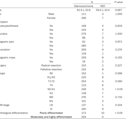
[image:5.612.92.521.69.567.2]Nine of the 10 patients (90.0%) showed TNM stage IV tumor. This percentage was higher compared with the 63.7% (240/377) gallblad-der adenocarcinoma patients that had stage IV tumors; however, the difference was not statis-tically significant. Nine of the 10 NEC patients (90.0%) had lymphatic metastases, represent-ing a higher rate than what obtained in patients with gallbladder carcinomas (73.7%, 278/377); however, the difference was not statistically significant (P > 0.05). Seven of the 10 patients
(70.0%) with NEC had N2 lymphatic metasta-ses, which was significantly higher than what observed in gallbladder carcinoma patients (34.0%, 128/377; P < 0.05) (Table 3).
Follow-up results
The minimal and longest postoperative survival times were 1.8 m and 23.3 m, respectively, with a median survival time of 3.0 m. The 1-, 2-, and 3-year cumulative survival rates were 20, 10, and 0%, respectively. For the patients with gallbladder adenocarcinoma treated during the same period, the follow-up rate was 83.8%, and median survival time of 6.0 m was obtained; the 1-, 2-, 3-, and 5-year survival rates were 38.0, 31.0, 30.1, and 28.4%, respectively. Figure 3 shows the Kaplan-Meier survival curves of the patients with different
follow-up outcomes and pathological classifica-tions. Log-rank test indicated that the differ-ence was statistically significant (P = 0.038). Furthermore, we found that survival time in the 10 patients varied considerably according to the different treatment options received. 3 patients who received postoperative radiother-apy and chemotherradiother-apy survived for 3.0, 23.3, and 12.7 m, respectively, median and mean survival times were 12.7 and 13.0 m, respec-tively; while for the remaining 7 patients that only received surgical treatments, median and mean survival times of 3 and 3.5 m were found, respectively.
Discussion
[image:6.612.102.521.85.481.2]GB-NEC is very rare in clinical practices, and only few case reports have been published to
Table 3. Comparision of clinical features between GB-NEC and gallbladder adenocarcinoma
N P value
Adenocarcinoma NEC
Age 61.9 ± 10.6 59.0 ± 10.0 0.667
Sex Male 111 3 1.000
Female 266 7
Symptom
Cholecystolithiasis No 158 5 0.633
Yes 219 4
Jaundice No 279 7 1.000
Yes 98 3
Epigastric pain No 92 3 0.973
Yes 285 7
Emaciation No 259 9 0.274
Yes 118 1
Epigastric mass No 259 8 0.155
Yes 18 2
Surgery Radical resection 153 2 0.327
Palliative resection 224 8
Margin R0 152 1 0.096
R1/R2 225 9
T T1-T3 254 4 0.090
T4 123 6
N N0-N1 249 3 < 0.05
N2 128 7
M M0 276 7 0.732
M1 101 3
TNM stage I-III 137 1 0.104
IV 240 9
Pathological differentiation Poorly differentiated 173 10 < 0.05
date. Two studies with relatively large sample sizes were from Korea, with 6 and 12 patients, respectively [8, 9]. Ten patients with GB-NEC were included in the present study, making it one of the largest in China. According to the sta-tistics of Surveillance, Epidemiology and End Result (SEER), GB-NEC accounts for 0.5% of all NEC and 2.1% of all gallbladder tumors [5]. In a study performed by Duffy et al [10], data from 435 patients with gallbladder cancers treated at the Memorial Sloan-Kettering Cancer Center (MSKCC) between 1995 and 2005 showed that 3% of them had NEC. In the present study, 10 patients were found with GB-NEC between 2007 and 2012, which accounted for 2.2% of all gallbladder cancers, which is similar to the findings published by SEER and MSKCC.
Most researchers agree that aged female patients are with higher risk of developing GB-NEC, an idea supported by the findings of the present study. Ahn et al [11] revealed that some functional GB-NEC could be found with specific presentations. However, the findings described here showed that clinical presenta-tions and signs of GB-NEC were of no specificity
As a highly malignant tumor, NEC progresses rapidly and induces early liver invasion and lymphatic metastasis. According to the patho-logical results of 41 gallbladder NEC cases (between 1973 and 2004) reported by SEER, 2.4% were highly differentiated tumors, 7.3% moderately differentiated tumors, and 89.7% poorly or undifferentiated tumors [5]. However, all the 10 gallbladder NEC cases described in the present study were poorly differentiated small cell NEC, and the level of malignancy was higher than what found for gallbladder adeno-carcinoma cases treated during the same peri-od. In addition, TNM staging showed that 90.0% of the 10 NECs were stage IV, which was not significantly different from the rates obtained for gallbladder adenocarcinoma cases treated during the same period; however, 70.0% of the gallbladder NEC cases had N2 lymphatic metastases, a rate significantly higher than that obtained in gallbladder adenocarcinoma patients treated during the same period (P < 0.05).
Surgery is the mostly used and preferred treat-ment method for GB-NEC. The surgical proce-Figure 3. Survival curves of GB-NEC and gallbladder adenocarcinoma.
as compared to gallbladder adenocarcinomas; most of them were non-functional NEC, in accordance with previous studies [12-14].
[image:7.612.91.395.69.367.2]dures vary from simple cholecystectomy to extensive radical resection (including local lymph node dissection and resection of metas-tases) [15]. In a study performed by MSKCC researchers, data from 13 patients with GB-NEC were analyzed, and a median patient survival time of 9.8 months was found, which was not significantly different from the median survival time obtained for the 435 patients with gallbladder carcinomas (10.3 months) [10]. In another study performed by Fujii et al, the 1- and 2-year survival rates of the 53 included patients with small cell gallbladder cancers were 28 and 0%, respectively [16]. In the pres-ent study, we found survival times from 1.8 m to 23.3 m (median, 3.0 m) for the 10 patients, while the 1-, 2-, and 3-year survival rates were 20, 10, and 0%, respectively; however, the median survival time of the 377 gallbladder adenocarcinoma patients treated during the same period was 6.0 m, with 1-, 2-, 3-, and 5-year survival rates of 38.0, 31.0, 30.1, and 28.4%, respectively, the difference was statisti-cally significant. These findings suggested a poorer prognosis for GB-NEC patients com-pared with patients with gallbladder adenocar-cinoma, this could be associated with the high-er phigh-ercentage of patients with advanced stage and lymphatic metastases.
Because GB-NEC cases are generally with high malignancy, and most NEC are highly invasive with high risk of lymphatic metastasis, most patients are diagnosed at late stages, which decrease the rate of radical resection. In the present study, only 2 of the 10 patients were treated with radical resection; however, adju-vant therapies including radiotherapy and che-motherapy also resulted in encouraging effects in patients with GB-NEC. Indeed, the median survival time of the 3 patients that received TACE, radiotherapy, and chemotherapy after surgical treatment was 12.7 m, while that of the remaining 7 patients solely treated with surgery was only 3.0 m; however, the difference was not statistically significant, which may be due to the small sample size. In a study per-formed by Elahi et al [17], the survival time of a patient with highly differentiated GB-NEC was 46 months after postoperative chemotherapy with combined application of gemcitabine, cis-platinum, docetaxel, and sunitinib. In addition, Okuyama et al [18] found that combined appli-cation of cis-platinum and docetaxel resulted in
patient survival time of 22 months. These find-ings and ours indicate that adjuvant therapy using radiotherapy and chemotherapy might substantially benefit the patients with GB-NEC. However, as only very limited number of patients with GB-NEC has been treated, no uni-versally accepted preferred radiotherapy or chemotherapy strategy is available to date. In conclusion, GB-NEC is a special type of gall-bladder carcinoma with low incidence rate. This disease has no specific clinical presentation, pathological and immunohistochemical exami-nations are needed for definite diagnosis. The malignancy rates of GB-NEC are generally high, and local invasion and lymphatic metastases can be found at early stage; the prognosis of GB-NEC is poorer than gallbladder adenocarci-noma. Combining surgical resection, radiother-apy, and chemotherapy could help increase patient survival. However, with a very low inci-dence and only few studies focused on this dis-ease, no universally accepted treatment is available, and further studies with larger sam-ple size are needed.
Acknowledgements
This study was supported by Shaanxi Province Natural Science Fund Project (2014JM4177).
Disclosure of conflict of interest
None.
Address correspondence to: Dr. Zhimin Geng, De-
partment of Hepatobiliary Surgery, The First Affili-ated Hospital of Xi’an Jiaotong University, 277 West Yanta Road, Xi’an 710061, P. R. China. Tel: +86-29-85323890; Fax: +86-29-85323473; E-mail: geng -zhimin@mail.xjtu.edu.cn
References
[1] Rothenstein J, Cleary SP, Pond GR, Dale D, Gallinger S, Moore MJ, Brierley J and Siu LL. Neuroendocrine tumors of the gastrointestinal tract: a decade of experience at the Princess Margaret Hospital. Am J Clin Oncol 2008; 31:
64-70.
[2] Oberg K. Diagnostic work-up of gastroentero-pancreatic neuroendocrine tumors. Clinics (Sao Paulo)2012; 67 Suppl 1: 109-112. [3] Modlin IM, Lye KD, Kidd M. A 5-decade
analy-sis of 13,715 carcinoid tumors.Cancer 2003;
[4] Modlin IM, Shapiro MD, Kidd M. An analysis of rare carcinoid tumors: clarifying these clinical conundrums. World J Surg 2005; 29: 92-101. [5] Yao JC, Hassan M, Phan A, Dagohoy C, Leary C,
Mares JE, Abdalla EK, Fleming JB, Vauthey JN, Rashid A and Evans DB. One hundred years after “carcinoid”: epidemiology of and prog-nostic factors for neuroendocrine tumors in 35,825 cases in the United States. J Clin Oncol 2008; 26: 3063-3072.
[6] Edge SB, Compton CC. The American Joint Committee on Cancer: the 7th edition of the AJCC cancer staging manual and the future of
TNM. Ann Surg Oncol 2010; 17: 1471-1474.
[7] Bosman FT, Carneiro F, Hruban RHand Theise ND. WHO classification of tumours of the
digestive system. International Agency for Research on Cancer. Lyon: IARC Press; 2010.
pp. 13-14.
[8] Kim J, Lee WJ, Lee SH, Lee KB, Ryu JK, Kim YT, Kim SW, Yoon YB, Hwang JH, Han HS, Woo SM and Park SJ. Clinical features of 20 patients with curatively resected biliary neuroendocrine tumours.Dig Liver Dis 2011; 43: 965-970.
[9] Lee JM, Hwang S, Lee SG, Lee YJ, Park KM, Kim KH, Ahn CS, Kim MH, Lee SK and Kim MW. Neuroendocrine tumors of the gallbladder: twelve cases in a single institution. Hepato-
gastroenterology 2010; 57: 1064-1068.
[10] Duffy A, Capanu M, Abou-Alfa GK, Huitzil D, Jarnagin W, Fong Y, D’Angelica M, Dematteo RP, Blumgart LH and O’Reilly EM. Gallbladder cancer (GBC): 10-year experience at Memorial Sloan-Kettering Cancer Centre (MSKCC). J
Surg Oncol 2008; 98: 485-489.
[11] Ahn JE, Byun JH, Ko MS, Park SH and Lee MG. Case report: neuroendocrine carcinoma of the gallbladder causing hyperinsulinaemic hypo-glycaemia.Clin Radiol 2007; 62: 391-394.
[12] Shimizu T, Tajiri T, Akimaru K, Arima Y, Yoshida H, Yokomuro S, Mamada Y, Taniai N, Mizuguchi Y, Kawahigashi Y and Naito Z. Combined neu-roendocrine cell carcinoma and adenocarci-noma of the gallbladder: report of a case. J Nippon Med Sch 2006; 73: 101-105.
[13] Shimono C, Suwa K, Sato M, Shirai S, Yamada K, Nakamura Y and Makuuchi M. Large cell neuroendocrine carcinoma of the gallbladder: long survival achieved by multimodal
treat-ment. Int J Clin Oncol 2009; 14: 351-355. [14] Iida Y, Tsutsumi Y. Small cell (endocrine cell)
carcinoma of the gallbladder with squamous and adenocarcinomatous components. Acta
Pathol Jpn 1992; 42: 119-125.
[15] Modlin IM, Kidd M, Latich I, Zikusoka MN and Shapiro MD. Current status of gastrointestinal carcinoids.Gastroenterology 2005; 128: 1717-1751.
[16] Fujii H, Aotake T, Horiuchi T, Chiba Y, Imamura Y and Tanaka K. Small cell carcinoma of the gallbladder: a case report and review of 53 cases in the literature. Hepatogastroenterology
2001; 48: 1588-1593.
[17] Elahi F, Ahmadzadeh A, Yadollahzadeh M, Hassanpour K and Babaei M. Neuroendocrine tumor of the gallbladder.Arch Iran Med 2013; 16: 123-125.