Original Article
The role of miR-222 and
miR-298 in breast cancer drug resistance
Jinyin Yan*, Yang Zhang*, Lu Wang, Hao Dai, Ning Li, Wanning Hu, Haifeng Cai
Department of Surgical Oncology, Tangshan People’s Hospital, Tangshan, China. *Equal contributors. Received July 15, 2016; Accepted August 30, 2016; Epub October 1, 2016; Published October 15, 2016
Abstract: Docetaxel is widely applied in the clinic for the treatment of breast cancer. However, drug resistance might appear for the long-term usage. It was considered that breast cancer drug resistance was related to the aberrant expressions of some miRNAs. This study explored the role of miR-222 and miR-298 in the development of docetaxel resistance in the treatment of breast cancer. A total of 45 cases of breast cancer patients with drug resistance were enrolled in this study. RT-PCR and in situ molecular hybridization were used to determine the expression of miR-222 and miR-298 in breast cancer tissues. Mimic and inhibitor transfections were performed on breast cancer cell line MCF-7 to manipulate the expressions of miR-222 and miR-298. MTT assay was adopted to evaluate MCF-7 cell drug resistance. Western blot was performed to analyze the expression of PTEN and MDR1. Our results showed that
miR-222 expression was significantly increased in breast cancer tissue with drug resistance, while miR-298 was obvi -ously downregulated (P<0.05). Drug susceptibility test showed that miR-222 overexpression or miR-298 inhibition markedly enhanced drug resistance. In addition, miR-222 overexpression reduced PTEN level, whereas miR-298 inhibition elevated MDR1 expression. In conclusion, miR-222 was upregulated while miR-298 was declined in drug resistant breast cancer cells and they may regulate the development of drug resistance through targeting PTEN and MDR1.
Keywords: Breast cancer, miR-222, miR-298, PTEN, MDR1
Introduction
The incidence of breast cancer occupies the first among female malignant tumors. There are about 230,000 females who are diagnosed as breast cancer in America, accounting for 29% of all female malignant tumors [1]. At pres-ent, breast cancer is mainly treated by surgery, radiotherapy, chemotherapy, and endocrine treatment methods, of which chemotherapy is still an important approach [2]. Various com-monly used chemotherapy drugs may induce drug resistance, including docetaxel and adria-mycin, etc, thereby significantly reduces the curative effect of breast cancer treatment and increase the burden of patients [3, 4]. Thus, further in depth study on the mechanism of breast cancer drug resistance is of great signifi-cance to improve the effect of breast signifi-cancer treatment as well as the life quality of patients. MiR-222 and miR-298 are small non-coding RNA molecules belonging to the miRNA family [5, 6]. It is reported that miRNAs can affect
vari-ous cell behaviors [6]. MiRNA expression profile analysis revealed that miRNAs play an impor-tant role in the regulation of malignant tumor cell behaviors [7]. Recently, it has been shown that miR-222 and miR-298 are associated with drug resistance in a large variety of malignant tumors, such as colorectal cancer, breast can-cer, and liver cancer [8-10]. This study intends to investigate the role and related mechanism of miR-222 and miR-298 in the development of drug resistance in breast cancer cells.
Materials and methods
Subject selection
axel resistance after chemotherapy. Tissue samples were obtained from surgery before and after drug resistance and stored in liquid nitrogen for miRNA detection and paraffin em- bedding for histopathologic analysis as well as for in situ molecular hybridization. This study was approved by the ethics committee and obtained informed consents from all patients.
RT-PCR
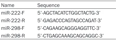
RT-PCR was performed to measure the expres-sion of miR-222 and miR-298 in breast can- cer and hyperplasia of mammary glands. The primers used were designed according to their sequences (GeneBank: AJ550426 and NR_030580) (Table 1). Total RNA was extract-ed from tissues using RNAprep pure Tissue Kit (QIAGEN). RT-PCR test was applied using mirVanat qRT-PCR miRNA detection kit (Am- bion). U6 was used as an internal refer- ence and the results were analyzed by 2-ΔΔCt
method [11].
In situ hybridization detection
Antisense locked nucleic acid modified miR-222 and miR-298 oligonucleotide probes were used for analysis of in situ molecular hybridization (Boster) using paraffin tissue section. The sequences used were as follows: miR-222, 5’-AGCTACATC-TGGCTACTGGGTCTC- 3’; miR-298, 5’-TGCTGCTTT-GCTCAGGAGTG-3’. Specially, paraffin section was routinely dew- axed and blocked. After washed by 0.5 M PBS, the section was treated by pre-hybrid liquid at 65°C for 4 h followed by addition of probe and incubated at 65°C for 15 h. Then, the section was washed by SSC solution to block nonspecific binding site. After treated with rabbit anti digoxin antibody at 37°C for 0.5 h, the section was developed and sealed for observation.
Results judgment: Violet particles in the cells indicate positive expression of miR-222 and miR-298 [12].
MiR-222 and miR-298 transfection
Mimic and inhibitor were adapted to upreg- ulate or downregulate miR-222 and miR-298 expression in breast cancer cells. The mimic and inhibitor were purchased from Geneph- arma and transfected using INTERFERinTM transfection kit (Polyplus transfection). Breast cancer cell line MCF-7 was purchased from the cell bank of Chinese academy of scie- nces. The cells were maintained in DMEM supplemented with 10% FBS, 5000 U penicil-lin, and 5000 g/ml streptomycin (HyClone) at 37°C and 5% CO2. The cells were seeded into 96-well plate for 24 h before transfection according to the manufacturer’s instructions.
Drug resistance assay
MCF-7 cells were seeded into 96-well plates and incubated overnight at 37°C. The cells were incubated with different concentrations of docetaxel for 48 h at 37°C. After addition of MTT to each well for 4 h at 37°C, 150 μl DMSO was added into each well. Absorbance of each well at 550 nm (A550) was read us- ing a spectrophotometer. The concentration of each drug producing 50% inhibition of gro- wth (IC50) was estimated from the relative survival curves [13].
Western blot
Proteins were extracted from MCF-7 cells in logarithmic phase, separated by SDS-PAGE and transferred to PVDF membrane. After blo- cked by 5% skim milk, the membrane was incubated with anti-PTEN, and MDR1 anti- bodies. β-actin was selected as an internal reference. The membrane was detected by chemiluminiscence and the image was ana-lyzed by using Image J.
Statistical analysis
All the data analysis was performed on SPSS 20.0 software. The data were represented as mean ± standard deviation (SD). T test was applied for data comparison. P<0.05 was con-sidered as statistical significance.
Results
RT-PCR
[image:2.612.91.288.83.152.2]RT-PCR was applied to test 222 and miR-298 expression in breast cancer tissues be- Table 1. Primers for RT-PCR
Name Sequence
IC50 of MCF-7 after miR-222 overexpression achieved 1.61 μM, which was obviously hig- her than that of 1.32 μM in negative control (P<0.05), while it was 1.27 μM in miR-222 inhibitor group. After miR-298 inhibition, IC50 for docetaxel in MCF-7 cells was 1.83 μM, which showed statistical difference to neg- ative control which was 1.31 μM (P<0.05). It was only 1.25 μM in miR-298 mimic group. Thus, we speculated that miR-222 overex- pression or miR-298 inhibition can increase MCF-7 cell proliferation in docetaxel, leading to drug resistance.
The effect of miR-222 overexpression on PTEN in breast cancer cells
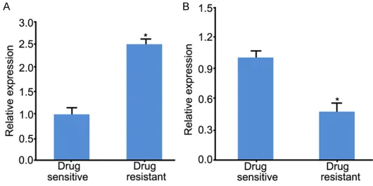
Western blot was used to determine PTEN expression in breast cancer cells after miR-222 fore and after drug resistance. As shown in
Figure 1, miR-222 expression was signifi- cantly upregulated (around 2.5-fold change) (P<0.05), whereas miR-298 expression was reduced (around 2.1-fold change) in drug res- istant group compared with drug sensitive group (P<0.05).
In situ hybridization detection
[image:3.612.91.356.73.205.2]MiR-222 and miR-298 specific probes were used to detect miRNAs expression in breast cancer tissues by in situ molecular hybridiza-tion. Blue granules appeared in the cytoplasm when positive expression of 222 or miR-298 was observed. It was found that miR-222 expression in the drug resistant breast cancer tissues was obviously higher than that in the
[image:3.612.91.359.276.462.2]Figure 1. Expression of miR-222 and miR-298 in drug sensitive or resis-tance tissues. A: 222 expression in breast cancer tissue. B: miR-298 expression in breast cancer tissue. *P<0.05, compared with drug sensitive group.
Figure 2. Analysis of the expression of miR-222 and miR-298 in drug sensitive or resistant tissues by in situ hybridization (×400).
drug sensitive group. On the con-trary, miR-298 presented lower staining level in drug resistant group compared with drug sensi-tive group (Figure 2). It was dem-onstrated that miR-222 expres-sion was upregulated while miR-298 was reduced in drug resistant breast cancer tissue, which was consistent with the results of RT-PCR.
miR-222 and miR-298 transfec-tion
Mimic and inhibitor were adapted to manipulate the expression of miR-222 or miR-298 in MCF-7 cells. RT-PCR was applied to test miRNA relative expression. As shown in Figure 3, miR-222 expression was markedly incre- ased, while miR-298 was obvio- usly declined in MCF-7 cells after transfection (P<0.05), indicating successful manipulation of the expression of miRNA by mimic and inhibitor.
Drug resistance changes
PTEN protein level in MCF-7 cells compared with blank control or negative control (P<0.05).
The effect of miR-298 inhibition on MDR1 in breast cancer cells
Western blot was adopted to detect MDR1 expression in breast cancer cells after miR-298 expression was inhibited. It was demon-strated that MDR1 expression was obviously upregulated after miR-298 inhibitor transfec-tion compared with control (P<0.05) (Figure 6). Discussion
At present, numerous studies have confirmed that there are many miRNA expression differ-ences between breast cancer cells and nor- mal cells, including miR-21, miR-10b, and miR-200 [14-16]. miRNA-mediated aberrant cell proliferation and differentiation has been mimic transfection. As shown in Figure 5,
[image:4.612.93.521.75.257.2]miR-222 mimic transfection apparently reduced
[image:4.612.91.526.316.435.2]Figure 3. Expression of miR-222 and miR-298 after mimic or inhibitor transfection in MCF-7 cells. A: miR-222 ex-pression in MCF-7. B: miR-298 exex-pression in MCF-7. *P<0.05, compared with drug sensitive group.
Figure 4. Cell proliferation after transfection.
[image:4.612.98.275.480.645.2]express miR-222 or inhibit miR-298 in drug sensitive breast cancer cell line MCF-7. Our results showed that MCF-7 proliferation was enhanced in docetaxel after transfection, suggesting increased drug resistance. Mean- while, western blot analysis demonstrated that miR-222 upregulation declined PTEN expression, whereas miR-298 reduction elev- ated MDR1 level, indicating that miR-222 and miR-298 may regulate cell proliferation and drug resistance through mediating the expr- ession of PTEN and MDR1.
The role of tumor suppressor gene PTEN exerts its function through PI3K/Akt signal- ing pathway. It can inhibit PI3K activation and maintain the activity of Akt, leading to the regulation of normal cell biological behaviors under physiological condition [19]. It was found that miR-222 can regulate cell prolife- ration and division by targeting PTEN [20]. Mardente et al. reported that miR-222 overex-pression can enhance cell proliferation and invasion in thyroid cancer cells. It was further found that such process was completely impaired after inhibiting cell cycle regulatory protein PTEN [21]. Bao et al. found that doce- taxel resistant breast cancer cell line MDA-MB-231 presented drug resistance through overexpressing MDR1/p-gp. MDR1 elevation may prevent the entry of docetaxel to nucl- eus, which is the mechanism of MDA-MB-231 cell line drug resistance [22]. Our study demonstrated that miR-298 can target MDR1 gene and inhibiting miR-298 expression can promote MDR1 expression, leading to the occurrence of docetaxel resistance.
Breast cancer is the leading malignant tumor in female that seriously affects the life quality of patients [1]. Docetaxel is a commonly used drug for the treatment of breast cancer in clinic. The presence of drug tolerance has a strong impact on the curative effect of breast cancer treatment. This study found that 222 expression was upregulated, while miR-298 was reduced in drug resistant breast cancer cell. Cell transfection, drug resistance assay, and Western Blot experiments deter-mined a close relationship between miR-222 and miR-298 expression with breast cancer drug resistance, which can be used in the future for drug resistance judgment in clinic, thus providing better treatment strategies and observed in breast cancer cells. For example,
miR-10b participates in the process of breast cancer cell invasion and metastasis [15]. MiR-21 also plays an important role in breast can-cer cell proliferation [14]. It was also reported that some miRNAs expression was closely related to the drug resistance of breast can- cer cells [8]. This study explored the role of miR-222 and miR-298 in the development of drug resistance in breast cancer cells. RT-PCR and in situ molecular hybridization detection revealed that miR-222 expression was upregu-lated, while miR-298 was declined in drug resistant breast cancer tissues compared with drug sensitive tissues.
It is still controversial on how miRNAs are dif-ferentially expressed in malignant tumor cells. Some scholars believe that cell growth factor signaling pathway, such as EGF and FGF act- ivation can promote malignant tumor cell divi-sion, while this process is achieved by sup-pressing a part of miRNAs [17]. Some other scholars consider that miRNA expression changes in malignant tumor cells were cau- sed by the irreversible modification of transc- riptional regulatory sequences. It was found that methylation reagent 5’-mixed nitrogen-2’-deoxidization cytidine may upregulate miR-NAs expression in glioma cells. MiRNA tran-scription regulatory sequences analysis has also confirmed this conclusion [18].
In order to further explore the impact of miR-222 and miR-298 on breast cancer drug resis-tance, we applied transfection method to
[image:5.612.97.285.71.227.2]mycin treatment through inhibition of Raf-1. Oncol Rep 2013; 30: 877-889.
[5] Liu X, Xiao J, Zhu H, Wei X, Platt C, Damilano F, Xiao C, Bezzerides V, Bostrom P, Che L, Zhang C, Spiegelman BM, Rosenzweig A. miR-222 is necessary for exercise-induced cardiac growth and protects against pathological cardiac re-modeling. Cell Metab 2015; 21: 584-595. [6] Zhao H, Zhao D, Tan G, Liu Y, Zhuang L, Liu T.
Bufalin promotes apoptosis of gastric cancer by down-regulation of miR-298 targeting bax. Int J Clin Exp Med 2015; 8: 3420-3428. [7] Nikitina EG, Urazova LN, Stegny VN.
MicroR-NAs and human cancer. Exp Oncol 2012; 34: 2-8.
[8] Robertson NM, Yigit MV. The role of microRNA in resistance to breast cancer therapy. Wiley Interdiscip Rev RNA 2014; 5: 823-833. [9] Xu K, Liang X, Shen K, Sun L, Cui D, Zhao Y,
Tian J, Ni L, Liu J. MiR-222 modulates multi-drug resistance in human colorectal carcino-ma by down-regulating ADAM-17. Exp Cell Res 2012; 318: 2168-2177.
[10] Liu K, Liu S, Zhang W, Ji B, Wang Y, Liu Y. miR222 regulates sorafenib resistance and enhance tumorigenicity in hepatocellular carci-noma. Int J Oncol 2014; 45: 1537-1546. [11] Yan J, Zhang N, Qi C, Liu X, Shangguan D.
One-step real time RT-PCR for detection of microR-NAs. Talanta 2013; 110: 190-195.
[12] Yin H, Zhou Y, Zhang H, Meng X, Ai S. Electro-chemical determination of microRNA-21 based on graphene, LNA integrated molecular bea-con, AuNPs and biotin multifunctional bio bar codes and enzymatic assay system. Biosens Bioelectron 2012; 33: 247-253.
[13] Zhong S, Li W, Chen Z, Xu J, Zhao J. MiR-222 and miR-29a contribute to the drug-resistance of breast cancer cells. Gene 2013; 531: 8-14. [14] Escobar-Hoyos LF, Shah R, Roa-Pena L, Vanner
EA, Najafian N, Banach A, Nielsen E, Al-Khalil
R, Akalin A, Talmage D, Shroyer KR. Keratin-17 Promotes p27KIP1 Nuclear Export and Degra-dation and Offers Potential Prognostic Utility. Cancer Res 2015; 75: 3650-3662.
[15] Kato J, Zhu J, Liu C, Stylianou M, Hoffmann V, Lizak MJ, Glasgow CG, Moss J. ADP-ribosylargi-nine hydrolase regulates cell proliferation and tumorigenesis. Cancer Res 2011; 71: 5327-5335.
[16] Li X, Roslan S, Johnstone CN, Wright JA, Brack-en CP, Anderson M, Bert AG, Selth LA, Ander-son RL, Goodall GJ, Gregory PA, Khew-Goodall Y. MiR-200 can repress breast cancer metas-tasis through ZEB1-independent but moesin-dependent pathways. Oncogene 2014; 33: 4077-4088.
[17] Santos MC, Tegge AN, Correa BR, Mahesula S, Kohnke LQ, Qiao M, Ferreira MA, Kokovay E, improving the curative effects. It can also
pro-vide certain theoretical basis for the applica-tion of RNA interference technology to reverse the drug resistance in the treatment of breast cancer in the future.
In conclusion, elevated miR-222 expression or reduced miR-298 expression may lead to docetaxel resistance in breast cancer cells which might be through mediating the expr- ession of PTEN and MDR1.
Acknowledgements
Research supported by the Tangshan City Science and Technology Plan Item (NO.11- 1302138a).
Disclosure of conflict of interest
None.
Address correspondence to: Dr. Haifeng Cai, De- partment of Surgical Oncology, Tangshan People’s Hospital, 65 Shengli Road, Tangshan 063000, China. Tel: 2877120; Fax: +86-315-2877120; E-mail: haifengcaizxc@sina.com
References
[1] Lyman GH, Temin S, Edge SB, Newman LA, Turner RR, Weaver DL, Benson AB 3rd, Bosser-man LD, Burstein HJ, Cody H 3rd, HayBosser-man J, Perkins CL, Podoloff DA, Giuliano AE; American Society of Clinical Oncology Clinical Practice. Sentinel lymph node biopsy for patients with early-stage breast cancer: American Society of Clinical Oncology clinical practice guideline up-date. J Clin Oncol 2014; 32: 1365-1383. [2] Khatcheressian JL, Hurley P, Bantug E,
Esser-man LJ, Grunfeld E, Halberg F, Hantel A, Henry NL, Muss HB, Smith TJ, Vogel VG, Wolff AC,
Somerfield MR, Davidson NE; American Soci -ety of Clinical Oncology. Breast cancer follow-up and management after primary treatment: American Society of Clinical Oncology clinical practice guideline update. J Clin Oncol 2013; 31: 961-965.
[3] Swain SM, Baselga J, Kim SB, Ro J, Semiglazov V, Campone M, Ciruelos E, Ferrero JM, Schnee-weiss A, Heeson S, Clark E, Ross G, Benyunes MC, Cortés J; CLEOPATRA Study Group. Pertu-zumab, trastuPertu-zumab, and docetaxel in HER2-positive metastatic breast cancer. N Engl J Med 2015; 372: 724-734.
Adria-Penalva LO. miR-124, -128, and -137 Orches-trate Neural Differentiation by Acting on Over-lapping Gene Sets Containing a Highly Con-nected Transcription Factor Network. Stem Cells 2016; 34: 220-232.
[18] Chakrabarti M, Ray SK. Direct transfection of miR-137 mimics is more effective than DNA demethylation of miR-137 promoter to aug-ment anti-tumor mechanisms of delphinidin in human glioblastoma U87MG and LN18 cells. Gene 2015; 573: 141-152.
[19] Ferraldeschi R, Nava Rodrigues D, Riisnaes R, Miranda S, Figueiredo I, Rescigno P, Ravi P, Pe-zaro C, Omlin A, Lorente D, Zafeiriou Z, Mateo J, Altavilla A, Sideris S, Bianchini D, Grist E, Thway K, Perez Lopez R, Tunariu N, Parker C, Dearnaley D, Reid A, Attard G, de Bono J. PTEN protein loss and clinical outcome from castra-tion-resistant prostate cancer treated with abi-raterone acetate. Eur Urol 2015; 67: 795-802.
[20] Li W, Guo F, Wang P, Hong S, Zhang C. miR-221/222 confers radioresistance in glioblas-toma cells through activating Akt independent of PTEN status. Curr Mol Med 2014; 14: 185-195.
[21] Mardente S, Mari E, Consorti F, Di Gioia C, Ne-gri R, Etna M, Zicari A, Antonaci A. HMGB1 in-duces the overexpression of 222 and miR-221 and increases growth and motility in papillary thyroid cancer cells. Oncol Rep 2012; 28: 2285-2289.