Original Article
Serum miRNA-21, miRNA-153 and miRNA-22
levels were identified as diagnostic biomarkers
of patients with hepatocellular carcinoma:
a Chinese population-based study
Hongchen Zhang1*, Yanwei Yang2*, Mingren Ma3, Yinong Qiu2, Jianwei Qin4
1Department of Clinical Nursing, School of Nursing, Fourth Military Medical University, Xi’an, Shaanxi, China;
Departments of 2Stomatology, 4Hepatobiliary Surgery, Lanzhou General Hospital, Lanzhou Military Area
Command, PLA, Lanzhou, Gansu, China; 3Experimental Center of Medicine, Lanzhou General Hospital, Lanzhou
Military Area Command, PLA & Key Lab of Stem Cells and Gene Drugs of Gansu Province, Lanzhou, Gansu, China.
*Equal contributors.
Received December 4, 2016; Accepted February 14, 2017; Epub March 1, 2017; Published March 15, 2017
Abstract: Background: Hepatocellular carcinoma (HCC) is the most common primary malignant tumor worldwide. The aim of this study is to identify the diagnostic values of serum miRNA-21, miRNA-22 and miRNA-153 of patients with HCC in a large Asian group. Methods: We retrospectively reviewed 65 patients with HCC and 65 patients with hepatic cirrhosis and HBV, respectively. The expression of serum miRNA-21, miRNA-22 and miRNA-153 was de-tected by real-time PCR. The comparison of diagnostic performance of serum miRNAs and AFP levels was measured using the area under ROC curve (AUC). Results: In the present study, we compared expression levels of miRNA-21, miRNA-153 and miRNA-22 in plasma among HCC, cirrhotic and the HBV groups. We found expression of serum
miRNA-21, miRNA-153 and miRNA-22 were significantly higher in patients with HCC compared with cirrhotic and HBV groups. Secondly, we found serum miRNAs had better diagnostic significance for patients compared with AFP
levels in all patients. Conclusions: In this study, we found expression of serum miRNA-21, miRNA-153 and miRNA-22
were significantly higher in patients with HCC compared with cirrhotic and HBV groups, and we confirmed the serum miRNA-21, miRNA-153 and miRNA-22 could be defined as novel diagnostic biomarkers for patients with HCC.
Keywords: miRNA-21, miRNA-153, miRNA-22, HCC
Introduction
Hepatocellular carcinoma (HCC) is the fifth most frequently diagnosed cancer worldwide and the second most frequent cause of cancer death [1], with the highest incidence in Asian and especially in China [2]. Partial hepatectomy remains the most commonly used curative therapy modality for HCC [3]. Although the prog-nosis of patients with HCC has been improved recently, the survival outcomes of patients with HCC following surgical resection may vary, as several factors are associated with the progno-sis of HCC, including completeness of tumor removal, serum alpha-fetoprotein (AFP) levels, tumor size, tumor multifocality and distant metastases, etc [4, 5]. The poor prognosis of
patients with HCC is attributed to the lack of an effective means of early diagnosis. Only 30% to 40% of patients are candidates for potentially curative hepatectomy at the time of diagnosis [6]. Discovery of an effective and reliable tool for early diagnosis of HCC would play a pivotal role in improving the prognosis of patients with HCC.
immu-nity, inflammation, and tumorigenesis [8, 9]. MiRNAs are ideal candidates for biomarkers because of their resistance to endogenous RNase and high stability under different stor-age conditions. Recent studies have shown that human serum miRNAs are aberrantly expressed in many malignancies such as liver [10, 11], colorectal cancer [12], and pancreatic cancer [13].
Several studies had demonstrated that expres-sion of microRNA-21 (miRNA-21) was
[image:2.612.91.331.83.545.2]signifi-definitive therapy. The tumor type and the grade of cell differentiation were diagnosed based on the criteria of World Health Organization (WHO), whereas the pathological stage of each tumor was determined by the International Union Against Cancer (UICC) TNM classification. Blood samples were also collected from 65 patients with HBV infection and matching ages and gen-ders to the patients group with HCC and cirrho-sis. Written consents were obtained from all subjects prior to the recruitment. The study protocol was approved by the Institutional
Table 1. Patient and tumor characteristics (N=195)
Variable HBV group Cirrhosis group HCC group
Case, n 65 65 65
Age 61.5±4.6 61.0±8.6 62.3±6.1 Sex
Female 22 25 23 Male 43 40 42 HBsAg
Positive 65 50 48 Negative 0 15 17 HBeAg
Positive 34 42 45 Negative 31 23 20 Liver cirrhosis
Yes 0 65 53
No 65 0 12
TBL (µmol/l) 12.5±8.3 15.1±7.3 16.1±8.2 ALB (g/dl) 39.4±6.6 38.9±6.5 37.9±4.6 ALT (U/L) 25.7±14.1 50.4±30.2 79.4±66.5 AFP at diagnosis (ng/ml)
≤ 400 65 42 40
> 400 0 23 25 Tumor size (cm)
> 5 cm _ _ 28
≤ 5 cm _ _ 37
Microvascular invasion
Yes _ _ 20
No _ _ 45
TNM staging
I _ _ 10
II _ _ 17
III-IV _ _ 38 Metastases
Yes _ _ 21
No _ _ 44
Abbreviations: TBL: total bilirubin; ALB: albumin; ALT: alanine amino-transferase; AFP: alpha-fetoprotein; HBV: Hepatitis B Virus.
cantly different in many human cancers compared with the healthy people, and miRNA-21 level was identified as a promising biochemical marker [14-16]. Similarly, miRNA-22 had been found to be ubiquitously expressed in various tis-sues [17], and previous studies suggest-ed that miRNA-22 functionsuggest-ed in multiple cellular processes such as proliferation, differentiation, apoptosis, senescence, and its deregulation is a hallmark of cancer [18-20]. miRNA-153 was first discovered as one of the several brain-specific miRNAs, based on analysis of expression profile of over one hundred miRNAs in adult organs [21]. Recent evi-dences have indicated that miRNA-153 was dramatically down-regulated in sev-eral cancer cells [22, 23].
However, few studies focused on the diagnostic significance of serum miRNA-21, miRNA-22 and miRNA-153 in patients with HCC. In this study, we aimed to explore the diagnostic values of serum miRNA-21, miRNA-22 and miRNA-153 in patients with HCC com-pared with HBV and cirrhotic patients. These results of our study shed new light on the identification of new diag-nostic and progdiag-nostic biomarkers for HCC patients.
Materials and methods
Patients
Review Board of Hospital Ethics Committee. The clinical characteristics of the subjects are listed in Table 1.
Total RNA isolation
Total RNA was isolated from 300 μl of serum using the mirVana PARIS Kit (Ambion, Austin, TX, USA) according to the manufacturer’s instructions. Briefly, for each sample, total RNA was extracted from 300 μl of serum with 2× denaturing solution, acid-phenol: chloroform, and 100% ethanol. After several washing and centrifugation, the RNA was eluted into 60 μl of preheated (95°C) elution solution. RNA quantity and purity were determined using a Nanodrop Spectrophotometer ND-1000 (Thermo Scientific, Waltham, MA, USA). RNA purity was considered satisfactory with A260/ A280 of 1.9-2.1. The RNA samples were stored at -80°C until reverse transcription.
Real-time quantitative PCR
We typically extracted 2 μg to 9 μg of total RNA, and OD260/280 ratios typically ranged from 1.8 to 2.0, indicating high RNA purity. 10 ng of total RNA was used for each miRNA quantifica-tion. miRNA detection was performed run on the Eppendorf Mastercycler EP Gradient S (Eppendorf, Germany) using commercial assays (TaqMan microRNA assays; Applied Biosystems, Foster City, CA, USA) for miRNAs. Relative quantification was calculated using 2-ΔΔCt, where Ct is cycle threshold. Normali-
zation was performed with universal small nuclear RNA U6 (RNU6B). Each sample was examined in triplicate, and the mean values were calculated. mRNA levels in tumor sam-ples/nontumorous samples of 0.5-fold was defined as under-expression of the gene, whereas a ratio of 2.0-fold was defined as over-expression.
Diagnosis and treatment
After a detailed history and a complete physical examination, the hepatitis B and C serology, liver function test and tumor markers examina-tion which included alpha-fetoprotein (AFP), carbohydrate antigen 19-9 (CA19-9), and carci-noembryonic antigen (CEA) was routinely per-formed. Other routine investigations were chest X-ray, upper gastrointestinal endoscopy,
abdominal ultrasound, contrast-enhanced com- puterized tomography (CT) and/or magnetic resonance imaging (MRI). A clinical diagnosis of HCC was based on the criteria of the American Association for the Study of Liver Diseases (AASLD) [24].
The type of partial hepatectomy carried out was based on the tumor size, number, location, presence/absence of cirrhosis and estimated volume of future liver remnant. As far as possi-ble, anatomical liver resection was carried out basing on Couinaud’s liver segments, sectors and hemilivers.
Histopathological study of the resected speci-mens was carried out independently by three pathologists who came to a consensus by dis-cussion if there was any discrepancy.
Statistical analysis
Continuous variables were expressed as mean ± SD (standard deviation) and compared using a two-tailed unpaired Student’s t test; categori-cal variables were compared using χ2 or Fisher analysis. The predictive performance of serum miRNAs were measured using the area under ROC curve (AUC). AUCs were also used to com-pare serum miRNAs and AFP level using the Hanleyand McNeil method [25]. Statistical analyses were conducted with the SPSS for Windows version 18.0 release (SPSS, Inc., Chicago, IL) and ROC curve analysis were computed using MedCalcV.11.0.3.0 (MedCalc software, Mariakerke, Belgium). A value of P < 0.05 was considered significant in all the analysis.
Results
Characteristics of the patients
The characteristics of patients with HCC, cir-rhotic and HBV groups enrolled in this study were shown in Table 1.
Comparing expression levels of serum miR-NA-21, miRNA-153 and miRNA-22 among three groups
1B). Expression of serum miRNA-153 was sig-nificantly higher in patients with HCC com- pared with cirrhotic and HBV groups (P < 0.001,
Figure 1C, 1D). Expression of serum miRNA-22
was significantly higher in patients with HCC compared with cirrhotic and HBV groups (P < 0.001, Figure 1E, 1F).
Comparison of diagnostic significance for pa-tients with HCC between serum miRNAs and AFP levels among different groups
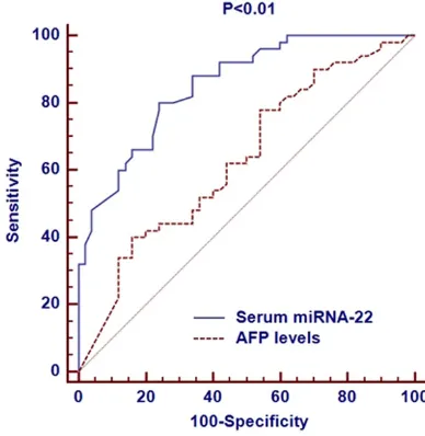
[image:5.612.324.518.147.346.2]Among the three groups, we performed ROC curves to compare the diagnosis values of dif-ferent miRNAs with AFP levels. Serum miRNA-21 was significantly more accurate in diagnos-ing HCC than the AFP level. The AUC of serum miRNA-21 was 0.862 (95% CI: 0.775 to 0.896), which was larger than that of AFP level (0.654, 95% CI: 0.587 to 0.736, Figure 2); Serum miRNA-153 was significantly more accurate in diagnosing HCC than the AFP level. The AUC of serum miRNA-153 was 0.802 (95% CI: 0.711 to 0.875), which was larger that of AFP level (0.654, 95% CI: 0.587 to 0.736, Figure 3); Serum miRNA-22 was significantly more accu-rate in diagnosing HCC than the AFP level. The AUC of serum miRNA-22 was 0.832 (95% CI: 0.726 to 0.903), which was larger than that of AFP level (0.654, 95% CI: 0.587 to 0.736,
Figure 4).
Figure 1. Serum miRNA-21, miRNA-153 and miRNA-22 were significantly higher in HCC patients. A, B. Serum miR
-NA-21 levels were significantly higher in patients with HCC compared with cirrhotic and HBV groups (P < 0.001); C, D.
Serum miRNA-153 levels in were significantly higher in patients with HCC compared with cirrhotic and HBV groups
(P < 0.001); E, F. Serum miRNA-22 levels in were significantly higher in patients with HCC compared with cirrhotic
[image:5.612.93.282.148.342.2]and HBV groups (P < 0.001).
Figure 2. Comparison of diagnostic significance for
patients with HCC between serum miRNA-21 and AFP levels.
Figure 3. Comparison of diagnostic significance for
[image:5.612.91.282.411.605.2]patients with HCC between serum miRNA-153 and AFP levels.
Figure 4. Comparison of diagnostic significance for
Discussion
The outcomes for patients with HCC have improved markedly over the last 30 years due to the presence of various therapeutic modali-ties and advances in surgical treatment [26]. Many Asian studies, however, included a high proportion of patients with advanced disease, including those categorized as TNM III or IV, and patients with primary refractory disease and metastatic disease [27]. Early diagnosis could be “a sense of urgency” for improving the prog-nosis and reducing the burden of patients with HCC.
Many studies showed that miRNAs could be used as diagnostic indicator and prognostic factor in various cancers. Increasing evidence suggested that unique serum miRNAs expres-sion signatures might serve as new noninvasive biomarkers for cancer diagnosis including HCC [28]. Previous studies showed miRNA-21 increased in several types of cancers, such as breast cancer, colon cancer and lung cancer [29-31]. Hu et al found that miRNA-21 were expressed at higher levels in the laryngeal squamous cell carcinoma samples compared to the normal samples; furthermore, they indi-cated that patients with high miRNA-21 expres-sion in tumor tissues had poorer prognosis compared to patients with lower miRNA-21 expression [32]. With respect to miRNA-153, mechanistic investigations indicated that miRNA-153 promoted invasiveness indirectly by inducing matrix metalloprotease enzyme 9 (MMP9) production and miRNA-153 played an important role in promoting proliferation of human prostate cancer cells and presents a novel mechanism of miRNA-mediated direct suppression of PTEN expression in prostate cancer [33]. While miRNA-22 was also an important factor associated with cancer patho-genesis, evolutionary clustering suggested that miRNA-22 was highly conserved in vertebrate evolution, indicating its functional importance in vertebrate species. It had been deduced from the statistical analysis of 3’-UTR in tran-scriptome that miRNA-22 is involved in the regulation of many target genes [34]. Mean- while, miRNA-22 was found to be associated with both diagnosis and prognosis of various cancers [35]. In the present study, we chose to evaluate the diagnostic significance of three distinctly differentially expressed miRNA-21,
miRNA-153 and miRNA-22. Firstly, we com-pared expression levels of 21, miRNA-153 and miRNA-22 in plasma among HCC, cir-rhotic and the HBV groups. We found expres-sion of serum miRNA-21, miRNA-153 and miRNA-22 were significantly higher in patients with HCC compared with cirrhotic and HBV groups. Secondly, we found serum miRNAs had better diagnostic significance for patients compared with AFP levels in all patients. This study proposed new direction of diagnostic biomarker research in patients with HCC in the future.
However, there are limitations of this study: (1) the sample size is too small in this study, and further larger sample study is needed to confirm the present experimental results. (2) whether the three miRNAs have the optimal specificity and sensitivity for liver cancer diag-nosis also needs future confirmation.
In conclusion, we found expression of serum miRNA-21, miRNA-153 and miRNA-22 were significantly higher in patients with HCC com-pared with cirrhotic and HBV groups, and we confirmed the serum miRNA-21, miRNA-153 and miRNA-22 could be defined as novel diag-nostic biomarkers for patients with HCC.
Acknowledgements
This work was supported by Medical Scientific Research Project of Lanzhou Military Area Command of PLA (CLZ13JB13).
Disclosure of conflict of interest
None.
Address correspondence to: Dr. Yinong Qiu, De- partment of Stomatology, Lanzhou General Hos- pital, Lanzhou Military Area Command, PLA, 333 South Riverside Road, Lanzhou 730050, Gansu, China. E-mail: qiuyn77@126.com; Dr. Jianwei Qin, Department of Hepatobiliary Surgery, Lanzhou General Hospital, Lanzhou Military Area Command, PLA, 333 South Riverside Road, Lanzhou 730050, Gansu, China. E-mail: qinjw77@126.com
References
[2] Ferlay J, Shin HR, Bray F, Forman D, Mathers C and Parkin DM. Estimates of worldwide burden of cancer in 2008: GLOBOCAN 2008. Int J Cancer 2010; 127: 2893-2917.
[3] Arii S, Yamaoka Y, Futagawa S, Inoue K, Kobayashi K, Kojiro M, Makuuchi M, Nakamura Y, Okita K and Yamada R. Results of surgical and nonsurgical treatment for small-sized he-patocellular carcinomas: a retrospective and nationwide survey in Japan. The Liver Cancer Study Group of Japan. Hepatology 2000; 32: 1224-1229.
[4] Tateishi R, Yoshida H, Matsuyama Y, Mine N, Kondo Y and Omata M. Diagnostic accuracy of tumor markers for hepatocellular carcino-ma: a systematic review. Hepatol Int 2008; 2: 17-30.
[5] Nathan H, Schulick RD, Choti MA and Pawlik TM. Predictors of survival after resection of early hepatocellular carcinoma. Ann Surg 2009; 249: 799-805.
[6] Llovet JM, Di Bisceglie AM, Bruix J, Kramer BS, Lencioni R, Zhu AX, Sherman M, Schwartz M, Lotze M, Talwalkar J and Gores GJ. Design and endpoints of clinical trials in hepatocellular carcinoma. J Natl Cancer Inst 2008; 100: 698-711.
[7] Bartel DP. MicroRNAs: genomics, biogenesis, mechanism, and function. Cell 2004; 116: 281-297.
[8] Gao F, Sun M, Gong Y, Wang H, Wang Y and Hou H. MicroRNA-195a-3p inhibits angiogene-sis by targeting Mmp2 in murine mesenchymal stem cells. Mol Reprod Dev 2016; 83: 413-423.
[9] Shenoy A and Blelloch RH. Regulation of mi-croRNA function in somatic stem cell prolifera-tion and differentiaprolifera-tion. Nat Rev Mol Cell Biol 2014; 15: 565-576.
[10] Vlassov AV, Magdaleno S, Setterquist R and Conrad R. Exosomes: current knowledge of their composition, biological functions, and diagnostic and therapeutic potentials. Biochim Biophys Acta 2012; 1820: 940-948.
[11] Kharaziha P, Ceder S, Li Q and Panaretakis T. Tumor cell-derived exosomes: a message in a bottle. Biochim Biophys Acta 2012; 1826: 103-111.
[12] Nonaka R, Nishimura J, Kagawa Y, Osawa H, Hasegawa J, Murata K, Okamura S, Ota H, Uemura M, Hata T, Takemasa I, Mizushima T, Okuzaki D, Yamamoto H, Doki Y and Mori M. Circulating miR-199a-3p as a novel serum biomarker for colorectal cancer. Oncol Rep 2014; 32: 2354-2358.
[13] Tu MJ, Pan YZ, Qiu JX, Kim EJ and Yu AM. MicroRNA-1291 targets the FOXA2-AGR2 pathway to suppress pancreatic cancer cell
proliferation and tumorigenesis. Oncotarget 2016; 7: 45547-45561.
[14] Tomimaru Y, Eguchi H, Nagano H, Wada H, Kobayashi S, Marubashi S, Tanemura M, Tomokuni A, Takemasa I, Umeshita K, Kanto T, Doki Y and Mori M. Circulating microRNA-21 as a novel biomarker for hepatocellular carcinoma. J Hepatol 2012; 56: 167-175. [15] Sheng WZ, Chen YS, Tu CT, He J, Zhang B
and Gao WD. MicroRNA-21 promotes phospha-tase gene and protein kinase B/phosphati-dylinositol 3-kinase expression in colorectal cancer. World J Gastroenterol 2016; 22: 5532-5539.
[16] Zhang X, Wang C, Shan S, Liu X, Jiang Z and Ren T. TLR4/ROS/miRNA-21 pathway under-lies lipopolysaccharide instructed primary tumor outgrowth in lung cancer patients. Oncotarget 2016; 7: 42172-42182.
[17] Chen B, Tang H, Liu X, Liu P, Yang L, Xie X, Ye F, Song C, Xie X and Wei W. miR-22 as a prognostic factor targets glucose transporter protein type 1 in breast cancer. Cancer Lett 2015; 356: 410-417.
[18] Qiu K, Huang Z, Huang Z, He Z and You S. miR-22 regulates cell invasion, migration and proliferation in vitro through inhibiting CD147 expression in tongue squamous cell carcinoma. Arch Oral Biol 2016; 66: 92-97. [19] Xiong F, Hu L, Zhang Y, Xiao X and Xiao J.
miR-22 inhibits mouse ovarian granulosa cell apop-tosis by targeting SIRT1. Biol Open 2016; 5: 367-371.
[20] Chen H, Lu Q, Fei X, Shen L, Jiang D and Dai D. miR-22 inhibits the proliferation, motili-ty, and invasion of human glioblastoma cells by directly targeting SIRT1. Tumour Biol 2016; 37: 6761-6768.
[21] Sempere LF, Freemantle S, Pitha-Rowe I, Moss E, Dmitrovsky E and Ambros V. Expression
pro-filing of mammalian microRNAs uncovers a
subset of brain-expressed microRNAs with possible roles in murine and human neuronal differentiation. Genome Biol 2004; 5: R13. [22] Gaur A, Jewell DA, Liang Y, Ridzon D, Moore
JH, Chen C, Ambros VR and Israel MA. Characterization of microRNA expression lev-els and their biological correlates in human cancer cell lines. Cancer Res 2007; 67: 2456-2468.
[23] Xu J, Liao X and Wong C. Downregulations of B-cell lymphoma 2 and myeloid cell leukemia sequence 1 by microRNA 153 induce apopto-sis in a glioblastoma cell line DBTRG-05MG. Int J Cancer 2010; 126: 1029-1035.
[25] Hanley JA. Receiver operating characteristic (ROC) methodology: the state of the art. Crit Rev Diagn Imaging 1989; 29: 307-335. [26] Gallicchio R, Nardelli A, Mainenti P, Nappi A,
Capacchione D, Simeon V, Sirignano C, Abbruzzi F, Barbato F, Landriscina M and Storto G. Therapeutic strategies in HCC: radia-tion modalities. Biomed Res Int 2016; 2016: 1295329.
[27] Fu S, Chen S, Liang C, Liu Z, Zhu Y, Li Y and Lu L. Texture analysis of intermediate-ad-vanced hepatocellular carcinoma: prognosis and patients’ selection of transcatheter arterial chemoembolization and sorafenib. Oncotarget 2016; [Epub ahead of print]. [28] Yu W, Shen Q, Jiang QF, Wang YX, Li K and
Xue HZ. Decreased levels of miR-34a and miR-217 act as predictor biomarkers of aggressive progression and poor prognosis in hepatocellular carcinoma. Minerva Med 2016; [Epub ahead of print].
[29] Qian B, Katsaros D, Lu L, Preti M, Durando A, Arisio R, Mu L and Yu H. High miR-21 expres-sion in breast cancer associated with poor disease-free survival in early stage disease and high TGF-beta1. Breast Cancer Res Treat 2009; 117: 131-140.
[30] Slaby O, Svoboda M, Fabian P, Smerdova T,
Knoflickova D, Bednarikova M, Nenutil R and
Vyzula R. Altered expression of 21, miR-31, miR-143 and miR-145 is related to clinicopathologic features of colorectal cancer. Oncology 2007; 72: 397-402.
[31] Gao W, Shen H, Liu L, Xu J, Xu J and Shu Y. MiR-21 overexpression in human primary squa-mous cell lung carcinoma is associated with poor patient prognosis. J Cancer Res Clin Oncol 2011; 137: 557-566.
[32] Hu A, Huang JJ, Xu WH, Jin XJ, Li JP, Tang YJ, Huang XF, Cui HJ and Sun GB. miR-21 and miR-375 microRNAs as candidate diagnostic biomarkers in squamous cell carcinoma of the larynx: association with patient survival. Am J Transl Res 2014; 6: 604-613.
[33] Wu Z, He B, He J and Mao X. Upregulation of miR-153 promotes cell proliferation via down-regulation of the PTEN tumor suppressor gene in human prostate cancer. Prostate 2013; 73: 596-604.
[34] Huang ZP and Wang DZ. miR-22 in cardiac remodeling and disease. Trends Cardiovasc Med 2014; 24: 267-272.