Original Article
Characteristics of core fucosylation changes of
TGF-β1-induced pericyte tranformation in mouse lungs
Wei Sun1*, Lili Gao1*, Haiying Tang1, Xiuna Sun1, Jia Liu1, Weidong Wang2, Taihua Wu1, Hongli Lin2
Departments of 1Respiratory Medicine, 2Nephrology, The First Affiliated Hospital of Dalian Medical University, Dalian, Liaoning, P.R. China. *Equal contributors and co-first authors.
Received January 23, 2017; Accepted February 23, 2017; Epub May 1, 2017; Published May 15, 2017
Abstract: Recent studies have shown that during the development of pulmonary fibrosis, pericytes are a significant source of myofibroblasts. However, the specific mechanism involved in the transformation of pericytes into myofi -broblasts remains unknown. Moreover, there is evidence that pericyte transdifferentiation is a complex process in-volving the activation of a multi-signaling pathway. Our previous studies found that core fucosylation (CF) alleviates
the development of organ fibrosis by regulating the TGF/Smad2/3 signaling pathway. The objective of this study was to treat pericytes with TGF-β1 for different time to observe whether pericytes transform into myofibroblasts. Moreover, we sought to detect the surface expression of PDGFβR on pericytes and the changes in CF. We found that during pericyte transdifferentiation, α-1,6 core fucosyltransferase (FUT8) catalyzed CF, which was necessary for this process. Thus, we adopted a FUT8 small interference RNA technique to inhibit the expression of CF to block the activation of the PDGFβ/Erk signaling pathway. Furthermore, the inhibition of CF expression is an optional method for inhibiting pericytes from transdifferentiating into myofibroblasts. Our findings indicate that CF plays an important role in the transformation of pericytes into myofibroblast. This study provides a theoretical basis for understanding
the penetration and intersection of glycobiology and pneumonopathy through in vitro experimental analysis.
Keywords:Pericyte, myofibroblasts, core fucosylation, FUT8
Introduction
Pulmonary fibrosis is the final stage of all inter -stitial lung diseases, resulting an average
life-time of less than five years after the diagnosis due to efficient treatment measures [1, 2]. Although the causes of pulmonary fibrosis are diverse, their final presentation is character
-ized by the aggregation and increment of myofi
-broblasts [3-6]. Therefore, the analysis of the major source of myofibroblasts in pulmonary fibrosis and the search for novel efficient tar -gets for prevention of the disease will have a
significant effect on mortality reduction [7].
The source of myofibroblasts remains unclear
and is increasingly controversial. The main-stream opinion over the past decade has been challenged because epithelial-mesenchymal transition (EMT) could not be demonstrated in
vivo [8, 9]. Recently, research supports the fact that pericytes are a major source of myofibro
-blasts through Genetic Fate Mapping [9].
Distributed between endothelial cells of the microvascular system and basement mem-brane, pericytes exhibit the physiological func-tion of maintaining the stability of blood vessels
[10]. Once stimulated, pericyte separate from
the endothelial cells and migrate into the
pul-monary interstitium and transform into myofi
-broblasts [10, 11]. It was suggested that peri -cyte activation was an important factor in-
volved in the generation of myofibroblasts [10].
However, the mechanism of pericyte transfor-mation remains unknown. It was reported in the literature that pericyte transformation involved a network pathway for the activation of
PDGFβ/Erk and TGF-β/Smad2/3 multi-signal
-ing pathways [12]. Find-ing their common target
-ing points is an efficient method of block-ing
pericyte transdifferentiation.
Core fucosylation (CF) is an important protein
glycosylation modification, which has the physi
-4942 Int J Clin Exp Pathol 2017;10(5):4941-4954
ase (FUT8) to target protein [13, 14]. Our previ
-ous work demonstrated that TGF-β receptor (TGF-βR) was modified by CF such that inhibit -ing CF expression was an optional method of
inhibiting the activation of the TGF-β/Smad2/3
pathway to inhibit the development of renal
fibrosis [15, 16]. Thus, we considered that CF
might be a potential target for regulating these signaling pathways and preventing pericyte transdifferentiation.
This study further determined the mechanism of pericytes transformation by observing
whether pericytes could transform into myofi -broblasts and change the characteristics of CF during the regulation of pericyte
transforma-tion. This is achieved via a typical fibrosing cytokine transforming growth factor-β1 (TGF-β1)-induced pericyte transformation model. This study is the first to propose and verify CF
as the key factor involved in pericyte transdifferentiation.
Materials and methods
Pericyte isolation and culture
All experiments were performed in accordance with the Research Ethics Committees of Dalian
Medical University, China. We isolated primary pericytes from C57BL/6J mice. Briefly, the
fresh lung tissue was washed with Hanks’ bal-anced salt solution three times; the lung tis-sues were removed and cut 0.5 × 0.5 mm small piece and the lung tissues were digested with a liberate TM (Roche Applied Science) for about 50 min at 37°C on an orbital shaker. The
digested lung tissue was filtered through a 100 μm and 200 μm nylon mesh (Falcon, BD, US).
Mouse lung pericytes were collected in a 50 mL centrifuge tube and centrifuged. We added 4
mL rabbit anti-PDGFβR antibody (ab93563, Abcam, US) for 15 min at 4°C. Then, the lung
pericytes were suspended in 320 mL D-Hanks balanced salt solution and incubated them in
80 mL anti-mouse IgG beads (MiltenyiBiotec)
for 15 min at 4°C. After washing three times, the bead-bound pericytes were seeded in gela-tin-coated cell culture plates containing
peri-cyte medium (Science Inc., US).
siRNA design, preparation, and transfection
Chemically synthesized FUT8-siRNAs were
designed to target the FUT8 gene. The siRNA
sequence was identified using the mouse
genome database to assess any potential
cross-reactivity. Once the siRNAs (5’ GCUA-CUGAUGAUCCUACUU dTdT3’; 3’dTdT CGAUGA-CUACUAGGAUGAA 5’) were synthesized, the
dried siRNA were reconstituted in DEPC-treated H2O to a final concentration of 20 nM and
stored at 20°C until use. The pericytes were plated in 12-well culture plates and incubated for 24 h. The siRNA and transfection reagents were complexed and added to the cell culture
wells. The transfection efficiency of FUT8siRNA
was evaluated by a Western blot.
Immunofluorescence of pericytes in mice
Pericytes cultured on cover slides were fixed
with 4% paraformaldehyde and incubated in 0.3% H2O2 and 0.1% triton X-100 to quench
endogenous peroxidase activity and penetrate the cytomembrane. Then, the cells were incu-bated in 3% blocking goat serum for 1 h and
then incubated with anti-PDGFβR, CD73, des
-min, and α-SMA overnight at 4°C. The following
day, the pericytes were incubated with the
rel-evant fluorescence conjugated secondary anti -body for 2 h at room temperature.
Pericytes cultured on cover slides were fixed
with 4% paraformaldehyde and incubated in 0.3% H2O2 and 0.1% triton X-100 to quench
endogenous peroxidase activity and penetrate the cytomembrane. Then, the cells were incu-bated in 3% blocking goat serum for 1 h and
then incubated with anti-PDGFR-β, CD73, des
-min, and α-SMA overnight at 4°C. The following
day, the pericytes were incubated with the
rel-evant fluorescence conjugated secondary anti -body for 2 h at room temperature, and then
analyzed with a laser confocal microscope.
Immunofluorescence
Immunofluorescence was used to test CF and PDGFβR, α-SMA expression. The cells were applied with a rabbit anti-PDGFβR antibody (1:200), rat anti-α-SMA antibody (1:200), a rab
-bit anti-FUT8 antibody (1:200) and LCA-FITC
Lectin blotting
Immunoprecipitated PDGFβR was separated
by 10% SDS-PAGE and electroblotted onto
polyvinylidenedifluoride (PVDF) membranes
(Bio-Rad). The membranes were blocked with 5% BSA (w/v) at 4°C overnight and then incu-bated for 2 h at 23°C in TBST containing LCA-Biotin (1:200), which preferentially
recog-nizes Fuc-1, 6GlcNAc. After washing with PBST
four times for 10 min, the lectin-reactive pro-teins were detected using an ECL kit (Amer- sham, Pittsburgh, PA).
Western blotting analysis
The protein samples were heated to 100°C for
5 min, and equal quantities of protein samples
were separated via sodium dodecyl sulfate-polyacrylamide gel electrophoresis (SDS-PAGE)
and blotted onto polyvinylidenedifluoride mem
-branes (Millipore, Bedford, MA, USA). The mem -branes were incubated with primary antibodies
against FUT8, PDGFβR, p-Erk, and α-SMA
(1:200) overnight at 4°C. After washing, the membranes were incubated with an appropri-ate horseradish peroxidase-conjugappropri-ated sec-ondary antibody (1:5000 diluted; Zhongshan Biotechnology, Beijing, China) at room tem- perature for 2 h. A positive band on the mem-branes was detected using an ECL kit
(Amersham). The band density was valued by
using image analysis software (UVP, Upland,
CA). The results are expressed as a ratio to
β-actin.
Results
Verification of magnetic activated cell sorting of mouse lung pericytes
Currently, no specific marker has been identi
-fied for pericytes [17, 18]. Due to irregular cell expression, pericytes were verified by adopting various labels (PDGFβR, CD73, and Desmin). The immunofluorescent staining results dem -onstrate that pericytes were stained positively
via monoclonal antibodies for PDGFβR, CD73,
and Desmin (Figure 1).
FUT8siRNA inhibits the up-regulation of core fucosylation on the surface of pericytes by in-hibiting TGF-β1
Since it remains unclear whether CF exists on
the surface of lung pericytes in mice, to confirm the existence of CF, we used red fluorescence to label FUT8 and fluorescent Lens culinaris
agglutinin-fluorescein complex (LCA-FITC) by
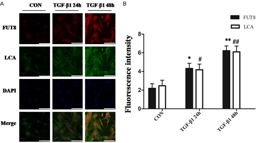
[image:3.612.91.524.71.288.2]using laser scanning confocal microscope. The results revealed that CF were expressed with moderate intensity and distributed on the peri-cyte membrane and endochylema, indicating
Figure 1. Verification of mice lung pericytes. Conduct double staining to detect the expression of pericyte markers through immunofluorescence with the amplification factor multiplied by 400. The experiment was repeated three
4944 Int J Clin Exp Pathol 2017;10(5):4941-4954 that core fucosylation expression exist in
mouse pericytes. The level of CF modification
gradually increased in the group of pericytes
stimulated with TGF-β1.
FUT8siRNA successfully silences the endog -enous FUT8 gene in pericytes
To understand the function of CF modification in the mechanism of TGF-β1-induced pericyte
transdifferentiation, we adopted
chemosyn-thetic FUT8siRNA interference fragments to
transiently transfect pericytes for 24 h with 20
nM. We applied immunofluorescence to deter -mine the expression of pericyte surface markers for each group. We applied Western
blot method to detect FUT8siRNA interference effect and the results showed that FUT8siRNA could reduce FUT8 expression in pericytes, indicating that FUT8siRNA successfully inter -fered with the pericytes.
FUT8siRNA inhibits TGF-β1-induced pericyte morphological changes
During the process of stimulating lung pericyte
transdifferentiation by TGF-β1, the pericyte
morphology transformed from a multi-star
shape into a fusiform myofibroblast shape. The
results demonstrated that the pericytes in the control group were multi-star or medusiform shaped with a plump endochylema and clear
outline. Following stimulation with TGF-β1 for
24 h, some of the cells began to stretch and separate from peripheral cells to induce a dis-ordered arrangement. Following stimulation
with TGF-β1 for 48 h, the shape of the pericytes changed significantly and formed into a strip. After a 20-nM concentration of FUT8siRNA
was applied to the pericytes and the cells were
incubated for 24 h, TGF-β1 was applied to stim
-ulate pericytes for 24 h and 48 h; the majority
of the pericytes under an optical microscope remained in a normal cell shape without obvi-ous stretching or changes in the strip shape. This indicates that inhibiting core fucosylation
by FUT8siRNA could suppress the develop-ment of the pericyte shape caused by TGF-β1
stimulation.
FUT8siRNA inhibits PDGFβR core fucosylation modification but not the expression of PDGFβR
Platelet-derived growth factor beta receptor
(PDGFβR) is the primary protein involved in
pericyte transformation following stimulation
with TGF-β1 for 24 h and 48 h. The relationship between the core fucosylation and PDGFβR in
pericytes remains unclear. The results of the
immunofluorescence co-staining method found that CF and PDGFβR were expressed in peri -cytes, and started to increase after 24 h,
peak-ing after 48 h. Moreover, the two markers
overlapped completely, which indicated that
PDGFβR was modified by core fucosylation. FUT8siRNA at a concentration of 20 nM was
applied to the incubating pericytes for 24 h,
and TGF-β1 was applied to stimulate the peri
-cytes for 24 h and 48 h respectively. The core fucosylation expression of PDGFβR was inhibit
-ed; however, the PDGFβR protein was not. To
verify this phenomenon, we adopted an immu-noprecipitation and lectin blot method to detect
the relationship between PDGFβR and the core
fucosylation in pericytes, the results indicates
that FUT8siRNA could not affect receptor pro
-tein expression. While the immunofluorescence result also revealed that FUT8siRNA inhibited the core fucosylation modification of PDGFβR
but had no effect on the receptor protein expression.
FUT8siRNA inhibited PDGFβR core fucosyl -ation and the activ-ation of TGF-β1-induced PDGFβ/Erk signaling in pericytes
P-Erk indicates the activation of the PDGFβ/Erk pathway. To detect whether the PDGFβ/Erk pathway was activated by TGF-β1 during
TGF-β-induced pericyte transformation, we applied
immunoblotting and immunofluorescence
me-thods to detect p-Erk changes. The results indicated that p-Erk expression increased
significantly in the TGF-β1 24 h and 48 h. This indicates that the activation of PDGFβ/Erk
involved pericyte transdifferentiation. In the
above experiment, we found that FUT8siRNA
did not affect receptor protein expression after interfering pericytes; thus, we further studied whether inhibiting core fucosylation by
FUT8siRNA could inhibit the expression of phosphorylated protein p-Erk in the PDGFβ/
Erk pathway. Moreover, the Western blot and
immunofluorescence results showed that com
-pared with the TGF 24 h and 48 h group, p-Erk expression in the pericytes decreased signifi
-cantly after being treated with FUT8siRNA. This indicates that FUT8siRNA could inhibit p-Erk expression during TGF-β1-induced pericyte
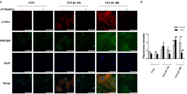
FUT8siRNA inhibits the transformation of TGF-β1-induced pericytes into myofibroblasts
The existence of myofibroblasts may cause α-SMA expression to increase. To determine whether myofibroblast transdifferentiation occurred and the effect of FUT8siRNA on α-SMA expression, we conducted western blot and immunofluorescence and found that stimulating the pericytes, the α-SMA protein
expression level increased and peaked after
48 h. After incubating the pericytes for 24 h by applying 20 nM FUT8siRNA, then stimulating the pericytes with TGF-β1, the level of α-SMA
protein expression obviously decreased. This indicates that after stimulating the pericyte
for 48 h with TGF-β1, the pericytes obtained myofibroblast markers and FUT8siRNA could
inhibit the transformation of pericytes into
myofibroblasts.
Modification changes of core fucosylation dur -ing pericyte transdifferentiation
The above experimental results have shown that CF plays an important role in pericyte transdifferentiation; however, the changes in CF before and after pericyte transformation
remain unclear. We adopted immunofluores -cence and immunoprecipitation methods to
detect the changes in FUT8, PDGFβR and α-SMA before and after pericyte transforma -tion. The results revealed that the protein
expression level of FUT8 was consistent with that of PDGFβR and α-SMA, increased gradu
-ally in TGF-β1 24 h group, reached the peak value in TGF-β1 48 h group; In addition, after applying FUT8siRNA before stimulating the pericytes with TGF-β1, the protein expression levels of FUT8 and α-SMA were obviously decreased. This indicates that FUT8, PDGFβR and α-SMA are important during the transfor
-mation of TGF-β1-induced pericytes into myofi
-broblasts. Moreover, FUT8siRNA could inhibit the transformation of pericytes into myofibro -blasts after silencing CF expression.
Discussion
The key factor involved in the development of
pulmonary fibrosis is the aggregation and acti
-vation of myofibroblasts [19]. Growing evidence
supports pericytes as the major source of
myo-fibroblasts [8, 9]. Currently, the specific mecha -nism of the transformation of pericytes into
myofibroblasts remains unclear and it was
reported in relevant literature that pericyte transdifferentiation is an activating process
involving PDGFβ/Erk and TGF-β/Smad2/3 multi-signal pathways [20]. Moreover, PDGFβ and TGF-β chemokine were also found to be
critical factors for the development of organ
fibrosis [21, 22]. Therefore, a common target -ing point for regulat-ing the above signal path-ways is an effective method of blocking peri-cyte transdifferentiation and thereby treats
pulmonary fibrosis.
Glycosylation is one of the most important
post-translational modifications of protein that
generally occurs in an extracellular
environ-ment [15, 23]. In particular, N-linked glycosyl -ation is involved in the pathological process of multiple diseases, plays an important role in
protein folding, transportation, and localization [24, 25]. Furthermore, it participates in recep -tor activation, signal transduction, and other
important biological processes [26]. Relevant
literature reports and a previous study by our research group have demonstrated that the
TGF-βR protein N-oligosaccharide was modified by CF [15, 16, 27]. Moreover, it further played a
decisive role in the development of pulmonary
fibrosis; therefore, we concluded that during pericyte transdifferentiation, PDGFβR and TGF-βR were also modified by CF. Currently, there
have been no studies that have investigated the relationship between CF and pericyte activation in pneumonopathy. This study is the
first to investigate CF modifications and the
mechanism of pericyte transdifferentiation in the lung.
Currently, the literatures involving the in vitro study of lung pericytes are extremely rare since
pericytes do not have specific markers, such
that the in vitro isolation and cultivation of
these cells are difficult [17]. The relevant
literature reports that pericytes are a type of unstably expressing cells and there are a
vari-ety of antibodies used to analyze the expres -sion of molecular markers on pericyte during
different stages [28]. Pericytes have been
found to express platelet-derived growth factor
receptor-β (PDGFR-β), desmin, CD73, NG-2 and others [28]. The expression level of these
labeled molecules changes in accordance with pericyte development, pathological reaction, and culture in vitro. Therefore, we
4946 Int J Clin Exp Pathol 2017;10(5):4941-4954 to verify the expression of various markers
after successfully extracting lung pericytes through the method of magnetic activated cell sorting (Figure 1).
After successfully extracting lung pericytes in
mice, we first sought to determine if the surface
of lung pericyte expresses core fucosylation.
We utilized an immunofluorescence method to
determine whether both the cytoplasm and cell surface exhibit the expression of the medium level of the core fucosylation (Figure 2). Next,
we used 5 ng/mL TGF-β1 to stimulate mice lung pericytes for 24 h and 48 h respectivelyto establish a model of pericyte
transdifferentia-tion and found that 24 h after TGF-β1 stimula
-Figure 2. TGF-β1 induced the up-regulation of pericyte core fucosylation expression. A: The immunofluorescence
method was applied to determine the expression of mouse lung pericyte core fucose links in each group with
ampli-fication times multiplied by 400. B: Quantitative analysis of the immunofluorescence results. All data was expressed
[image:6.612.93.523.72.310.2]as the mean ± standard deviation, each experiment was repeated three times. *P < 0.05, **P < 0.01 versus Control group, #P < 0.05, ##P < 0.01 versus TGF group.
Figure 3. FUT8siRNA successfully silencing the FUT8 gene in pericytes. A: Apply immunoblotting to detect the ef
-ficiency of interference. B: Immunoblotting for a quantitative analysis of the interference ef-ficiency results. All data
[image:6.612.93.520.392.551.2]tion, part of the cell shape began to change in
TGF-β1 24 h group and shape of 80% of the
pericytes changed from a multi-star shape to a
long strip shape in TGF-β1 48 h (Figure 4).
Immunofluorescence also revealed that the
pericytes began to increase the expression of
α-SMA while core fucosylation expression level
also began to rise (Figure 7). Moreover, the
immunofluorescence demonstrated that the pericytes exhibit a significant expression of α-SMA and core fucosylation (Figure 8). This
indicates that TGF-β1 may be the factor caus -ing the transformation of pericytes and
sug-gests that the core fucosylation modification may also function in promoting the TGF-β1
induction of pericytes transdifferentiation. To further study this mechanism, we inhibited the expression of pericytes surface core
fucosylation. Since core fucosylation requires the α-1,6-fucosyltransferase (FUT8) catalytic
protein N-oligonucleotide chitosan to complete
the core fucosylation modification [29], we designed and synthesized a FUT8siRNA se-quence and transfected pericytes. The experi -mental results demonstrate that the synthetic
FUT8siRNA sequence successfully reduced the pericytes FUT8 protein expression (Figure 3). After we silenced FUT8 using gene silencing, 24 h and 48 h after TGF-β1 stimulation, it was found that over 80% of the pericytes did not change. In addition, the immunofluorescence test revealed that the expression of α-SMA as a marker of the transformation from to myofi
-broblasts was significantly lowered (Figures 4,
7).
Since the relevant literature reports that
PDGFβR and TGF-βRI are the key receptor pro -teins involved in the pericytes
activation-relat-ed signaling pathways [30], we usactivation-relat-ed the west
-ern blotting and precipitation methods to test
whether these two proteins were modified by
core fucosylation. Foreign Wang and our previ-ous study conducted by our research team
confirmed that TGF-βR is modified by core fucosylation [15, 16, 31]. However, our research
team found that, in the percytes
transdifferen-tiation process, PDGFβR is also modified by
core fucosylation (Figure 5). We also found that
during this process, TGF-β1 can induce peri -cytes transdifferentiation and the expression
of PDGFβR was higher than in the control
group. This indicates that the PDGF/Erk signaling pathway plays an important role in pericytes transdifferentiation. Interestingly, we
found that FUT8siRNA can inhibit the core fucosylation modification level of PDGFR but does not influence the expression of the PDGFβR receptor itself (Figure 5). This means
that the modification of the protein after trans -lation is independent of the receptor protein expression.
The phosphorylation of Erk is an indicator of
the activation of the PDGFβ/Erk signaling pathway [32]. To determine whether inhibiting
core fucosylation can abolish periytes
transdif-ferentiation by inhibiting PDGFβ/Erk signaling
pathway, we assessed the expression of
phosphorylated Erk. Our findings indicate that FUT8siRNA can inhibit the activation of the PDGFβ/Erk signaling pathway (Figure 6),
indi-cating that core fucosylation modification is essential in the activation of the PDGFβ/Erk
signaling pathway.
The experiment verified that the FUT8siRNA
stops the lung pericytes from transforming into
myofibroblasts by inhibiting the activation of the PDGFβ/Erk signaling pathway. To further
[image:7.612.92.521.75.179.2]clarify the characteristics involved in the CF
4948 Int J Clin Exp Pathol 2017;10(5):4941-4954
Figure 5. PDGFβR was modified by core fucosylation and FUT8siRNA blocked the core fucosylation modification of PDGFβR. A: An immunol
-ogy double staining method to detect PDGFβR and core fucosylation expression in each group. B: Quantitative analysis of the immunofluo -rescence results. *P < 0.05, **P < 0.01 versus Control group, #P < 0.05, ##P < 0.01 versus TGF group. All data were expressed as the mean ± standard deviation, each experiment was repeated three
times, with amplification times multiplied by 400. C: The immunopre
-cipitation and lectin blotting methods were applied to detect PDGFβR
and core fucosylation expression in each group. D: An
immunopre-cipitation and lectin blotting method were applied for a quantitative analysis on PDGFβR protein and core fucosylation expression in each
Figure 6. During the TGF-β1 induced pericyte transformation, p-Erk protein expression was up-regulated and FUT8siRNA blocked p-Erk protein expression. A: Im
-munofluorescence was used to detect changes in p-Erk protein expression levels in each group. B: Quantitative analysis of the im-munofluorescence results. All
data was expressed as the mean ± standard deviation, each experiment was repeated three times. *P < 0.05, **P < 0.01 versus Control group, #P < 0.05, ##P < 0.01 versus TGF group. C: Immunoblotting was used to detect the changes in the level of p-Erk protein expression in each group. D: Immunoblotting was used for a
quantitative analysis on p-Erk protein expression in each group. All data were expressed as the mean ± standard deviation. *P < 0.05, **P < 0.01 versus Control
4950 Int J Clin Exp Pathol 2017;10(5):4941-4954
Figure 7. Effect of FUT8siRNA on TGF-β1-induced pericyte transformation. A: An immunofluorescence double staining method was used to detect PDGFβR and α-SMA protein expression in each group. The experiment was repeated three times, with the amplification times multiplied by 400. B: Quantitative analysis of the immunofluorescence results. All data were expressed with the mean ± standard deviation. *P < 0.05, **P < 0.01 versus Control group, #P < 0.05, ##P < 0.01
4952 Int J Clin Exp Pathol 2017;10(5):4941-4954 changes that occur before and after pericytes
transformation, this experiment involved
change of PDGFβR as well as α-SMA and FUT8 before and after transformation. The results revealed that α-SMA exhibited mini-mal expression before the TGF-β1-induced transformation, and that PDGFβR and FUT8 are moderately expressed. After 24 and 48 h of TGF-β1 stimulation of the pericytes, the
above index gradually increased and peaked at
48 h; however, FUT8siRNA cannot inhibit the increase in PDGFβR following stimulation with TGF-β1. Rather, it can inhibit the high expres
-sion of α-SMA after stimulation with TGF-β1
(Figure 8). Even during the process of pericytes transdifferentiation in which a high level of
PDGFβR expression exists, if the PDGFβR was not modified by core fucosylation, the pericytes could not transform into myofibroblasts.
Therefore, the change in CF may be an impor-tant mechanism involved in the pericytes trans-formation process.
TGF-βR and PDGFβR are also important pro
-teins causing fibrosis [33-36]; thus, we specu
-lated that the core fucosylation modification
could regulate pericytes transdifferentiation and plays an important role in the process of
pulmonary fibrosis. Our study shows lung peri
-cytes transdifferentiation mechanisms through
core fucosylation modifications for the first
time, which provides a novel perspective for studying the molecular mechanisms of the
pericytes that participate in pulmonary fibrosis,
and indicates that the control of core fucosyl-ation may be a potential target for the
treat-ment of pulmonary fibrosis.
Acknowledgements
This work was supported by National Natural Science Foundation of China (NSFC) (No.
81273924 and 8153000127).
Disclosure of conflict of interest
None.
Address correspondence to: Taihua Wu, Depart-
ment of Respiratory Medicine, The First Affiliated Hospital of Dalian Medical University, 222#
Zhongshan Road, Dalian 116011, Liaoning, P.R.
China. Tel: 83635963; Fax: +86-411-83635963; E-mail: wutaihuadoc@126.com; Hongli Lin, Department of Nephrology, The First Affiliated Hospital of Dalian Medical University, 222#
Zhongshan Road, Dalian 116011, Liaoning, P.R.
[image:12.612.247.520.80.205.2]China. Tel: 83635963; Fax: +86-411-83635963; E-mail: linhonglidoc@126.com
Figure 8. Changes in CF modification during pericyte transformation. A, B: An immunofluorescence double staining method was applied to detect FUT8 and PDGFβR, α-SMA protein expression in each group. The test was repeated three times, with the amplification multiplied by 400. C: Quantitative analysis of the immunofluorescence results.
All data were expressed with the mean ± standard deviation. *P < 0.05, **P < 0.01 versus Control group, #P < 0.05, ##P < 0.01 versus
TGF group. D: Immunoblotting was used for a quantitative analysis on
p-Erk protein expression in each group. E: Immunoblotting was used
to perform a quantitative analysis on the level of FUT8, PDGFβR, and α-SMA protein expression in each group. All data is expressed as the
References
[1] Wakamatsu K, Nagata N, Kumazoe H, Oda K,
Ishimoto H, Yoshimi M, Takata S, Hamada M,
Koreeda Y, Takakura K, Ishizu M, Hara M, Ise S, Izumi M, Akasaki T, Maki S, Kawabata M,
Mukae H, Kawasaki M. Prognostic value of se-rial serum KL-6 measurements in patients
with idiopathic pulmonary fibrosis. Respir Investig 2017; 55: 16-23.
[2] Li LC, Kan LD. Traditional Chinese medicine for
pulmonary fibrosis therapy: progress and fu -ture. J Ethnopharmacol 2017; 198: 45-63. [3] Hofmann K, Fiedler S, Vierkotten S, Weber J,
Klee S, Jia J, Zwickenpflug W, Flockerzi V, Storch U, Yildirim AÖ, Gudermann T, Königshoff
M, Dietrich A. Classical transient receptor
po-tential 6 (TRPC6) channels support myofibro -blast differentiation and development of
ex-perimental pulmonary fibrosis. Biochim
Biophys Acta 2017; 1863: 560-568.
[4] Bindu S, Pillai VB, Kanwal A, Samant S, Mutlu GM, Verdin E, Dulin N, Gupta MP. SIRT3 blocks
myofibroblast differentiation and pulmonary fibrosis by preventing mitochondrial DNA dam -age. Am J Physiol Lung Cell Mol Physiol 2017;
312: L68-L78.
[5] Wang C, Gu S, Cao H, Li Z, Xiang Z, Hu K, Han X. miR-877-3p targets Smad7 and is associat
-ed with myofibroblast differentiation and bleo
-mycin-induced lung fibrosis. Sci Rep 2016; 6: 30122.
[6] Xie T, Liang J, Liu N, Huan C, Zhang Y, Liu W,
Kumar M, Xiao R, D’Armiento J, Metzger D, Chambon P, Papaioannou VE, Stripp BR, Jiang
D, Noble PW. Transcription factor TBX4
regu-lates myofibroblast accumulation and lung fi -brosis. J Clin Invest 2016; 126: 3063-79.
[7] Zhao H, Bian H, Bu X, Zhang S, Zhang P, Yu J, Lai X, Li D, Zhu C, Yao L, Su J. Targeting of discoidin domain receptor 2 (DDR2)
pre-vents myofibroblast activation and neovessel formation during pulmonary fibrosis. Mol Ther 2016; 24: 1734-1744.
[8] Rock JR, Barkauskas CE, Cronce MJ, Xue Y, Harris JR, Liang J, Noble PW, Hogan BL. Multiple stromal populations contribute to
pulmonary fibrosis without evidence for epi -thelial to mesenchymal transition. Proc Natl
Acad Sci U S A 2011; 108: E1475-83.
[9] Hung C, Linn G, Chow YH, Kobayashi A, Mittelsteadt K, Altemeier WA, Gharib SA,
Schnapp LM, Duffield JS. Role of lung
peri-cytes and resident fibroblasts in the pathogen
-esis of pulmonary fibrosis. Am J Respir Crit
Care Med 2013; 188: 820-30.
[10] Yuan K, Orcholski ME, Huang NF, de Jesus
Perez VA. In vivo study of human endothelial-pericyte interaction using the matrix gel plug assay in mouse. J Vis Exp 2016.
[11] Stefanska A, Eng D, Kaverina N, Duffield JS,
Pippin JW, Rabinovitch P, Shankland SJ. Interstitial pericytes decrease in aged mouse kidneys. Aging (Albany NY) 2015; 7: 370-82. [12] Pozdzik AA, Giordano L, Li G, Antoine MH,
Quellard N, Godet J, De Prez E, Husson C, Declèves AE, Arlt VM, Goujon JM,
Brochériou-Spelle I, Ledbetter SR, Caron N, Nortier JL.
Blocking TGF-β signaling pathway preserves
mitochondrial proteostasis and reduces early
activation of PDGFRβ+ pericytes in aristolochic
acid induced acute kidney injury in wistar male rats. PLoS One 2016; 11: e0157288.
[13] Takamatsu S, Shimomura M, Kamada Y, Maeda H, Sobajima T, Hikita H, Iijima M, Okamoto Y, Misaki R, Fujiyama K, Nagamori S,
Kanai Y, Takehara T, Ueda K, Kuroda S, Miyoshi
E. Core-fucosylation plays a pivotal role in hep-atitis B pseudo virus infection: a possible
impli-cation for HBV glycotherapy. Glycobiology
2016; 26: 1180-1189.
[14] Vainauskas S, Duke RM, McFarland J, McClung
C, Ruse C, Taron CH. Profiling of core fucosyl -ated N-glycans using a novel bacterial
lectin that specifically recognizes α1,6
fucosylated chitobiose. Sci Rep 2016; 6: 34195.
[15] Shen N, Lin H, Wu T, Wang D, Wang W, Xie H, Zhang J, Feng Z. Inhibition of TGF-β1-receptor
posttranslational core fucosylation attenuates
rat renal interstitial fibrosis. Kidney Int 2013;
84: 64-77.
[16] Lin H, Wang D, Wu T, Dong C, Shen N, Sun Y, Sun Y, Xie H, Wang N, Shan L. Blocking core
fucosylation of TGF-β1 receptors downregu -lates their functions and attenuates the epi-thelial-mesenchymal transition of renal tubular cells. Am J Physiol Renal Physiol 2011; 300: F1017-25.
[17] Meguro S, Akamatsu T, Matsushima S, Kosugi I, Kawasaki H, Arai Y, Baba S, Tsuchida T, Shido Y, Suda T, Iwashita T. Phenotypic
charac-terization of perivascular myoid cell neo
-plasms, using myosin 1B, a newly identified
human pericyte marker. Hum Pathol 2017; 11:
S0046-8177.
[18] de Souza LE, Malta TM, Kashima Haddad S,
Covas DT. Mesenchymal stem cells and peri-cytes: to what extent are they related? Stem
Cells Dev 2016; 25: 1843-1852.
[19] Hofmann K, Fiedler S, Vierkotten S, Weber J, Klee S, Jia J, Zwickenpflug W, Flockerzi V, Storch U, Yildirim AÖ, Gudermann T, Königshoff
M, Dietrich A. Classical transient receptor
po-tential 6 (TRPC6) channels support myofibro -blast differentiation and development of
ex-perimental pulmonary fibrosis. Biochim
Biophys Acta 2017; 1863: 560-568.
4954 Int J Clin Exp Pathol 2017;10(5):4941-4954 player in vascular pathobiology. Pharmacol
Ther 2017; 171: 30-42.
[21] Dadrich M, Nicolay NH, Flechsig P, Bickelhaupt
S, Hoeltgen L, Roeder F, Hauser K, Tietz A, Jenne J, Lopez R, Roehrich M, Wirkner U, Lahn
M, Huber PE. Combined inhibition of TGFβ
and PDGF signaling attenuates
radiation-in-duced pulmonary fibrosis. Oncoimmunology 2015; 5: e1123366.
[22] Deng X, Jin K, Li Y, Gu W, Liu M, Zhou L. Platelet-derived growth factor and transforming growth
factor β1 regulate ards-associated lung fibro -sis through distinct signaling pathways. Cell Physiol Biochem 2015; 36: 937-46.
[23] Cuccui J, Terra VS, Bossé JT, Naegeli A, Abouelhadid S, Li Y, Lin CW, Vohra P, Tucker
AW, Rycroft AN, Maskell DJ, Aebi M, Langford PR, Wren BW; BRaDP1T Consortium. The N-linking glycosylation system from Actino-
bacillus pleuropneumoniae is required for ad -hesion and has potential use in glycoengineer-ing. Open Biol 2017; 7.
[24] Beriault DR, Dang VT, Zhong LH, Petlura CI,
McAlpine CS, Shi Y, Werstuck GH. Glu- cosamine induces ER stress by disrupting lip-id-linked oligosaccharide biosynthesis and N-linkedprotein glycosylation. Am J Physiol
Endocrinol Metab 2017; 312: E48-E57. [25] Gusakov AV, Dotsenko AS, Rozhkova AM,
Sinitsyn AP. N-linked glycans are an important component of the processive machinery of cel-lobiohydrolases. Biochimie 2017; 132:
102-108.
[26] Matoba K, Mihara E, Tamura-Kawakami K,
Miyazaki N, Maeda S, Hirai H, Thompson S,
Iwasaki K, Takagi J. Conformational freedom of the LRP6 ectodomain is regulated by N-glycosylation and the binding of the Wnt an-tagonist Dkk1. Cell Rep 2017; 18: 32-40. [27] Gu W, Fukuda T, Isaji T, Hashimoto H, Wang Y,
Gu J. α1,6-Fucosylation regulates neurite for -mation via the activin/phospho-Smad2 path-way in PC12 cells: the implicated dual effects
of Fut8 for TGF-β/activin-mediated signaling. FASEB J 2013; 27: 3947-58.
[28] Navarro R, Compte M, Álvarez-Vallina L, Sanz
L. Immune regulation by pericytes: modulating innate and adaptive immunity. Front Immunol
2016; 7: 480.
[29] Yang Q, Wang LX. Mammalian α-1,6-fucosyltransferase (FUT8) is the sole enzyme
responsible for the N-acetylglucosaminyl- transferase I-independent core fucosylation of high-mannose N-glycans. J Biol Chem 2016; 291: 11064-71.
[30] Armulik A, Abramsson A, Betsholtz C. En- dothelial/pericyte interactions. Circ Res 2005; 97: 512-23.
[31] Wang X, Gu J, Miyoshi E, Honke K, Taniguchi N.
Phenotype changes of Fut8 knockout mouse:
core fucosylation is crucial for the function of
growth factor receptor(s). Methods Enzymol
2006; 417: 11-22.
[32] Guo S, Yu L, Cheng Y, Li C, Zhang J, An J, Wang H, Yan B, Zhan T, Cao Y, Zheng H, Li Z.
PDGFRβ triggered by bFGF promotes the prolif -eration and migration of endothelial progenitor cells via p-ERK signalling. Cell Biol Int 2012; 36: 945-50.
[33] Walraven M, Akershoek JJ, Beelen RH, Ulrich
MM. In vitro cultured fetal fibroblasts have myofibroblast-associated characteristics and produce a fibrotic-like environment upon stimulation with TGF-β1: is there a thin line be
-tween fetal scarless healing and fibrosis? Arch Dermatol Res 2017; 309: 111-121.
[34] Mueller AA, van Velthoven CT, Fukumoto KD,
Cheung TH, Rando TA. Intronic polyadenylation
of PDGFRα in resident stem cells attenuates muscle fibrosis. Nature 2016; 540: 276-279.
[35] Tang H, He H, Ji H, Gao L, Mao J, Liu J, Lin H, Wu T. Tanshinone IIA ameliorates
bleomycin-induced pulmonary fibrosis and inhibits trans
-forming growth factor-beta-β-dependent epi -thelial to mesenchymal transition. J Surg Res 2015; 197: 167-75.
[36] He H, Tang H, Gao L, Wu Y, Feng Z, Lin H, Wu T. Tanshinone IIA attenuates bleomycin-induced