TRINITY COLLEGE DUBLIN
The
Effects
of
Low-Dose
Methotrexate
on
Metabolism in Primary Human PBMCs.
Laura O'Farrell
A dissertation submitted to the University of Dublin in candidature for the degree of Doctor of Philosophy
July 2017
Supervisor:
Declaration
I declare that this thesis has not been submitted as an exercise for a degree at this or any other university and it is entirely my own work.
I agree to deposit this thesis in the University’s open access institutional repository or allow the library to do so on my behalf, subject to Irish Copyright Legislation and Trinity College Library conditions of use and acknowledgement.
i
Summary
Low-dose Methotrexate (MTX) is a powerful anti-arthritic drug used in the treatment of a variety of inflammatory diseases. It is an economical drug and is easily administered, but incomplete knowledge into its mechanism of action limits its therapeutic capability. As an anti-folate drug, MTX has been shown to have a variety of effects including effects on the one-carbon metabolism pathway, including purine and thymidine nucleotide synthesis. MTX has also been shown to reverse the aberrant glycosylation patterns in Rheumatoid arthritis (RA) patients. It is not known exactly which effect of MTX has therapeutic potential nor is it known which aspect of this anti-folate is responsible. Elucidating new effects of MTX and whether purine and thymidine inhibition is the mechanism by which these effects occur, would provide new insight into the workings of this drug and expand its therapeutic potential. Using a model of activated T cells, the effects of MTX on a variety of biochemical processes were investigated and compared to unactivated PBMCs to see if MTX could preferentially affect activated PBMCs, such as the over-active T cells in RA.
Flow cytometric analysis revealed that MTX displayed activation-dependent effects on cell viability, proliferation, transferrin receptor expression and blastogenesis, but dissipated the mitochondrial inner-membrane potential (∆Ѱm) in both activated and
unactivated PBMCs. The MTX-mediated effects were either partially or completely reversed upon purine and thymidine rescue, which indicated that insults to this biosynthesis pathway were the mechanism in which these effects occurred.
No direct effect of low-dose MTX on cellular glycosylation has ever been shown. In this study, flow cytometry was also employed to quantify ManNAz incorporation on the-cell-surface, using click chemistry and was used to show how changes to surface sialylation can occur during activation which can thus be used to evaluate the effects of drug treatment that limit cellular activation. MTX was found to significantly affect cell-surface sialylation, which was reversed by purine and thymidine rescue.
ii
nucleotides in activated PBMCs but no changes were found in the sugar-nucleotides detected. NAD/NADH did not increase during activation, which suggested that MTX could exert effects which were activation-independent. Purine and thymidine inhibition was also shown to be the mechanism in which MTX reduced the levels of NAD/NADH nucleotides.
Combined, the findings shown here suggest that the MTX-mediated inhibition of purine and thymidine synthesis has far-reaching consequences, encompassing effects on cellular activation and on cell-surface sialylation. A state of folate-deficiency was found under these MTX-treated conditions, illustrating the powerful effect of this anti-folate, even at low-doses. This finding has implications for any future effects shown by MTX whereby a consequence of folate-deficiency would have to be disproven.
i
v
Acknowledgements
First and foremost, I would like to thank Dr. Anne Molloy for her encouragement, support and guidance over the last two years, without which this thesis would never have been possible. It is impossible to put into words how much I learned, but I can say that I was blessed to have you as a mentor. I only hope this thesis does all your help justice, Anne.
The second person who deserves a mention is Dr. Jerrard Hayes for the 4 years of work you put in helping me with this project (the last year doesn’t count). Your unpolished advice and expertise saw me through the first three years. I hope you realise what your help meant, because I will never forget.
Thanks to Dr. Olga Abramczyk, whose technical expertise got me my final results chapter and made me eligible for this doctoral degree.
A special thanks goes to Barry Moran, the flow cytometry expert, whose help was gladly given for the entirety of this project and who has taught me everything I know about flow cytometry. I hope I can return the favour in the future.
To Dr. Cliona O’Farrelly, Dr. Amir Khan and Dr. Jean Fletcher. I want to thank each and every one of you for the help and advice you gave at a time when I really needed it. To all the PhD students in the fifth-floor reading room,; thank you for always being there, for the amazing science conversations at lunch, for the great laughs we had, for being the astounding people I have gotten to know and for hopefully being some of my future life-long friends (if you’ll have me). In particular, to Darren, Aisling, Natalie, Niki, Ryan and Andy, it’s been an absolute blast! A second (I may regret this later), thanks to Jer for all our arguments over a pint!! I wouldn’t trade them for anything.
To my parents Jacinta and Martin, who have put up with me and more importantly, have given me the strength and support I needed to continue over the past five years. I hope you enjoy reading this thesis; you’ve most certainly earned it.
vii
Contents
Summary ... i
Acknowledgements ... i
Abbreviations ... xi
Chapter 1: ... i
General Introduction ... i
1.1 Rheumatoid Arthritis ... 1
1.2. Low-dose Methotrexate ... 2
1.2.1 Cellular uptake of MTX ... 3
1.2.2 Polyglutamation of MTX ... 4
1.2.3 An overview of the mechanism of action of MTX ... 4
1.3 Folate ... 7
1.4 Folate and MTX ... 9
1.5 Anti-CD3-activation of T cells-a model of RA ... 13
1.6 One-Carbon metabolism ... 16
1.7 The anti-folate MTX effecting 1C metabolism ... 20
Table 1.1. The effects of low-dose MTX on 1C metabolism ... 21
1.7.1 The effect of MTX on methionine synthesis ... 21
1.7.2 The effect of MTX on thymidylate synthesis ... 22
1.7.3 The effect of MTX on uracil ... 25
1.7.4 The inhibitory effect of MTX on purine synthesis ... 27
1.7.5 Enzyme inhibition by unreduced folate ... 29
1.8 The anti-proliferative effect of MTX... 29
1.8.1 Megaloblastic anaemia ... 30
1.9 The apoptotic effect of MTX ... 32
1.9.1 DNA damage in response to MTX ... 33
1.10 Mitochondrial function ... 36
1.10.1 The mitochondrial membrane potential and MTX ... 36
1.10.2 ROS ... 38
1.11 Signalling pathways activated by MTX ... 43
1.12 Nucleotide salvage... 44
1.12.1 Pyrimidine salvage ... 44
1.12.2 Purine salvage ... 45
viii
1.14 Purine rescue from MTX ... 48
1.15 Folate rescue from MTX ... 51
1.16 MTX and immunosuppression by adenosine ... 53
1.17 Glycosylation ... 54
1.17.1 The structure of glycans ... 54
1.17.2 Sialic acids ... 61
1.17.3 Methods used to determine surface-sialic acid expression ... 63
1.17.4 The Sialome ... 64
1.17.5 Siglecs ... 65
1.17.6 The activation-dependence of glycosylation ... 66
1.17.7 Regulation of glycosylation ... 67
1.18 Antibodies ... 68
1.18.1 Aberrant Glycosylation patterns in IgG and serum proteins in RA ... 71
1.18.2 MTX and the effect on glycosylation in IgG and serum proteins in RA ... 73
1.19 Aims of the project... 75
Chapter 2 ... 78
Materials and General Methods ... 78
2.1 General methods ... 79
2.1.1 Antibodies and reagents ... 79
2.1.2 Preparation of solutions ... 80
2.1.3 Centrifugation ... 80
2.2 Statistical analysis ... 80
2.3 Cell culture ... 81
2.3.1 Isolation of human PBMCs from whole blood ... 81
2.3.2 Extraction and culture of murine dendritic cells from femur and tibia of wild-type mice... 82
2.3.3 Activation of CD3+ PBMCs using anti-CD3 ... 82
2.3.4 Use of FACS buffer ... 85
Chapter 3: ... 86
The toxicity of low-dose Methotrexate in human PBMCs associated with purine and thymidine inhibition ... 86
3.1 Introduction ... 87
3.1.1 Antigen-dependent toxicity ... 87
3.1.2 Rescue from the apoptotic effects of MTX ... 88
ix
3.1.4 Mitochondrial toxicity ... 90
3.2 Aims of the chapter ... 91
3.3 Methods... 92
3.3.1 Cell viability assays ... 92
3.3.1.2 Immortalised cell lines ... 92
3.3.1.3 Unactivated and LPS-activated human PBMCs and mouse DCs ... 92
3.3.1.4 Partially-activated PBMCs ... 92
3.3.2 Cell proliferation assays ... 93
3.3.2.1 Cell counts ... 93
3.3.2.2 Carboxyfluorescein succinimidyl ester (CFSE) staining ... 93
3.3.3 Measurement of viability, transferrin receptor expression and lymphoblast formation ... 94
3.3.4 Measurement of ∆Ѱm with JC-1 ... 95
3.3.5 The measurement of cellular redox status using H2DCF-DA... 99
3.4 Results ... 102
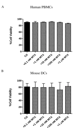
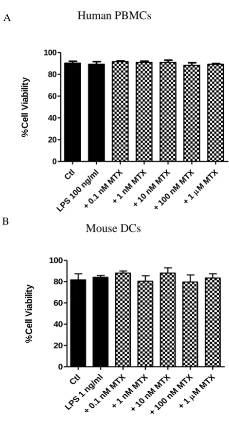
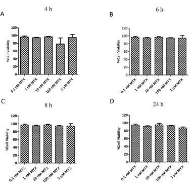
3.4.1 MTX has differential cytotoxic effects on immortalised cell lines compared to primary murine DCs or human PBMCs ... 102
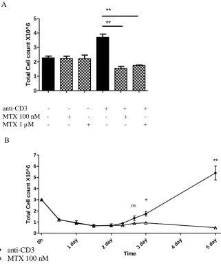
3.4.2 MTX exerts an anti-proliferative effect in anti-CD3-activated PBMCs ... 109
3.4.3 MTX treatment of anti-CD3-activated PBMCs results in reduced levels of transferrin (CD71) receptor expression on CD4+ and CD8+ T cells ... 117
3.4.5 MTX lowers the ∆Ѱm in both unactivated and anti-CD3-activated PBMCs ... 126
3.4.6 MTX increases ROS production in anti-CD3-activated but not in unactivated PBMCs ... 126
3.5 Discussion ... 134
Chapter 4: ... 148
To develop a quantitative method for the evaluation of the effect of Methotrexate treatment on the cell-surface sialome ... 148
4.1 Introduction ... 149
4.1.1 Labelling and quantifying the cell-surface sialome using click chemistry ... 150
4.1.2 The toxicity associated with copper-catalysed click chemistry ... 150
4.1.3 The effect of drug treatment on cell-surface sialyl glycans ... 152
4.2 Aims of the chapter ... 153
4.3 Material and methods ... 154
4.3.1 Click IT reaction ... 154
4.3.2 Preparation of PBMCs for confocal microscopy ... 154
x
4.3.4 Confocal microscopy ... 156
4.3.5 Quantification using flow cytometry ... 156
4.4 Results ... 160
4.4.1 The cell-surface sialome of CD4+ T cells and CD8+ T cell subsets can be visualised using confocal microscopy ... 160
4.4.2 The incorporation of ManNAz on the cell-surface sialome of PBMCs can be quantified using flow cytometry ... 163
4.4.3 MTX treatment reduces ManNAz incorporation on the cell-surface sialome ... 167
4.4.4 MTX has differential effects on the incorporation of ManNAz on the cell-surface sialome of distinct populations of activated PBMCs ... 172
4.5 Discussion ... 190
Chapter 5: ... 202
To elucidate the effects of Methotrexate on intracellular sugar-nucleotides and nucleotides in unactivated and anti-CD3-activated PBMCs ... 202
5.1 Introduction ... 203
5.1.1 The effect of MTX on intracellular nucleotides ... 205
5.1.2 The effect of MTX on intracellular sugar-nucleotides ... 206
5.1.3 Activation-dependent effects on nucleotides and sugar-nucleotides ... 207
5.2 Aims of the chapter ... 208
5.3 Materials and methods ... 209
5.3.1 Extraction of intracellular nucleotides and nucleotide sugars ... 209
5.3.2 Ion-pair RP-HPLC analysis ... 209
5.3.3 Determination of metabolite concentrations from standards ... 211
5.3.4 Standard recovery ... 214
5.4 Results ... 216
5.5 Discussion ... 242
Chapter 6: ... 250
Conclusion ... 256
Future Perspectives ... 258
Appendix ... 260
Supplementary material ... 260
Metabolite extraction for NMR analysis: ... 260
xi
Abbreviations
5-FU 5-flurouracil
7-OH-MTX 7-hydroxymethotrexate
DAPA Diamino-2, 4-N-10-methylpteroic acid
ACN Acetonitrile
ACPA Anti-citrullinated protein antibody
ADA Adenosine deaminase
ADP Adenosine diphosphate
AICD Activation induced cell death
AICAR Amino-imidazolecarboxamide ribosyl-5-phosphate
AICARFT AICAR formyltransferase
AMP Adenosine monophosphate
AO Aldehyde oxidase
APRT Adenine phosphoribosyltransferase
APS Ammonium persulfate
ATP Adenosine triphosphate
BER Base-excision repair
BSA Bovine serum albumin
CA Candida albicans
CAD Caspase-activated deoxyribonuclease
CFSE Carboxyfluorescein succinimidyl ester
CHO Chinese hamster ovary
CIA Collagen induced arthritis
CMP NeuAc Cytidine monophosphate neuraminic acid
CNT Concentrative nucleoside transporters
cSHMT Cytoplasmic serine hydroxymethyltransferase
CTP Cytidine triphosphate
DAMPA Diamino-2, 4-N-10-methylpteroic acid
DC Dendritic cell
DHF Dihydrofolate
DHFR Dihydrofolate reductase
DMG Dimethylglycine
DMSO Dimethyl sulfoxide
DNA Deoxyribonucleic acid
dTMP Deoxythymidine monophosphate (thymidylate)
dTTP Deoxythymidine triphosphate
dUMP Deoxyuridine monophosphate
dUTPase Deoxyuridine triphosphate nucleotidohydrolase
ENT Equilibrative nucleoside transporters
ETC Electron transport chain
FA Folic acid
FACS Fluorescence activated cell sorting
Fab Fragment antigen binding
FBS Foetal calf serum
xii
FCCP Carbonyl cyanide 4-(trifluoromethoxy)phenylhydrazone
FCS Foetal bovine serum
FDH 10-formyl THF dehydrogenase
FITC Fluorescein isothiocyanate
FPGS Folylpolyglutamate synthase
FR Folate receptor
FSC Forward scatter
FTHFS 10-formyl THF synthase
GalNAc N-acetylgalactosamine
GAR Glycinamide ribonucleotide
GARFT GAR formyltransferase
GCS Glycine cleavage system
GDP fucose Guanosine diphosphofucose
GDP mannose Guanosine diphosphomannose
GGH γ-glutamyl hydrolase
GlcNAc N-acetylglucosamine
GTP Guanosine triphosphate
Hcy Homocysteine
Hep-2 Human laryngeal squamous carcinoma cells
HGPRT Hypoxanthine-guanine phosphoribosyltransferase
HLA Human leukocyte antigen
HPLC High-performance liquid chromatography
HRP Horse radish peroxidase
Hx Hypoxanthine
iBMDM Immortalised bone marrow derived macrophages
ICAM-1 Intercellular adhesion molecule 1
ICMT Isoprenylcysteine carboxymethyltransferase
Ig Immunoglobulin
IMP Inosine monophosphate
JC-1 5,5’6,6’-tetrachloro-1,1’3,3’-tetraethylbenzimidazolylcarbocyanine iodide
LPS Lipopolysaccharide
ManNAc N-acetylmannosamine
MAPK Mitogen-activated protein kinase
MAT Methionine adenosyltransferase
MEFs Mouse embryonic fibroblasts
MFT Mitochondrial folate transporter
MHC Major-histocompatibility complex
MMR Mismatch repair
MS Methionine synthase
mSHMT Mitochondrial serine hydroxymethyltransferase
MTHFC Methenyl THF cyclohydrolase
MTHFD MethenylTHF dehydrogenase
MTHFR 5, 10-methylenetetrahydrofolate reductase
mTOR Mammalian target of rapamycin
xiii
MTXPG Methotrexate polyglutamate
NAC N-acetyl-L-cysteine
NADH Nicotinamide adenine dinucleotide
NADPH Nicotinamide adenine dinucleotide phosphate
NeuAc Neuraminic acid
NF-kB Nuclear factor-kappa B
NK Natural Killer cell
NTDs Neural tube defects
NS Non-significant
OCM One carbon metabolism
ODC Ornithine decarboxylase
PBL Peripheral blood lymphocytes
PBMC Peripheral blood mononuclear cell
PBS Phosphate buffered saline
PCFT Proton-coupled folate transporter
PFA Paraformaldehyde
PHA Phytohaemagglutinin
PI Propidium iodide
PKC Protein kinase C
PMA phorbyl myristate acetate
PRPP phosphoribosyl pyrophosphate
PS Phosphatidyl serine
RA Rheumatoid arthritis
RBC Red blood cell
RFC Reduced folate carrier
ROS Reactive oxygen species
RPMI Roswell Park Memoria Institute 1640 medium
RNA Ribonucleic acid
SAH S-adenosyl homocysteine
SAM S-adenosyl methionine
SF Synovial fluid
SHMT Serine hydroxymethyltransferase
Siglecs Sialic acid immunoglobulin-type lectins
SSC Side scatter
SLC Solute carrier
TCR T cell receptor
TT Tetanus toxoid
Th Helper T-lymphocyte
THF Tetrahydrofolate
Thy Thymidine
TLD Thymineless death
TLR Toll-like receptor
TNF Tumour necrosis factor
Treg Regulatory-T-lymphocyte
TS Thymidylate synthase
xiv
UDP-Gal Uridine diphosphate galactose
UDP-GalNAc Uridine diphosphate N-acetylgalactosamine
UDP-Glc Uridine diphosphate glucose
i
Chapter 1:
1
1.1 Rheumatoid Arthritis
Rheumatoid Arthritis (RA) is a chronic inflammatory disease characterised by inflammation of the joints and overlying synovium (Rossi et al., 2015). RA affects approximately 1% of the population, particularly the elderly, however early onset can occur with some patients presenting symptoms in their mid-twenties (McInnes and Schett, 2007). Left untreated, the inflammation can destroy the cartilage and underlining bone, causing suffering to the individual.
The aggravated synovial joint is infiltrated by a complex network of mononuclear cells, cytokines and chemokines, which exert both localised and systemic effects (Shinde et al., 2014). The RA joint contains plasma cells, mast cells, activated macrophages, as well as B and T cells (Brennan and McInnes, 2008). Autoantibodies including rheumatoid factor and anti-citrullinated protein antibody (ACPA), are present in 60% of cases (Bax et al., 2014). Citrullination is a post-translational modification of proteins occurring under a variety of conditions, including inflammation (Makrygiannakis et al., 2006). In autoimmune conditions, immune responses are mounted to these modified proteins, called ACPA’s. The acute inflammation perpetuates the generation of these auto-reactive antibodies by causing tissue and bone destruction thereby exposing self-particulates not usually exposed to the immune system. These neo-antigens, can be degraded further into multiple neo-antigens, with each capable of activating an inflammatory response.
2
synovial fluid mononuclear cells (Burns et al., 1992). Another CD8+ T cell subset, containing a naïve surface maker, CD45RA, was shown to be decreased in both the synovial fluid and peripheral blood of RA patients (Kremer et al., 2016). The frequency of naturally occurring regulatory T cells was also reduced in early RA (Ponchel et al., 2014). Certain T cell subsets have a direct involvement in RA pathogenesis. Correlations have recently been shown between disease activity and CD4+CD29+ memory T cells (Kremer et al., 2016). A particular type of T helper cell, Th17 was shown to instigate much of the inflammation through the secreted cytokine IL-17 (McInnes and Schett, 2011).
1.2. Low-dose Methotrexate
Methotrexate (MTX) is often used as a chemotherapeutic drug. High doses of MTX exert anti-cancer effects by perturbing purine and thymidine incorporation into DNA, killing highly active cancerous cells (Wessels et al., 2008). Low-dose MTX is now the frontline treatment in RA and psoriatic arthritis. However, disparities exist as to what constitutes low-dose MTX. The concentration of MTX considered to be low-dose was originally based on the final serum concentration of MTX in patients. Variation in the methods used to accurately detect and measure MTX in serum samples, as well as inter-patient variability in renal clearance of the drug, led to discrepancies in the literature as to what was considered low-dose therapy. In addition, the wide range of treatment (5 mg-25 mg per week) considered low-dose MTX, potentiated this variability (Cronstein, 2005). The reported low-dose concentration in serum was also dependent on the time samples were taken after administration of MTX. Ingestion of a 20 mg MTX tablet was shown to lead to plasma concentrations of ~0.5 µM after 1 h and ~0.1 µM after 10 h (Spurlock et al., 2011). At 52 h, serum concentrations were found to be less than one hundredth the original value measured 5 h after ingestion (0.58 µM to 0.005 µM) (Kremer et al., 1986). Leeb et al,1989, the most widely cited study in this regard, found that serum concentrations ranged from 0.1 nM- 1 µM (Paillot et al., 1998, Annussek et al., 2014). Other reports found that the lowest serum concentrations were found to be 1.3 nM (Seideman et al., 1993).
3
working concentration. These concentrations ranged from 50 nM-340 nM over the course of 9 days, an average of 180 nM (Kremer et al., 1986).
The current consensus is that low-dose treatment is considered to be ~1 µM for at least 24 h (Cohen and Wolff, 2013). However, much research has been conducted using 1 mg/ml of MTX, which corresponds to 2.2 µM thought to be considered low-dose treatment at the time of study. Although this is now considered high-dose treatment, these studies highlight possible mechanisms of action of MTX, in which parallels can be drawn with low-dose treatment. Indeed, it was acknowledged that there may be overlapping mechanisms of action of high-dose MTX used in chemotherapy and low-dose used in RA (Blits et al., 2013). For this reason, the effects of MTX in this region have been included in this study. Furthermore, it was suggested that the duration of treatment was a more important measurement of efficacy than concentration as severe toxicity was found during high-dose MTX treatment, when 8 g MTX was given for 48 h but not when 20 g MTX was given over the course of 24 h (Cohen and Wolff, 2013). This finding potentiates the need to include these higher-than-low-dose-concentrations of MTX, which are still far from the high-doses used in chemotherapy (1-5 g/week) (Cronstein, 2006).
1.2.1 Cellular uptake of MTX
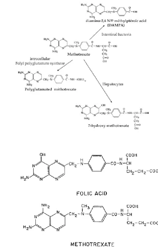
In serum, MTX is metabolised to 7-hydroxy-MTX (7-OH-MTX) and diamino-2, 4-N-10-methylpteroic acid (DAMPA) with the former comprising 3-12% of MTX in circulation (Cutolo et al., 2001). Each metabolite of MTX can compete for cellular uptake. Both metabolites are less effective inhibitors of DHFR and have an increased affinity for the folate transporters as well as FPGS, which means their accumulation minimises MTX-induced toxicity. 7-OH-MTX was also found to be a less efficacious catabolite in rat adjuvant arthritis than MTX (Baggott and Morgan, 2009).
4
was shown to be taken up by the PCFT in particular as the receptor was found to function optimally at the acidic pH of the small intestine (Blits et al., 2013).
1.2.2 Polyglutamation of MTX
Cellular uptake was shown to result in the addition of glutamate residues (polyglutamation) to MTX by folylpolyglutamate synthase (FPGS) which increased the intracellular retention (Walling, 2006). In serum, degradation occurred between 8-15 h or between 8-10 h following administration (van Ede et al., 1998, Patane et al., 2013). It was undetectable in serum after 24 h (Chan and Cronstein, 2013). However, a significant proportion of the MTXglu-4 and MTXglut-5 residues were found 24 h after removal of MTX (Genestier et al., 2000). The same authors showed that the extent of glutamation influenced retention, as a significant fraction of MTX containing 2 and 3 glutamate residues were lost. Thus, MTX can either be metabolised or polyglutamated which increases the cellular retention and resultant half-life (Fig 1.1A).
1.2.3 An overview of the mechanism of action of MTX
5
Figure 1.1 The structure of MTX, its metabolites and folic acid.
(A) Extracellularly MTX is degraded to two metabolites, DAMPA and 7-OH-MTX; the latter by the action of liver aldehyde oxidase. Intracellularly, MTX can be polyglutamated to increase its cell retention. Taken from Genestier et al. (2000). (B) The antagonistic effects of MTX on folate metabolism are in part due to the fact that MTX is a structural analog. Taken from Morgan et al. (2004).
A
6
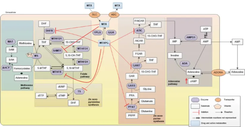
Figure 1.2 An overview of the action of MTX which depends on both uptake and intracellular
retention.
Uptake of MTX is mainly mediated by solute carriers (SLCs), part of the family of RFC and efflux occurs
by adenosine triphosphate (ATP)-binding cassette (ABCs) transporters. Retention of MTX is dependent on
the sum of the addition of glutamate molecules, mediated by FPGS and their removal by
γ-glutamylhydrolase (GGH). MTX and its polyglutamated derivative (MTXPG) inhibit multiple enzymes
involved in folate metabolism. These include enzymes involved in de novo thymidylate (dTTP) and purine
synthesis as well as methionine synthesis, inhibiting nucleotide production and effecting methylation
reactions. As a result of purine inhibition, adenosine also increases. MTX inhibits DHFR, reducing the
availability of reduced folates (THF). MTX also inhibits serine-hydroxymethyltransferase (SHMT),
inhibiting the formation of the 1C donor, 5,10-THF (5, 10-MTHF). MTXPG inhibit
methylene-THF-reductase (MTHFR), which also prevents the synthesis of: 5,10-methylene-THF from 5-methyl-THF
(5-MTHF). This reduction in 5,10-methylene-THF, affects the other folate derivatives 5,10, methenyl-THF
(5, 10-CH-THF) and 10-formyl-THF (10-CHO-THF), the latter crucial to purine synthesis. This inhibition
of MTHFR also affects 5-methyl-THF from 5,10-methylene-THF, the activated folate carrier for
methionine synthesis and methylation reactions by way of the universal methyl donor,
S-adenosylmethionine (SAM). Methionine inhibition raises the levels of the methionine precursor,
homocysteine. MTXPG also inhibits de novo purine synthesis by way of AICAR-formyl-transferase
(AICARFT) and glycinamide-aicar-formyl-transferase (GARFT), the former denoted by the dual enzyme
complex, 5-aminoimidazole- 4-carboxamide ribonucleotide transformylase (ATIC). The resultant increase
in the undepleted substrate, AICAR, inhibits two enzymes in the salvage purine synthesis pathway,
adenosine deaminase (ADA) degrading adenosine to inosine and AMP deaminase (AMPD1) converting
AMP to IMP. The conversion of AMP to adenosine contributes to the increase in adenosine. MTXPG also
inhibits phosphoribosyl pyrophosphate amidotransferase (PPAT), the first committed step in de novo purine
[image:28.595.100.512.76.290.2]7
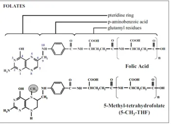
1.3 Folate
Folate is the general name for soluble vitamin B9 and its derivatives which are provided in the diet from leafy vegetables (Danenberg et al., 2016). The main dietary natural folate is 5-methyl-THF and is also the main folate found in blood (Visentin et al., 2012). These ‘natural folates’ are composed of a pteridine ring, a p-aminobenzoic acid and a glutamate moiety (Fig 1.3) (Assaraf, 2007). They are converted to the functional reduced folates that are utilised in biochemical pathways by the action of dihydrofolate reductase (DHFR) (Wibowo et al., 2013).
There are a variety of receptors responsible for uptake of both the oxidised (folic acid) and reduced (tetrahydrofolate moieties) forms of folate. The RFC also known as the folate-binding protein is the main carrier for reduced folates which binds and exchanges these folates for organic phosphates such as adenine nucleotides (Walling, 2006, Assaraf, 2007). The RFC has a much lower affinity for oxidised folate (Km of 200-400 µM)
compared to reduced folates, (Km of 1-3 µM) and thus preferentially takes up the oxidised
form. The RFC are highly expressed in a variety of tissues including peripheral blood leukocytes (Inoue and Yuasa, 2014). Oxidised and reduced folates are taken up by the PCFT also called SLC46A1, which is a member of the SLC family and plays a pivotal role in folate uptake as loss of function leads to hereditary folate-malabsorption (Inoue and Yuasa, 2014). The folate receptors (FRs) also mediate folate uptake via receptor-mediated endocytosis but are only expressed on certain cells (Assaraf, 2007).
Folates are present in the blood at ~10-50 nM (Assaraf, 2007). Intracellularly, folate is present at a much higher concentration of 1-10 µM (Walling, 2006). This is achieved following its polyglutamation by FPGS (Blits et al., 2013). In this reaction, successive glutamate molecules are added at the γ-carboxyl moiety to form a peptide chain with up to eight glutamate molecules, resulting in increased intracellular retention (Visentin et al., 2012).
8
Figure 1.3 The basic structure of the oxidised form of folate, folic acid, and the reduced folate, 5-methyl-THF.
Folic acid contains a pteridine ring attached to a p-aminobenzoic acid moiety which is attached to a variety of glutamyl residues. 5-methyl-THF (5-CH3-THF) contains an
[image:30.595.114.467.105.360.2]9
substituents at the 6 carbon have different functions within these pathways (Fig 1.4). As such folate-deficiency, either diet or drug-induced has far reaching consequences.
1.4 Folate and MTX
In the clinic, folate supplementation, either folinic acid or folic acid proved effective in lowering the MTX-induced side effects (Ortiz Z, 2009, Shea et al., 2014). In addition, it also prevented the discontinuation of treatment (Chladek et al., 2008). In one study it was shown to extend the course of treatment to 48 weeks for 83% of patients compared to 62% on MTX treatment alone (Whittle and Hughes, 2004). Folate supplementation could also increase the dose of MTX used (Cohen and Wolff, 2013). As a result, folate supplementation is now widely used with both low- and high-dose MTX.
10
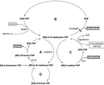
Figure 1.4. The direct and indirect involvement of tetrahydrofolate-derivatives in 1C metabolism.
11
A putative role for folic acid in increasing efficacy of MTX was a result of the competitive inhibition demonstrated for aldehyde oxidase (AO) which catabolises MTX to 7-OH- MTX, a less efficacious catabolite in rat adjuvant arthritis than MTX (Baggott and Morgan, 2009). This suggested that folic acid could increase MTX efficacy by preventing its oxidative degradation. Folinic acid, as a reduced form of folate was thought to work by displacing MTX from THF binding sites. In theory, rescuing the reactions using reduced folate cofactors which include the majority of the folate-dependent pathways would seem to have a greater impact on the anti-folate, if competitive enzyme inhibition was a mechanism of action. However, most studies have found no differential effect of folic acid or folinic acid rescue in reducing the side effects of MTX treatment (Ortiz Z, 2009). This would suggest that perhaps folate rescue is not mediated by overcoming the MTX mediated enzyme inhibition, but rather competition for cellular uptake of MTX by these folates. Indeed, the folate receptor (FR) was shown to have a much lower affinity for MTX than both reduced folates and folic acid (Walling, 2006). Overall, this would suggest that the main mechanism of action of folate rescue is lowering the effective dose of MTX. In practise, this is achieved by adjusting the dose of folate to levels where side effects are reduced, whilst efficacy is still maintained.
12
proliferation of some cells were rescued, minimising side effects whilst others were susceptible to the effects of MTX, maintaining efficacy.
The only real evidence supporting this hypothesis was reports that found no diminution in T cell count in RA patients on MTX treatment, suggesting that MTX was not exerting an anti-proliferative effect. One such report found no change in circulatory T cell counts following MTX treatment (Olsen et al., 1987). Other reports found that treatment with a CD4 monoclonal antibody, which depleted CD4 cell numbers, did not correlate with therapeutic efficacy when given as monoclonal therapy or in conjunction with MTX treatment (Moreland et al., 1995, van der Lubbe et al., 1995). This suggested that a reduction in CD4+ T cell numbers in those studies were not associated with therapeutic efficacy, however, the effect of MTX alone was not investigated. A frequently cited report which demonstrated that long term treatment with MTX actually increased the proportion of CD3+ and CD4+ T cells in circulation further confused the matter (Chan and Cronstein, 2002). However, these authors failed to report an inherent contradiction in this increase in CD4+ T cells as the CD8+ T cell numbers did not increase nor was there a change in the ratio of CD4:CD8 T cells. Furthermore, many reports cite the absence of a reduction in the proliferative responses of T cells from MTX treated RA patients following mitogenic activation ex vivo, as evidence that the efficacy of MTX is not linked to the anti-proliferative effect (Olsen et al., 1987, Furst and Kremer, 1988). One such study measured thymidine incorporation on mitogen-stimulated lymphocyte proliferation after a 2 yr MTX treatment, a finding that implicates the activation state of cells more than the anti-proliferative capacity of MTX (Weinblatt et al., 1988).
13
There are reports documenting cases of folate over-rescue and cases of reduced efficacy, incorrectly cited as over-rescue with high-dose MTX but are now what is understood as low-dose treatment (Cohen, 2013). Folinic acid was shown to normalise 5-Aminoimidazole-4-carboxamide (AICA) urine levels following low-dose MTX treatment suggesting that it had prevented the MTX-mediated inhibition of aica-ribotide-formyl-transferase (AICARFT) (Morgan et al., 2004). It was also shown to reverse the ability of MTX to lower the levels of RF (Chan and Cronstein, 2010). Folic acid also demonstrated antagonistic effects. It reduced intracellular red blood cell (RBC) MTXGlu and plasma homocysteine levels (Chladek et al., 2008, Hazra et al., 2010). Cases of over-rescue were also found with folinic acid supplementation (Ortiz Z, 2009). This was explained as competition of folinic acid with MTX for uptake for the same folate transporter (Visentin et al., 2012).
Because it is not known whether folate supplementation acts by competing for uptake of MTX or limiting the effects of folate antagonism, experiments involving folate rescue from the inhibitory effects of MTX do not offer any clear explanation as to how MTX is exerting these differential effects. Competition for uptake could also explain how folate reduces side effects whilst maintaining efficacy in some cells because of differential intracellular concentrations of MTX. Thus, since cases of reduced efficacy and over-rescue have been reported, the proliferative effect of MTX may be crucial to its anti-inflammatory action.
1.5 Anti-CD3-activation of T cells-a model of RA
14
Some studies have looked at universal activation of cells, using phorbol myristate acetate (PMA), a cell permeable molecule, that directly activates protein kinase C, inducing signalling events resulting in universal cellular activation (Suzawa et al., 1984). Ionomycin, a calcium ionophore is used alongside PMA, converging on this same signalling pathway (Voet, 1990). Other activators such as phytohaemagglutinin (PHA) or concavalin A, both plant lectins, cause select cellular-activation through cross-linking of glycans on TCRs, or immunoglobulins which are present on certain cell types. Indeed the number of N-glycosylation sites on the TCRαβ chains and on the γϐ are extensive and the O-glycosylation sites on the T cell co-receptor, CD8 are also well characterised, reviewed in Hounsell and Davies (1993). Finally, superantigens can activate antigen presenting cells (APC) as well as the T cell it is presented to, causing polyclonal activation (Li et al., 1999). However this is not the best model of the inflammation characterising RA. The inflammation in RA is mainly attributed to overly-activated T cells, which recognise self-antigen. Thus, the use of PMA to universally activate all cells does not seem fitting in this study.
15
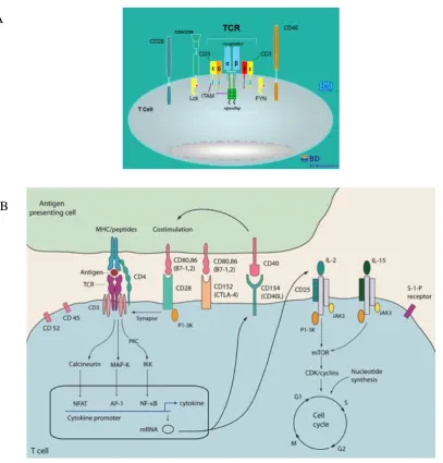
Figure 1.5. Signalling pathways leading to the induction of T cell-activation.
(A) The TCR synapse. (B) The signalling pathways initiated upon ligation of the TCR. The TCR ligates the antigen-bound antigen-presenting cells and the CD28 receptor ligates the CD80/86 ligand on the antigen-presenting cell. This binding transduces activation through calcineurin, RAS-mitogen-activated protein kinase (MAP-K), and protein kinase C (PKC) signalling pathways, leading to the activation of NFAT, AP-1 and the nuclear factor-kappa B (NF-kB). These transcription factors transcribe genes involved in other activation-dependent processes such as IL-2 production. The upregulation of receptors for IL-2 allows for this third signal to orchestrate cell proliferation through the induction of mammalian target of rapamycin (mTOR). Taken from (A) (P.Weller) and (B) Hunt and
Haddad (2008).
A
16
In vitro, this process is mimicked through artificial ligation of the CD3 receptor, part of the T cell receptor (TCR) complex and the CD28 receptor by supplying the reciprocal antibodies, anti-CD3 and CD80/86 respectively (Goronzy et al., 1987). However, the concentrations of anti-CD3 used (1 µg/ml) induce polyclonal T cell-activation, whereby nearly 100% of T cells are activated (Kleiveland, 2015). Thus, anti-CD3-activation amplifies what happens on a physiological level. Even reducing concentrations of anti-CD3 to induce sub-optimal activation is not conducive to in vivo conditions as the presentation of antigen to one T cell induces the select proliferation of that specific T cell, recognising that particular antigen. Despite this limitation, the use of anti-CD3 was the preferred activator for this study.
Activation of a mixed population of immune cells, such as those within the PBMC cohort, negates the need to supply the co-stimulatory molecule, which is already provided by the antigen-presenting cells present. It is also more representative of conditions in vivo. Within the PBMC population, activation with anti-CD3 occurs in cells carrying a CD3 receptor, of which ~60% of the PBMC cohort are CD3+ T cells, comprising CD4+ T cells and CD8+ T cells in a 2:1 ratio (Kleiveland, 2015). Following activation, a program of differentiation into distinct subsets of CD4+ T cells can be initiated in the presence of exogenous polarising cytokines, such as IL-4, IL-12 or IFN- γ, to differentiate these CD4+ T cells, into Th2 or Th1 respectively (Zhu et al., 2010). Even the production of endogenous cytokines by these T cells or other cells in the PBMC population could contribute to partial polarisation into either a Th1 or a Th2 phenotype (Swain, 1995). For this study however, only activation of T cells was required so PBMCs were activated in the absence of polarising cytokines (Th0). The effect, if any of endogenous cytokines on the differentiation into effector T cells was thus not a concern.
1.6 One-Carbon metabolism
17
component of 1C metabolism, specifically methionine synthesis acts to replenish the levels of the free reduced-folate, from 5-methyl-THF, the most abundant form of folate in serum (van Ede et al., 1998). 1C metabolism includes the synthesis of DNA and RNA precursors, S-adenosylmethionine (SAM), the universal methyl donor for methylation reactions and also serves to metabolise the amino acids serine, glycine, tryptophan, histidine as well as choline.
A schematic of the interplay of 1C pathways of the cytoplasm, mitochondria and nucleus is shown in Fig 1.6. It is an intricate pathway comprising three compartments, connected only by select one-carbon donors and sharing one fundamental reaction. Each compartment has its own de novo thymidylate synthesis pathway-via thymidylate synthase (TS) and its substrate, 5,10-methylene-THF, itself synthesised by compartment-specific isozymes of serine- hydroxymethyltransferase (SHMT). In the mitochondria this was found to be SHMT2 and was localised to the mitochondrial matrix (Anderson et al., 2011). This was discovered when isolated nuclei from mouse liver synthesized thymidylate, following the addition of dUMP, NADPH and serine (Anderson and Stover, 2009). Isolated mitochondria were also found to have de novo thymidylate synthesis enzymatic capability when CHO cells also produced thymidylate when provided with serine and NADPH (Anderson et al., 2011). The reduction of unreduced folate precursors is also a commonality between compartments, mediated by dihydrofolate reductase (DHFR).
18
Figure 1.6. The interplay of 1C donors (blue) and THF-derivatives (green) for methionine,
thymidylate and purine synthesis between the cytoplasm, nucleus and mitochondria.
Reduced folate (THF) is synthesised from DHFR and unreduced folate, reaction 13. Formate enters the 1C
metabolism pathway in both the cytoplasm and mitochondria, generating the one-carbon donor
5,10-methylene-THF (CH2-THF) by reactions 1-3; catalyzed by 10-formyl-THF synthase, 5,10-methenyl-THF
(CH+-THF) cyclohydrolase, and 5,10-methylene-THF dehydrogenase, respectively. This substrate is also
synthesised from the one-carbon donor glycine by the mitochondrial glycine cleavage system (GCS);
reaction 5. Dimethylglycine (DMG) derived from the betaine precursor, choline via betaine-homocysteine
methyltransferase, reaction 14; and sarcosine are also sources of 5,10-methylene-THF via dimethylglycine
dehydrogenase and sarcosine dehydrogenase; reactions 8 and 9 respectively. This one-carbon donor is also
generated by serine, via serine hydroxymethyltransferase (SHMT) in each compartment, with the nuclear
and mitochondrial compartments denoted by n and m, respectively. Thymidylate synthase (TS) synthesises
thymidylate using 5,10-methylene-THF and dUMP in the de novo thymidylate synthesis pathway in each
compartment. 5,10-methylene-THF can be converted to 5-methyl-THF via 5,10-methylene-THF reductase,
reaction 6. This one-carbon donor is utilized by methionine synthase for the synthesis of methionine from
homocysteine (Hcy), reaction 7. Methionine is the precursor for S-adenosylmethionine (AdoMet) which is
interconverted to S-adenosylhomocysteine (AdoHcy). Free THF is regenerated from formyl-THF by
10-formyl-THF dehydrogenase, reaction 11 and methylene-THF dehydrogenase, (MTHFD1L), reaction 1m
and methionyl-tRNA formyltransferase, reaction 12. End products of one-carbon metabolism are in red.
19
mitochondria has also been shown in yeast and in vitro (Barlowe and Appling, 1988, Garcia-Martinez and Appling, 1993). Glycine was also taken up by the mitochondria as demonstrated by 1-14C labelling experiments and converted to CO2 (Hampson et al.,
20
gene had two isoforms, one containing a mitochondrial leader sequence, which directed its fate to the mitochondria, affirming the impermeability of the mitochondria to polyglutamates (Lawrence et al., 2014).
The importance of adequate function of the 1C pathway, in particular the mitochondrial compartment, is demonstrated when deficiencies in this pathway can cause neural tube defects (NTDs). Inactivating mutations in the GCS, an enzyme localised to the mitochondria is a predisposition for neural tube defects, demonstrating the importance of glycine as a provider of 5,10-methylene-THF (Brosnan et al., 2015). These defects were prevented by replenishing the methyl donors, specifically formate which normalized the folate profile and prevented neural tube defects (Pai et al., 2015). Although the defects could not be completely reversed, as glycine levels in the plasma and urine were still elevated, it illustrates how important the 1C donors are, of which the mitochondria is the main receiver. These 1C donors and the reactions of their metabolism, have roles in DNA synthesis, and the synthesis of methionine, the precursor to the universal methyl donor SAM for reactions involving RNA, DNA, proteins, phospholipids and amino acids (Cronstein, 1996). Methylation is also crucial for polyamine synthesis, the proteins involved in cell proliferation, regulation, differentiation, protein synthesis and immune mediated cell reactions (van Ede et al., 1998). As such, perturbations of this 1C metabolism can have major consequences on cellular function.
1.7 The anti-folate MTX affecting 1C metabolism
21
was able to inhibit thymidylate production from isolated mitochondria in CHO cells (Anderson et al., 2011). Thus, the MTX-mediated effect on purine, thymidylate and methionine synthesis, as well as methylation reactions via the reduction of the methyl donor, SAM could permeate every 1C metabolism compartment.
Table 1.1. The effects of low-dose MTX on 1C metabolism
1.7.1 The effect of MTX on methionine synthesis
MTX as well its polyglutamate derivative, MTXGlu, inhibit methionine synthesis by reducing the levels of the 1C donor, 5-methyl-THF required for its synthesis. This has implications for cellular methylation, including polyamine synthesis and cell function. By inhibiting DHFR, MTX depletes the reduced folate THF, and the subsequent activated 1C donor carrier, 5-methyl-THF, inhibiting the formation of methionine from homocysteine (Cronstein, 2005). The increasing levels of DHF, were also shown to inhibit methionine synthase (MS) (Baggott et al., 1986). MTXGlu's were shown to inhibit 5,10 methylene-THF reductase, required for the conversion of 5,10-methylene-methylene-THF to 5-methyl-methylene-THF. Although it has also been suggested that MTX could inhibit MS directly, little has been shown in this regard (van Ede et al., 1998).
22
2012). It was also shown to have consequences on polyamine synthesis, which have multiple roles in proliferation and cell function (Cronstein, 1996).
Additional insults to cell methylation by MTX arise from the concurrent increase in homocysteine, which had not been depleted for methionine synthesis. Homocysteine has been shown to form s-adenosylhomocysteine (SAH), which is an inhibitor of methyltransferases including isoprenylcysteine carboxyl methyltransferase (ICMT), involved in the methylation of RAS, a crucial G protein involved in cell growth signalling pathways. Indeed, MTX (10 µM) caused a 90% reduction in Ras methylation in DK0B8 cells (Winter-Vann et al., 2003). Even artificially increasing SAH, by incubating cells with homocysteine and adenosine in the presence of adenosine deaminase inhibitor was shown to inhibit lymphocyte and mononuclear cell function (Cronstein, 1996).
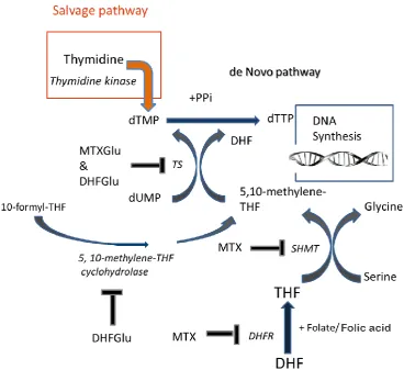
1.7.2 The effect of MTX on thymidylate synthesis
MTX (1µM) was shown to reduce TS activity by 20% but there was only a partial reduction in the TS substrate, 5,10-methylene-THF, which suggested that other mechanisms of inhibition were in place (Genestier et al., 2000). Inhibition of de novo thymidylate synthesis by MTX was thus found to be a multifactorial process, which included TS inhibition by both polyglutamated DHF (DHFGlu) and MTXGlu as well as partial-substrate depletion (Genestier et al., 2000). DHF which accumulated in response to DHFR inhibition, inhibited TS (Baggott et al., 1986). MTXGlu blocked TS, with the degree of binding increasing with polyglutamation. The Ki for MTX with one glutamate residue was found to be 13 µM, but for two and five glutamate residues it decreased to 0.17 µM and 0.047 µM respectively (Allegra et al., 1985a).
23
Figure 1.7. MTX enzyme inhibition and substrate depletion results in the inhibition of de novo thymidylate synthesis.
24
The importance of the one-carbon folate carrier 5,10-methylene-THF for TS was clear by its numerous carbon sources (Barlowe and Appling, 1988, Garcia-Martinez and Appling, 1993). Serine was found to be a major source of 5,10-methylene-THF, both for methionine synthesis as well as for thymidylate synthesis. C-13 labelled serine provided nearly all of the methyl groups required for homocysteine remethylation to methionine, via the interconversion of 5,10-methylene-THF to 5-methyl-THF (Brosnan et al., 2015). Knockdown of SHMT2, was found to prevent the generation of 5,10-methylene-THF from serine and diminished thymidylate levels (Anderson et al., 2011). Glycine was also shown to generate 5,10-methylene-THF via the GCS in the mitochondria (Hampson et al., 1983). The 1C donors sarcosine and dimethylglycine were also shown to be sources of 5,10-methylene-THF via sarcosine dehydrogenase and dimethylglycine dehydrogenase, respectively (Mitoma and Greenberg, 1952, Lewis et al., 1978, Barlowe and Appling, 1988).
1.7.2.1 The regulatory role of thymidylate
25
µM and 100 µM) exerted maximal negative feedback on this pathway, reducing dCTP levels by 80% and causing 50% inhibition of deoxycytidylate deaminase, respectively. Thymidylate was also shown to inhibit CDP reductase, upstream of dCMP preventing an increase in dCMP levels (Jackson, 1978).
1.7.3 The effect of MTX on uracil
This waning in the negative feedback mechanisms normally exerted by thymidylate leads to an increase in dUMP and other uracil moieties, such as deoxyuridine or uridine. MTX (10 µM) treatment caused deoxyuridine levels to increase ~10^3 fold and also caused an increase in dUMP concentration in cultured human lymphoblasts (Goulian et al., 1980b, 1980a). However, there is also evidence found where MTX did not lead to an increase in dUMP (FitzGerald and Wick, 1987). Although normal cell processes seek to minimise dUTP levels by the actions of UTPase, which hydrolyses dUTP to dUMP, increasing dUMP levels, the MTX-mediated increase in dUTP was suggested to outcompete the maximal workings of the dUTPase to keep dUTP levels low. Even at the rate of hydrolysis of UTPase equal to the rate of formation of dUTP, MTX treatment was suggested to lead to a gradual increase in dUTP levels (Goulian et al., 1980a).
1.7.3.1 The correlation of uridine moieties in circulation with uracil incorporation in DNA
26
indicating that preferential uptake of deoxyuridine had occurred. The group found that this was not because plasma uridine concentrations did not reflect the increase in uridine or deoxyuridine in the diet, as plasma levels of these nucleosides significantly increased. Other evidence was provided by depleting thymidine levels in mouse embryonic fibroblasts (MEF) using thymidylate synthase inhibitors, 5-flurouracil (5-FU) (1 µM) and 5-fluro-deoxyuridine (5-FdUrd) (50 nM), which resulted in a high dUMP:dTTP ratio, but did not lead to an increase in uracil content of DNA (Nielsen et al., 2007). Studies have also investigated whether folate treatment can reduce uracil incorporation in DNA, however, the level of uracil misincorporation in white blood cells was not reduced compared to those without supplementation (Hazra et al., 2010).
However, there are reports where increasing uracil concentrations have led to an increase in uracil incorporation in DNA. Deoxyuridine accumulated in mitochondrial DNA following impaired de novo dTMP synthesis (Anderson et al., 2011). The addition of deoxyuridine also increased uracil incorporation in HeLa cells, grown in folate-deficient or replete media (Duthie and McMillan, 1997). Elevated uracil in DNA has also been shown to occur in cells starved of folate, akin to incubation in the anti-folate drug MTX. At folate concentrations found in the human plasma of folate-deficient individuals, (12 nM), CD4+ and CD8+ T cells had significantly increased uracil content in DNA as compared to folate-replete cells (Courtemanche et al., 2004). Incubating human lymphocytes in folate-deficient media also increased the level of incorporated uracil (Duthie and McMillan, 1997).
The presence of uracil in DNA can occur spontaneously through the deamination of cytosine already present or through misincorporation. Although one molecule of dUMP is incorporated into DNA for every 10^5 dTMP in normal human lymphoblasts, the presence of uracil in DNA is dependent on the efficiency of the uracil removal repair mechanisms in place (Goulian et al., 1980a).
27
It is possible that in certain cell types, this removal mechanism is more efficient and thus can withstand elevated uracil levels in circulation, preventing a build-up in DNA.
1.7.4 The inhibitory effect of MTX on purine synthesis
28
Figure 1.8. MTX polyglutamates inhibit de novo purine synthesis, whilst AICAR metabolites inhibit the salvage pathway of purine synthesis.
29
which have been shown to inhibit ribonucleotide reductase, necessary for DNA synthesis (van Ede et al., 1998).
1.7.5 Enzyme inhibition by unreduced folate
Folate antagonism by the anti-folate, MTX is partly mediated by the antagonism of the unreduced (DHF) folate pool which increases following treatment. These unreduced folates with dihydro or oxidised pteridine rings were shown to inhibit TS, GARFT, MS, 5 forminotetrahydrofolate cyclodeaminase, 5, 10 methenyltetrahydrofolate cyclohydrolase and methylenetetrahydrofolate reductase (Baggott et al., 1986, Blits et al., 2013). Treatment of human MCF-7 breast cancer cells with MTX (1 µM) increased DHFGlu to about 20% of the total folate pool (Sant et al., 1992). Polyglutamation was shown to endow these unreduced folates with further inhibitory properties. DHFGlu was also shown to inhibit AICARFT (Allegra et al., 1985b). However, when DHF levels had built up, these unreduced folates were shown to antagonise the effects of MTX by competing for binding for the few available binding sites of DHFR needed to maintain THF cofactor pools within cells (Visentin et al., 2012).
1.8 The anti-proliferative effect of MTX
30
where both the number of antibody forming cells and a corresponding decrease in antibody production was found (Rosenthal et al., 1988). However, there has been a report which shows that low-dose MTX treatment has no anti-proliferative effect. This was shown following MTX (0.05 µM or 1 µM) treatment with concavalin A for 4 days (Tohyama et al., 2013).
MTX also inhibited the proliferation of immortalised cell lines. AML 193 human myeloid leukemic cells stimulated with the growth factor, GM-CSF, showed an increase in inhibition from 49-89% following MTX (2.2 µM) treatment (Ciaiolo et al., 1988). The growth of Erlich tumour ascites was also inhibited by MTX (2 µM) (Li and Kaminskas, 1984). So too was the growth of Human Leukemic-T lymphoblasts CCRF-CEM cell line and Human Jurkat T lymphoblastsinhibited by MTX (100 nM) (da Silva et al., 1996). The anti-proliferative effect of folate-deprivation has also been shown in cells cultured in folate-deficient media. Human mononuclear cells that were stimulated with IL-2 and PHA showed a dose-dependent reduction in proliferation when folate was reduced from (12 nM- 6 nM) (Courtemanche et al., 2004). Lymphocytes also failed to proliferate in response to stimulation with 0.5% PHA following a 6 day folate-deficient media incubation (Duthie and McMillan, 1997).
The inhibition of cell division does not necessarily prevent the increase in cytoplasmic volume accompanying the increased intake of cellular building blocks. This unbalanced cell growth leads to an increase in cell size and fragmented DNA, if the cells cannot divide. Indeed, MTX treatment of monocytes led to enhanced cellular fusion and the formation of multiple nucleated giant cells upon phorbol esters stimulation (Tian and Cronstein, 2007).
1.8.1 Megaloblastic anaemia
31
Figure 1.9. The clinical manifestations of folate or cobalamin deficiency.
32
cell growth without division. It is thought to occur as a result of a disturbance of DNA synthesis in the bone marrow (Hoffbrand, 1973). Although defects usually occur in the bone marrow, chromosome breaks have been observed in both bone marrow cells and leukocytes from patients with megaloblastic anaemia (Keller and Norden, 1967).
The anti-folate, MTX induces a state of folate-deficiency. As such, megaloblastic anaemia has been reported during low-dose MTX treatment in RA patients. The mean corpuscular volume was also found to increase in patients on low-dose MTX treatment. Low-dose MTX treatment has also been shown to result in DNA fragmentation (see Section 1.10.1) (Weinblatt and Fraser, 1989). However, megaloblastic anaemia was not shown to occur due to the reduction in thymidylate following folate-deficiency, as PHA-stimulated lymphocytes from untreated megaloblastic anaemic patients were not found to have decreased thymidylate concentrations (Hoffbrand, 1973). This suggested that megaloblast formation following low-dose MTX treatment may be due to a different consequence of folate-deficiency.
1.9 The apoptotic effect of MTX
The extent of apoptosis induced by MTX has been shown to depend on the activating stimulus, the growth phase of the cell and the dose and duration of MTX treatment. In immortalised cell lines it is generally accepted that MTX causes cell death but at various doses. MTX (100 nM) caused significant cell death in Human Leukemic T lymphoblasts and Jurkat T lymphoblasts (da Silva et al., 1996). MTX (1 µM) significantly reduced cell viability in A549 cells (Huang et al., 2011). MTX (2 µM) caused significant cell death in Ehrlich ascites tumour cells (Li and Kaminskas, 1984). Higher doses of MTX were required to cause cell death in Jurkat T cells as MTX (0.1 µM) did not significantly increase cell death after 48 h (Spurlock et al., 2011). 48 h also proved too short a duration in CaSki and NRK cells, as treatment with MTX (1 µM) did not induce cell death after this time (Mazur et al., 2009).
33
control (Strauss et al., 2002). The same concentration of MTX caused (30% and 60%) apoptosis in PMA- and ionomycin-activated PBMCs (Quemeneur et al., 2003). MTX (0.05 µM) also caused significant increases in cell death after 4 days treatment of PBMCs cultured in concavalin A or PHA (Tohyama et al., 2013). Up to 1 µM MTX was shown to induce apoptosis in PBMCs already activated with PHA (Herman et al., 2003, Swierkot et al., 2004). Folate-deficiency also increased the apoptosis of CD4+ and CD8+ T cells activated with PHA (Courtemanche et al., 2004).
The differential cytotoxic effect of MTX in non-dividing verses dividing cells was verified, when PBMCs stimulated with tetanus toxin (TT) or candida albicans (CA) showed no increase in apoptosis in non-dividing cells. In dividing cells however, an increase in the proportion of apoptotic cells was found following high-dose MTX (22 µM) treatment (Nielsen et al., 2007). This was also the case for dividing T-helper cells, stimulated with TT or CA, whereby MTX treatment caused a six-fold increase in the proportion of apoptotic cells (Herman et al., 2005). Further evidence was shown when peripheral blood lymphocytes (PBL) activated with either PHA or concavalin A underwent apoptosis following MTX (1 µM) treatment, but not in the resting lymphocytes (Genestier et al., 1998). The same authors also showed that lymphocytes exposed to MTX (1 µM) treatment following activation with anti-CD3, or PMA and ionomycin, also induced apoptosis but not in the resting lymphocytes. However, one report was found where MTX had no effect on cell viability which was more than 95% after whole blood-activation with CD28 and anti-CD3 (Haroon et al., 2008). A possible explanation for these findings is that these cultures were isolated from RA patients on low-dose MTX treatment, which may have become desensitised to the effects of MTX.
1.9.1 DNA damage in response to MTX
34
et al., 1997). Human hepatoma HepG2 cells also showed DNA fragmentation preceding apoptosis following folate-deficiency (Huang et al., 1999). Supplementation with deoxyuridine (100 µM) significantly increased the DNA damage in HeLa cells cultured in folate-replete conditions, suggesting that uracil moieties could damage even healthy cells (Duthie and McMillan, 1997).
MTX treatment was also shown to lead to DNA fragmentation (Jackson, 1978). MTX (2 µM) led to DNA strand breaks in NIH 3T3R cell and in Ehrlich ascites tumour cells (Li and Kaminskas, 1984, Lorico et al., 1988). MTX (1µM) induced DNA strand breaks in PHA-activated PBLs (Genestier et al., 1998). MTX (60 nM) was also found to induce DNA fragmentation in rat intestinal epithelial cells (Papaconstantinou et al., 2001).
However, DNA fragmentation does not necessarily correspond to cytotoxic effects on cell viability or proliferation. Uridine supplementation resulted in DNA fragmentation but did not correlate to cytotoxic effects on cell proliferation (Martiniova et al., 2015). The addition of deoxyuridine (100 µM) had no effect on cell proliferation in HeLa cells cultured in folate-replete conditions (Duthie and McMillan, 1997). A higher dose of deoxyuridine (10 mM) also had no effect on proliferation in mouse lung fibroblast CCL39 cells (Ingraham et al., 1986). Treatment with TS inhibitors 5-FU and 5 FrUrd, increased uracil in the DNA of Ung -/- MEFs leading to a reduction in cell proliferation but had no effects on cell viability (Andersen et al., 2005).
Indeed one form of uracil, deoxyuridine has even been shown to be protective. Deoxyuridine administered to folic acid-deficient mice, rescued these mice from neural tube defects (NTDs) resulting from DNA fragmentation (Martiniova et al., 2015). This was shown to be a result of increasing plasma folate levels as deoxyuridine supplementation raised folate levels compared to control mice. Indeed, deoxyuridine was shown to replenishing dUMP levels via thymidine kinase and thus enter the de novo pathway of thymidylate synthesis (Garavito et al., 2015).
35
DNA vulnerable to nucleases. Thus the apurigenic sites or DNA strand breaks induced by the repair mechanism were found to be toxic. Little difference was found in the cytotoxicity incurred in DNA repair knockouts compared to healthy controls. One such study treated MEFs with the thymidylate synthase inhibitor, 5-flurouracil (5-FU) (5 µM) and found only a slight difference in growth inhibition in Ung-/- compared to WT MEFs despite the increase in the levels of uracil in the genome (Andersen et al., 2005). Apoptosis following a 24 hr MTX (100 µM) treatment occurred more frequently in MMR-competent Hela cells and the human colon tumour cell line, HCT116 compared to those unable to remove uracil (Frouin et al., 2001). Cytotoxicity upon removal of uracil has been observed for several cytotoxic drugs, including alkylating and methylating agents as well as platinum compounds (Frouin et al., 2001). Even in bacteria, the toxicity of thymine starvation was reduced when the Uracil-DNA glycosylase was impaired (Ingraham et al., 1986). Of further note, supplementation with Uracil-DNA-glycosylase, significantly increased the DNA damage of lymphocytes cultured under folate-deficient conditions (Duthie and McMillan, 1997).
However, it is impossible to study the toxic effect of uracil alone, as it is still unclear how much uracil toxicity is attributed to the toxicity induced by thymidine-deprivation.
36
1.10 Mitochondrial function
The pathways of one-carbon metabolism (1C) and ATP synthesis by the electron transport chain (ETC) are interdependent. 1C metabolism was shown to fuel the ETC by supplying 50% of the NADPH reductive equivalents via the mitochondrial SHMT. This was shown to occur using the mitochondrial isozyme, methylene-THF dehydrogenase (MTHD2) which can produce either NADH/NADPH during the conversion of 5,10-methylene-THF to 5,10-methenyl-THF. The malic enzyme and the pentose phosphate pathway accounted almost equally for the remaining 50% (Brosnan et al., 2015). Adequate flux of these one-carbon donors in the mitochondria was shown to be dependent on the respiratory state of the mitochondria, maintained by the ETC (Garcia-Martinez and Appling, 1993). This process was found to be dependent on an adequate mitochondrial inner-membrane potential and a proton gradient to drive the synthesis of ATP from the ATP synthase (Fig 1.10) (Voet, 1990).
1.10.1 The mitochondrial inner-membrane potential and MTX
The importance of a functioning mitochondrial inner-membrane potential (∆Ѱm) is
demonstrated by the toxic effects of compounds that affect this potential. Dissipation of the ∆Ѱm is one of the first steps preceding apoptosis, prior to cytochrome c release.
Protonophores that dissipate this ∆Ѱm uncouple ATP synthesis from oxygen consumption,
so that the cell cannot utilise the energy contained in the reductive equivalents to make ATP. Matrix condensation, a step in the apoptotic pathway, can even be induced in mitochondria isolated from healthy cells, by either denying oxidisable substrates or by protonophores that dissipate the ∆Ѱm(Gottlieb et al., 2003). Oligomycin, an ATP synthase
inhibitor, prevents the utilisation of this ∆Ѱm, by hyperpolarising the ∆Ѱmand dissipating
cellular respiration, inhibiting the workings of the cell (Jeremy M.Berg, 2007).
In light of the interdependence of these processes, insults to 1C metabolism, drug- or damage-induced, can reflect changes in mitochondrial function; in particular the ∆Ѱm,
limiting energetic output and impeding cellular function. In addition to affecting 1C metabolism, MTX was also shown to affect the ∆Ѱm. PHA-activated PBL treated with
MTX (1 µM) had a reduced ∆Ѱm (Genestier et al., 1998). The same dose significantly
37
Figure 1.10. Electron transport and the production of ATP by the electron transport chain.
Oxidation of substrates malate and succinate result in the reduction of the cofactors, producing NADH and FADH2 whose electrons enter the ETC at complex I and II. The
electrons are transferred between complexes (arrows), promoting proton (H+) transport from the matrix to the intermembrane space. This creates an electrical and chemical gradient, called the mitochondrial inner-membrane potential ∆Ѱm, used to drive the
38
(Huang et al., 2005, Chang, 2013). Folate-deprivation depolarised the ∆Ѱm in cultured
embryonic cortical neurons and in DRG neurons (Ho et al., 2003, Tjiattas et al., 2004). Another study investigated the effect of MTX on the ∆Ѱmof peripheral lymphocytes from
South African RA patients, using the dimeric-potential dependent dye, JC-1 (Moodley et al., 2008). However, their results have not been taken into consideration since neither the concentration of JC-1, nor the wavelength us