E A continuous and large-scale process can be economically competitive to hexane-based opera-tions.
Exploiting the commonality of high pressure be-tween supercriticalSuid and extrusion processing op-erations, a hybrid unit operation called supercritical Suid extrusion (SCFX) has been recently developed. This new process permits generation of microcellular structure at low temperature by using SC-CO2 as a blowing agent instead of steam to puff the extru-date, thus decoupling the conventional dual role of water, which otherwise serves both as a blowing agent as well as a plasticizer. The use of super-criticalSuid also permits deposition of solute into the extrudate matrix.
SigniRcant progress has also been made in the anal-ysis of food and related materials using SFE with SFC. Sample preparation for analysis often requries orders of magnitude more time than the analysis itself and the use of supercritical Suids obviates the need for hazardous organic solvents with no additional treat-ment prior to identiRcation of the analyte by other techniques such as GC, GC-MS, FTIR, etc. The solu-bility of lipid-like materials in SC-CO2 ranges from 1 to 30 wt%, depending on the density of the Suid used, and therefore, SFE has become a method of choice for rapid extraction of fats and oils from a variety of food matrices such as animal, vegetable, grain and seafood products. Other successful applica-tions include extraction of fat-soluble vitamins, pesti-cides, sterols, and fatty acids. As an analytical tool, SFC has also made signiRcant progress over the past decades but has yet to prove its superiority over the more conventional techniques.
See also: II/Extraction: Supercritical Fluid Extraction. III/Food Technology: Supercritical Fluid Chromatogra-phy. On-Line Sample Preparation: Supercritical Fluid Extraction.
Further Reading
Brunner G (1994)Gas Extraction. New York: Springer. Chang AD and Randolph AD (1989) Precipitation of
microsize organic particles from supercritical Suids.
AICHE Journal35: 1876}1882.
Charpentier BA and Sevenantes MR (eds) (1988) Tech-niques and Applications.Supercritical Fluid Extraction and Chromatography. ACS Symposium Series 366. Washington DC: American Chemical Society.
Friedrich, JP (1984) Supercritical CO2extraction of lipids
from lipids containing materials. U.S. Patent 4,466,923. Lee BC, Kim JD, Hwang KY and Lee YY (1994) In: Rizvi SSH (ed.) Supercritical Fluid Processing of Food and Biomaterials. New York: Chapman and Hall.
King JW and Friedrich JP (1990) Quantitative correlations between solute molecular structure and solubility in supercritical Suids. Journal of Chromatography 517: 449}458.
McHugh M and Krukonis VA (1994)Supercritical Fluid Extraction. Boston: Butterworth-Heinemann.
Rizvi SSH, Mulvaney SJ and Sokey AS (1995) The com-bined application of supercritical Suid and extrusion technology.Trends in Food Science Technology 6(7): 232}240.
Rizvi SSH (ed.) (1984) Supercritical Fluid Processing of Biomaterial. New York: Blackie Academic and Profes-sional.
Stahl E, Quirin KW and Gerard D (1987)Dense Gases for Extraction and ReTning. New York: Springer-Verlag. Taylor LT (1996) Supercritical Fluid Extraction. New
York: John Wiley.
Williams DF (1981) Extraction with supercritical gases.
Chemical Engineering Science36(11): 1769.
Wong JM and Johnston KP (1986) Solubilization of bi-omolecules in carbon dioxide based supercriticalSuids.
Biotechnology Progress2: 29}39.
Yu ZR, Rizvi SSH and Zollweg JA (1992) Extraction of oil from evening primrose seed with supercritical carbon dioxide.Journal of Supercritical Fluids5: 114. Zosel K (1978) Separation with supercritical gases:
practi-cal applications. Angewandte Chemical International Edition English17: 702.
FORENSIC SCIENCES
Capillary Electrophoresis
J. Sadecka, Slovak Technical University, Bratislava, Slovak Republic
Copyright^ 2000 Academic Press
Several slab electrophoretic techniques have fre-quently been used to discriminate between red cell
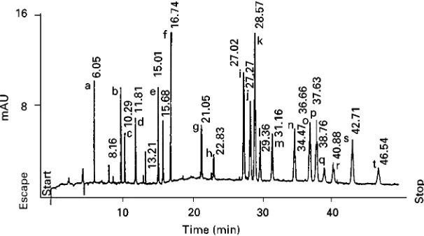
Figure 1 Typical example of a MEKC separation of the components of a drug mixture. Buffer: 25 mmol L\1borate (pH 9.24)}20% methanol}100 mmol L\1SDS. Capillary: bare fused silica, i.d. 50m, total length 55 cm (35 cm to detector). Potential 20 kV. UV detection at 200 nm. Peak identification: a, caffeine; b, barbital; c, pentobarbitone; d, morphine; e, narceine; f, 6-monoacetylmorphine; g, codeine; h, nalorphine; i, lidocaine; j, procaine; k, heroin; l, flunitrazepam; m, acetylcodeine; n, thebaine; o, papaverine; p, amphetamine; q, narcotine; r, cocaine; s, diazepam; t, tetracaine. (Reprinted with kind permission of Elsevier Science from Tagliaro F et al. (1996) Journal of Chromatography A 735: 227}235.)
year. Today, forensic applications of CE include anal-ysis of drugs of abuse, gunshot residues, explosives, pen inks and toxins as well as polymerase chain reaction (PCR) ampliRed DNA.
Drugs of Abuse
One of the major tasks for forensic laboratories is the analysis of illicit and controlled drugs, in both the seizure and biological samples.
Seizure Samples
Seizure samples are analysed in order to identify the major compounds. In addition, the determination of trace compounds permits the samples to be allocated to the source and production procedures. Seizure samples may consist of a mixture of acidic, neutral and basic compounds that may be nonpolar and/or polar. At least two independent analytical parameters should be used to establish the identity of the drug, and infrared spectroscopy and thin-layer chromatog-raphy (TLC) are widely used for this purpose. Quan-titation is usually carried out by gas chromatography (GC) and high performance liquid chromatography (HPLC). GC is a high resolution technique, but prob-lems can arise for thermally degradable, polar and nonvolatile substances. HPLC is less suited to drug proRling, because it is a relatively low resolution technique compared with GC.
CE is a relatively new technique is forensic drug analysis. The three main separation mechanisms have been used for seizure samples: (i) low pH to analyse basic compounds; (ii) high pH to analyse acidic
com-pounds; and (iii) MEKC to analyse neutral and/or charged compounds.
Most abused drugs are bases which are generally water-soluble and ionized as cations at low pH. The use of simple electrolyte solutions such as phosphate, citrate and formate at pH values of 2}3 gives a useful initial separation. Basic drugs can be analysed by TLC and HPLC, but interactions with the stationary phases can lead to peak tailing. This problem does not occur so frequently in CE. In addition, these simple electrolytes have low background UV absorbance and can be operated at low wavelengths of 190}200 nm, where many drugs have signiRcantly enhanced UV absorbance coefRcient. CE at low pH can be used to detect by-products in puriRed codeine, to investigate amphetamine derivatives in Ecstasy tablets, and for assay for various pharmaceutical formulations which contain 1,4-benzodiazepines and phenothiazines.
At high pH the migration direction of acidic com-pounds is against the electroosmotic Sow, which maximizes mobility differences. Operation with simple electrolytes such as phosphate pH 7 or borate pH 9.5 often leads to useful initial separation for acidic compounds.
barbiturates, benzodiazepines and cannabinoids. An electropherogram of a complex mixture of 20 drugs (acidic, neutral and basic) is shown inFigure 1.
It is clear that MEKC represents an excellent tech-nique for drug screening. In addition, photodiode-array UV, laser-inducedSuorescence (LIF) and mass spectrometry (MS) detection can greatly increase spe-ciRcity of the analysis. Greater speciRcity of screening could be obtained by using two complementary sep-aration techniques, e.g. MEKC with either GC or HPLC. The complementary nature of MEKC and CZE for the identiRcation of 17 illicit drugs and related compounds ionized at pH 2.35 has also been demonstrated. MEKC with sodium dodecyl sulfate (SDS) at pH 9.2 gave a highly noncorrelated separ-ation compared to that obtained on a CZE system at pH 2.35. MEKC was found to be signiRcantly, but inversely, correlated with a CZE system at pH 9.2. The reproducibility of migration times or relative migration times in MEKC is most important for screening applications. Migration time precision of 1% relative standard deviation (RSD) for repeated injection has been shown; this is essential to allow conRrmation of the identity of each individual com-pounds present. Relative migration times generally give better repeatability, with RSD values of less than 1%.
The results generated by MEKC are often com-pared with those of HPLC and/or GC. GC affords higher resolution than MEKC; however, derivatiz-ations are commonly required. MEKC offers signiR -cantly greater efRciency, selectivity, peak symmetry and speed compared to HPLC. In addition, the drugs that are poorly chromatographed by HPLC or not at all by GC exhibit good electrophoretic behaviour using MEKC. A recognized deRciency of MEKC is sensitivity, which is below that for HPLC-UV. Biological Samples
The analysis scheme of drugs of abuse in biological samples involves screening using an immunoassay test. This does not enable positive identiRcation to be made, but permits negative samples to be detected and discarded. Subsequently, the results must be con-Rrmed by a more speciRc method. Without doubt, GC-MS is the reference method to conRrm positive screening tests. At present, blood and urine represent most samples analysed for abused drugs and toxi-cants in most laboratories. If analyte concentrations are high enough, biologicalSuids, even those contain-ing high concentrations of ions and proteins, can be directly injected on to a CE system, after simple Rltration/centrifugation of the sample. Urine analysis can be very fast and simple, while SDS additive must be used with plasma to solubilize protein.
The important problem faced by CE in theReld of biological sample analysis is still its relatively low concentration sensitivity. Increased sensitivity can be obtained both instrumentally and by processing the sample before analysis. Liquid}liquid extraction (LLE) and solid-phase extraction (SPE) methods have been used as sample pretreatment for CE. After LLE and SPE, extracted mixtures can be dried and re-solved with a small volume of solvent, thus achieving detection limits of about 10 ng mL\1. The sensitivity can be gained from employing more sensitiveS uores-cence detection. When LIF can be applied, the sensi-tivity limit of CE can be improved by a factor of about 1000 or more over UV absorbance detection. Unfortunately, the choice of wavelength emitted by laser is limited and this is the main limitation of LIF application to drug analysis. Concentrating the sample on the capillary } ‘stacking’ } is a simple technique that overcomes the poor detection limits of CE. Three general stacking methods are used in CE: (i) low ionic strength buffer in the sample; (ii) stack-ing by includstack-ing acetonitrile in the sample; and (iii) isotachophoresis (ITP).
For forensic purposes it is often more appropriate to identify the metabolite rather than the parent drug, since additional information yielded by the full meta-bolic proRle of a drug may be important in ascertain-ing the route of administration. Drug metabolites are most often studied by HPLC; however, phase II metabolites, e.g. glucuronide and sulfate conjugates, are acidic and highly polar and elute with little resolu-tion in reversed-phase HPLC systems. Phase II metab-olites are ideal for direct analysis by CE without the need for previous derivatization or hydrolysis. MEKC with diode array detection has been used for the determination of morphine, morphine-3-glucuronide, morphine-6-glucuronide, normorphine, meclofena-mic acid and its metabolites in equine urine. SPE procedures were developed to concentrate and purify the analytes from post-administration urines. The low concentration sensitivity of MEKC in compari-son with HPLC and GC-MS can be overcome by using a suitable sample preparation procedure, in particular ofSine SPE.
moderate sensitivity 0.2 ng mL\1 for both analytes. Later, stacking techniques were developed in order to increase sensitivity (aboutRve times). MEKC has also been applied to hair analysis. The sensitivity was slightly worse }0.4 ng mL\1 }than with CZE, but selectivity was much higher. Good resolution and efRciency were obtained with both methods. The same-day RSDs of migration time were (1% in CZE and(2% in MEKC. Same-day precision RSD was 3}5%.
Among banned pharmaceutical substances, those which are of greatest relevance to sport medicine are anabolic agents, stimulants, diuretics, narcotics and -blockers. In addition to several methods for screen-ing (immunoassays, GC and HPLC), conRrmation by MS, if needed, is used. Also, CE is a useful technique for the simultaneous screening of different types of drugs, e.g.-blockers, anabolic steroids, diuretics and narcotics. LIF detection provides ultimate sensitivity while maintaining the extreme separation efRciency of CE. Both CE and MEKC have been applied. The main advantage of MEKC over CZE is that neutral and charged compounds as well as compounds which are insoluble in water can be separated in a single run.
Chiral Separations
The chiral resolution of drugs is of forensic signiR -cance for legal and intelligence purpose. In many instances, only one enantiomer is controlled under legal status, and proper identiRcation is therefore critical. For example, only the (#)-enantiomer of nerpseudoephedrine and the (!)-enantiomer of propoxyphene are controlled. In addition, the enan-tiomeric composition of seized samples can provide information on the possible different synthetic routes. The enantiomeric purity of drug detected in urine or other biological samples may exclude the possibility that the subject has taken that drug in a racemic preparation.
CE is a technique that has been shown to be ideally suited for chiral separations. When compared to other techniques, such as liquid chromatography, CE has the advantages of efRciency, resolution, selectiv-ity, speed and direct chiral separation. Therefore, CE has become a method of choice in the chiral analysis of therapeutic drugs, but the attention paid to con-trolled or illicit drugs is still low. As for chromatogra-phy, it is possible to separate enantiomers directly or after a pre-derivatization step.
In the separation of amphetamine analogues, 2,3,4,4-tetra-O-acetyl--D-glucopyranosyl
isothio-cyanate as a derivatization reagent was used and enantiomers were separated using MEKC. However, more applications are conducted by CE with
-cyc-lodextrin (-CD) as a chiral selector. Different-CD derivatives have been used with success to separate ephedrine and analogous compounds, amphetamines, methamphetamine and methylenedioxy-derivatives of amphetamines.
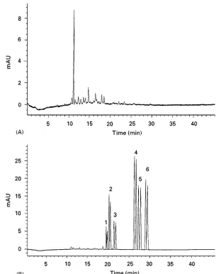
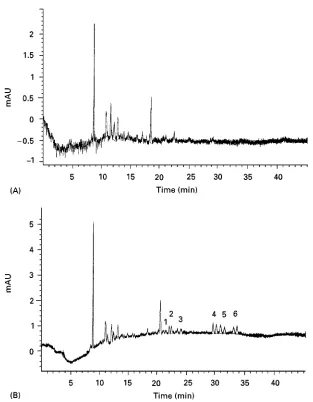
The versatility of modiRed uncharged and charged -CDs in the direct resolution of-agonists, -antag-onists, phenylethylamines, alcohol stimulants and thalidomide and its metabolites by CE was shown. A total of 42 compounds were optically resolved using hydroxypropyl--CD and 20 with sodium sulfobutyl ether--CD. The preliminary analysis of ephedrine, amphetamine, methamphetamine and methylenedioxy-derivatives of amphetamine in urine (Figure 2) and hair (Figure 3) showed that after a liquid}liquid extraction, urine samples could be analysed with a sensitivity below 500 ng mL\1. For hair analysis, it is necessary to increase sensitivity (0.1 ng mL\1) by applying a stacking procedure.
Forensic DNA Samples
DNA polymorphism analysis has recently been recog-nized as a source of identiRcation for individuals in criminal cases and unidentiRed human remains. The conventional technique for DNA typing based on restriction fragment length polymorphism (RFLP) has been replaced by more accurate, sensitive and faster PCR procedures. In contrast to RFLP, the PCR pro-cedures require less DNA and can be used on DNA which is degraded. There are several PCR-based pro-cedures under development; however, short tandem repeat (STR) sequences are currently of major im-portance in theReld of identiRcation of individuals in forensic cases. STRs are DNA segments, typically found in noncoding regions, which are composed of repeating units of 2}5 base pairs (bp). Co-ampliR ca-tion of two or more of these loci in one PCR provides an efRcient mechanism for typing multiple genetic loci simultaneously. The detection of STRs is based on the variation in the length of STR-containing PCR products. These PCR-ampliRed STRs must be separ-ated to determine the size, quantity and/or sequence of each fragment.
DNA restriction fragments and PCR products have traditionally been separated by slab gel electrophor-esis.
Figure 2 Typical electropherograms of: (A) blank human urine extract; (B) extract from blank human urine spiked with: 1, racemic ephedrine; 2, amphetamine; 3, methamphetamine; 4, 3,4-methylenedioxyamphetamine; 5, 3,4-methylenedioxymethamphetamine; 6, 3,4-methylenedioxyethylamphetamine, at concentrations of 1g mL\1for every racemic analyte. Conditions: buffer, 100 mmol L\1 phosphate, pH 2.5, containing 10 mmol L\1 -cyclodextrin. Capillary, uncoated fused silica, 45 cm;50m i.d. Potential, 10 kV. Detection, UV absorbance at 200 nm. (Reprinted with kind permission of Wiley-VCH from Tagliaro Fet al. (1998) Electrophoresis 19: 42}50.)
viscosity, which makes replacement of separation me-dium possible after each electrophoresis run. The polymer solution also has a broader effective DNA size range due to itsSexible and larger effective pore size structure.
The CE system produced results which were com-parable to those obtained on slab gel electrophoresis, with a level of precision of $0.1% bp (between instruments). This comparison is very important if a comparison is to be made of results obtained by different laboratories and to standardize available procedures.
DNA fragments cannot normally be separated in free solution. However, theRrst clinical experimental results demonstrated that adding an uncharged
mol-ecule at the end(s) of the DNA fragments could lead to efRcient separation of relatively large DNA frag-ments (100}900 bp) in free solution. Contrary to current electrophoretic methods, this method requires no sieving matrix, provides better results at high voltage and leads to shorter preparation time and faster separations.
Detection of PCR products has been achieved in three ways: (i) UV absorbance by the DNA fragment; (ii) LIF using intercalating dyes; and (iii)Suorescence of primers tagged withSuorescence dyes.
Figure 3 Typical electropherograms of: (A) blank human hair extract; (B) extract from blank human hair spiked with: 1, racemic ephedrine; 2, amphetamine; 3, methamphetamine; 4, 3,4-methylenedioxyamphetamine; 5, 3,4-methylenedioxymethamphetamine; 6, 3,4-methylenedioxyethylamphetamine, at concentrations of 1 ng mg\1for every racemic analyte. Conditions: Buffer, 100 mmol L\1 phosphate, pH 2.5, containing 15 mmol L\ -cyclodextrin. Capillary, uncoated fused silica, 45 cm;50m i.d. Potential, 10 kV. Detection, UV absorbance at 200 nm. (Reprinted with kind permission of Wiley-VCH from Tagliaro Fet al. (1998) Electrophoresis 19: 42}50.)
Denatured PCR products are then analysed by CE with in-lane size standard (DNA-fragments of known size labelled in a different colour dye) on slab gel or CE capable of real-time multicolourSuorescence de-tection. The collected data are then analysed by soft-ware which automatically determine allele size based on a standard curve for the in-line size standard. STR loci which overlap in size can be distinguished using different dyes thatSuoresce at different wavelengths. Results have indicated that the sizes obtained for STR alleles can differ depending on the gel and elec-trophoresis conditions and depending on the instru-ment used, however, high precision can be obtained in multiplex PCR analysis by using an in-line internal standard ((0.16 nucleotide SD).
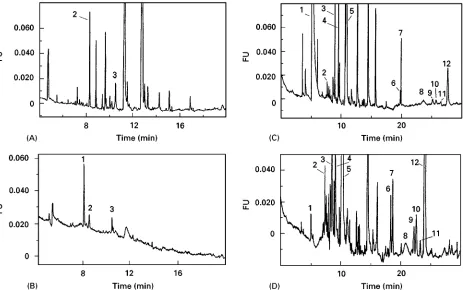
Figure 4 Electropherograms of extracts from dried blue and black inks see (see Table 1) and original inks diluted 100-fold in 5 mmol L\1borate buffer, pH 8.25. Capillary, fused silica 45 cm;50m i.d. Potential, 25 kV. Detection, laser-induced fluorescence at exc/em"320/436 nm. (A) Extract ink 1; (B) original ink 1 (diluted 1 : 100); (C) extract ink 2; (D) original ink 2 (diluted 1 : 100). possible; analysis time per sample is less than 30 min;
capillary life is at least 100 injections; and the run temperature is set at 603C to provide a highly de-naturing environment for the DNA samples.
The disadvantage of CE is that it is a serial tech-nique, making its total throughput no better than the long run times and parallel separations in convential slab gel electrophoresis. Several attempts to obtain faster and higher throughput separations have been reported. These include capillary array electrophor-esis in ultra thin slab gels. These two techniques are limited by difRculty in assembling the separation sys-tem and in carrying out sample introduction. The use of CE to provide continuous automated loading of PCR products on to ultra thin slab gels shows new potential for increasing sample throughput in STR analysis, although separation resolution still needs to be improved.
Over the years CE has become widely used as a power tool in post-PCR analysis. However, it is difRcult to introduce PCR to routine laboratories, because of the possibility of false-positive results. These false positives may be caused by sample-to-sample contamination or by the carry-over of pre-viously ampliRed PCR product. The online coupling of fused silica capillary as the microreactor for PCR
and CE for separation and detection can be recom-mended in order to avoid false-positive results.
Other
The analysis of inks as part of the detection of fraudu-lent documents is a small but important part in the operation of a forensic laboratory. TLC and HPLC have been extensively used to separate and distin-guish inks during the last decades. In comparison, CE has been applied only rarely. UV-Vis, Suorescence and particle-induced X-ray emission (PIXE) detection of electrophoretically separated diluted original inks and ink extracts (Figure 4) from substrate material provide sufRcient information for the comparison of different inks (Table 1). The possibility of compari-son of 50 forensic inks by MEKC has also been investigated. The separation patterns of individual dyes were compared with those obtained by HPLC and TLC, showing a much higher separation efR cien-cy for MEKC. Some inks, which cannot be dis-criminated by applying the HPLC and TLC method, can deRnitely be distinguished using MEKC.
Table 1 Listing of fountain pen inks investigated
Ink number
Colour Manufacturer Country of origin
1 Blue Cross USA 2 Black Cross USA 3 Royal blue Pelikan Germany 4 Brilliant black Pelikan Germany 5 Blue Pilot Japan 6 Black Pilot Japan 7 Blue Lamy Germany 8 Black Lamy Germany 9 Royal blue Geha Germany 10 Brilliant black Geha Germany 11 Blue-black Parker USA 12 Washable blue Parker USA 13 Permanent black Parker USA 14 Royal blue washable Parker France 15 Permanent blue Parker France 16 Black Waterman France 17 Royal blue Mont Blanc Germany
(Reprinted with kind permission of Wiley-VCH from Rohdeet al. (1998)Electrophoresis 19: 31}41.)
characterize tobacco products on their alkaloid pro-Rles for classiRcation purposes has also been demon-strated.
Marine phytotoxines present a major public health problem because they can contaminate seafood. CE and MEKC enable okadaic acid, microcystins and maitotoxin to be detected in the picogram range.
Since any case of mushroom intoxication may have legal consequences, the accurate determination of mushroom toxins is of primary importance for foren-sic pathologists and toxicologists. The analysis of amatoxins by CE instead of radioimmunoassay has several advantages: analysis is faster, less costly and it requires smaller amounts of sample. Rapid and sensi-tive CE method for the separation and determination of the psilocybin and baeocystin in hallucinogenic mushrooms has also been reported.
Many types of explosives consisting of inorganic and organic components have been used in criminal cases. It has been demonstrated that the original com-position of some explosive devices can be derived from the components of the post-blast residue. In short, CE offers a powerful tool which is suitable for both high and low explosive and gunshot residue analysis. CE is suitable for the determination of both inorganic and organic components, showing greater versatility than the traditional methods such as atomic absorption spectrometry and PIXE.
Conclusion
CE is a new technique in the forensic laboratory for the separation and quantitation of a wide variety of
molecules based not only on charge, but also on size, hydrophobicity and stereospeciRcity. CE offers cer-tain advantages for forensic analysis:
1. higher theoretical plate number than HPLC; 2. in many instances CE is faster than GC and HPLC; 3. with regard to sample preparation, CE is easier than GC and HPLC. In many instances the sample can be injected directly with little or no prepara-tion;
4. lower cost per analysis; 5. full automation;
6. CE is complementary to GC and HPLC;
7. two complementary techniques such as CE and MECC can be carried out with the same instru-ment.
Despite these features, the technique has not yet been widely accepted in the forensic community. This may be in part due to the legal system. Different countries have different standards to achieve legal defensibility of analytical results in court and forensic laboratories rely to a large extent on commercial instruments which are specially built and approved by a governmental agency for speciRc analysis. Even so, the US Drug Enforcement Agency is now using CE for general drug screening to quantitate heroin sam-ples.
Two drawbacks of CE are often stated: low repro-ducibility and low sensitivity. However, due to sev-eral recently presented results (e.g. detection limits in the region of ng mL\1, the precision of migration times(1% RSD, same-day and day-to-day repeata-bility characterized by RSD values in the range of 1}4%, when peak area ratios were used), these draw-backs seem not to be so critical.
As stated by Kuffneret al.: ‘The legal criteria of Daubert, as long as they are met by the scientiRc community, will allow CE into evidence as acceptable expert testimony’.
See also: II/Electrophoresis: Capillary Electrophoresis; Capillary Electrophoresis-Mass Spectrometry; Capillary Electrophoresis-Nuclear Magnetic Resonance. III /Clini-cal Chemistry: Thin-Layer (Planar) Chromatography. Forensic Sciences: Liquid Chromatography.
Further Reading
Kuffner CA Jr, Marchi E, Morgado JM and Rubio CR (1996) Capillary electrophoresis and Daubert: time for admission.Analytical Chemistry68 (7): 241A. Lurie IS (1997) Application of micellar electrokinetic
McCord BR (ed.) (1998) Volume symposium capillary electrophoresis in forensic science. Electrophoresis
19(1): 11.
Tagliaro F and Smith FP (1996) Forensic capillary elec-trophoresis. Trends in Analytical Chemistry 15 (10): 513.
Tagliaro F, Turrina S and Smith FP (1996) Capillary electrophoresis: principles and applications in illicit drug analysis. Forensic Science International 77: 211.
Tagliaro F, Smith FP, Turrina Set al. (1996) Complement-ary use of capillComplement-ary zone electrophoresis and micellar electrokinetic capillary chromatography for mutual
conRrmation of results in forensic drug analysis.Journal of Chromatography A735: 227.
Tagliaro F, Turrina S, Pisi Pet al. (1998) Determination of illicit and/or abused drugs and compounds of forensic interest in biosamples by capillary electrophoretic/ elec-trokinetic methods.Journal of Chromatography B713: 27.
Thormann W, Molteni S, Caslavska J and Schmutz A (1994) Clinical and forensic applications of capillary electrophoresis.Electrophoresis15: 3.
von Heeren F and Thormann W (1997) Capillary elec-trophoresis in clinical and forensic analysis. Electro-phoresis18: 2415.
Liquid Chromatography
L. A. Kaine, C. L. Flurer and K. A. Wolnik, Forensic Chemistry Center, US Food and Drug Administration, Cincinnati,
OH, USA
Copyright^ 2000 Academic Press
Forensic science is the application of the sciences to the court of law. Consequently, forensic science and the legal system are intimately intertwined. Results obtained from the examination and analysis of foren-sic samples and the forenforen-sic samples themselves com-prise evidence of a crime. It is the individualization of the sample, i.e. the singular association between the samples(s) and an illegal act, that is unique to forensic science. Because of the legal consequences, results require a high degree of certainty and the techniques used must be admissible in court. The Daubert rule, a 1993 decision upheld by the US Supreme Court, assigns to the judge the role of determining admissi-bility of scientiRc evidence. Among the factors con-sidered by the judge are: (i) whether the technique has been tested and subjected to peer review; (ii) whether error rates have been deRned; (iii) whether standards controlling the operation of a technique exist; and (iv) whether the technique has been widely accepted in the scientiRc community. Techniques used in a forensic laboratory may be applied to the investigation of a wide variety of crimes. Some exam-ples are illegal drug use, counterfeiting, arson, tam-pering, fraud, poisoning, terrorism and environ-mental crimes. It is this diversity of cases, variety of sample matrices and the staggering number of poten-tial analytes that necessitate a continuous evaluation of the testing that is needed to constitute proof in each situation.
Analytical requests in a forensics laboratory may be classiRed into four categories: (i) screen for the