Characterisation of Staphylococcal and Candida Populations
from Healthy and Diseased Oral and Subgingival Sites
Reveals a High Prevalence and Diversity of the Arginine
Catabolic Mobile Element ACME in Staphylococcus
epidermidis
A thesis submitted to the University of Dublin in fulfilment of the requirements for the degree of Doctor of Philosophy by
Aoife M. O’Connor September 2017
Microbiology Research Unit Division of Oral Biosciences Dublin Dental University Hospital
Declaration
I hereby declare that this thesis has not previously been submitted for a degree at this or any other university, and that it represents my own unaided work, except where duly acknowledged in the text. I agree to deposit this thesis in the University’s open access institutional repository or allow the library to do so on my behalf, subject to Irish Copyright Legislation and Trinity College Library conditions of use and acknowledgement.
Summary
The oral cavity harbours a diverse microbiological population, that exists mainly as plaque biofilm, the accumulation of which is associated with oral diseases such as periodontal disease and peri-implantitis. Traditionally, staphylococci were considered transient members of the oral flora, and not thought to contribute to plaque-associated diseases. In contrast, Candida species, particularly Candida albicans, are well-recognised oral commensals and opportunistic pathogens. Detailed investigations of oral staphylococcal and Candida populations in healthy individuals with and without dental implants and in patients with periodontal disease are currently lacking.
The aim of this study was to determine the prevalence and abundance of staphylococcal and Candida species in periodontal pockets, subgingival sites and oral rinses from patients with periodontal disease (n=20), healthy patients with dental implants (n=31) and orally healthy participants without implants (n=64). Participants were subjected to oral rinse, nasal, periodontal pocket and subgingival site sampling. Staphylococci were recovered on selective agar media and definitively identified by Matrix Assisted Laser Desorption Ionization Time-of-Flight analysis. Candida species were recovered on CHROMagar Candida™ medium (CHROMagar, France) and definitively identified by polymerase chain reaction (PCR) using species-specific primers.
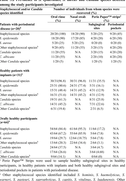
Staphylococci were prevalent in all three participant groups and Staphylococcus epidermidis predominated in all oral sites investigated. This species was most prevalent in oral rinses (18/20, 90%), periodontal pockets (6/20, 30%) and subgingival sites (4/20, 20%) of patients with periodontal disease, but was also highly prevalent in healthy patients with implants [oral rinses (25/31, 80.6%) and subgingival sites (5/31, 16.1%)] and in orally healthy participants [oral rinses (43/64, 67.2%) and subgingival sites (5/64, 7.8%)]. The average cell density of S. epidermidis was also significantly higher in oral rinses from patients with periodontal disease [82.4 ± 218.9 colony forming units (CFU)/ml] compared to orally healthy participants [20.4 ± 14.8 CFU/ml] (p = 0.0153). In contrast, Staphylococcus aureus was much less prevalent [patients with periodontal disease: oral rinses (5/20, 25%), periodontal pockets (0/20, 0%) and subgingival sites (0/20, 0%); healthy patients with implants: oral rinses (15/31, 48.4%), subgingival sites (4/31, 12.9%); orally healthy participants: oral rinses (19/64, 29.7%), subgingival sites (5/64, 7.8%)].
MLST scheme for C. albicans (www.pubmlst.org/calbicans) to identify genomic markers or clonal lineages that might be associated with a participant group, anatomical sites or oral disease state(s).
A diverse population of C. albicans isolates was identified among the three groups of participants by MLST. No clonal lineages were particularly associated with oral health status or anatomical site. Diverse populations of S. aureus and S. epidermidis were also detected in all three participant groups by microarray profiling. Genes encoding resistance to antimicrobial agents including macrolides, tetracycline, and methicillin were more prevalent in S. epidermidis, whereas genes encoding virulence factors were typically more prevalent in S. aureus.
Interestingly, microarray profiling revealed that the arc genes carried by the arginine catabolic mobile element (ACME) were highly prevalent in oral S. epidermidis. The prevalence of each of three previously described ACME types (I: arc and opp3 genes, II: arc genes only, and III: opp3 genes only) was investigated in 143 S. epidermidis isolates by multiplex PCR using ACME-arc- and ACME-opp3-specific primers. A total of 85/143 (59.4%) isolates harboured ACME, of which 60/85 (70.6%) harboured ACME II, 16/85 (18.8%) harboured ACME I and 9/85 (10.6%) harboured ACME III. ACME was significantly (p = 0.016) more prevalent among isolates from periodontal pockets (7/9, 83%) compared to subgingival sites of healthy participants (3/5, 60%).
The genomic diversity of 25 ACMEs from S. epidermidis isolates selected as representative of participant groups and oral sites [type I (n=2), type II (n=20) and type III (n=3)] was investigated by whole genome sequencing (WGS). This was the first detailed investigation of the structural organisation of ACME type III to date. Surprisingly, all three ACME III-positive isolates belonged to the extremely rare S. epidermidis sequence type (ST) ST329, suggesting that this ST may represent an ancestral strain of historic ACME rearrangements that retains ACME III as a remnant. The ACMEs characterised were frequently components of composite genetic elements, often co-located with staphylococcal cassette chromosome (SCC)-associated genes. Based on the size of the elements and presence or absence of SCC-associated genes, highly diverse composite elements were identified in 16/25 isolates in association with ACME types I-III. Typically copA is located at the 3' end of ACME, and the ars operon located downstream of ACME, however, these were internalised within ACME composites containing ACME I (n=2) and III (n=3) and directly into ACME II (n=2) highlighting the genomic plasticity of these elements.
The ACME-arc operon encodes an arginine deaminase pathway thought to enable staphylococcal persistence in nutrient and oxygen poor environments by metabolism of L-arginine for energy and pH regulation. Based on the high prevalence of ACME-arc and the low prevalence of ACME III lacking ACME-arc in periodontal pockets and subgingival sites, it is likely that this operon facilitates the adaptation of S. epidermidis to the semi-anaerobic environment of these sites.
Table of Contents
Acknowledgements ... i
Index of Tables... iii
Index of Figures ... v
Abbreviations ... vi
Publications ... xi
Chapter 1.
General Introduction ... 1
1.1 The oral cavity and its microenvironments ... 2
1.2 Periodontal disease ... 3
1.3 Peri-implantitis ... 5
1.4 Staphylococcal species ... 7
1.4.1 Identification of staphylococcal species ... 7
1.4.2 Genetic transfer among staphylococci ... 8
1.4.2.1 Methicillin-resistance and the staphylococcal chromosomal cassette (SCC) ... 8
1.4.3 Staphylococcusaureus ... 9
1.4.3.1 Adhesion, immune evasion and virulence ... 12
1.4.3.2 Antimicrobial Resistance ... 13
1.4.4 Staphylococcusepidermidis ... 14
1.4.4.1 Adhesion, immune evasion and virulence ... 14
1.4.4.2 Antimicrobial resistance ... 15
1.5 Candida species ... 15
1.5.1 Candida infection ... 15
1.5.2 Identification of Candida species ... 16
1.6 Microbial Typing Systems ... 17
1.6.1 Molecular typing of staphylococci ... 19
1.6.1.1 Multilocus sequence typing... 19
1.6.1.2 Staphylococcus aureus protein A (spa) typing ... 20
1.6.2.3 Multilocus sequencing typing (MLST) of diploid species ... 22
1.6.2.4 DNA microarray technology ... 23
1.6.2.5 Whole genome sequence based typing systems ... 23
1.7 Project aims ... 23
Chapter 2
.
General Materials and Methods ... 25
2.1 General microbiology methods ... 26
2.1.1 Culture and storage ... 26
2.1.2 Chemicals, enzymes, and oligonucleotides ... 26
2.1.3 Buffers and solutions ... 27
2.1.4 Nucleic acid extraction ... 28
2.1.4.1 Bacterial DNA extraction... 28
2.1.4.2 DNA extraction for DNA microarray analysis ... 28
2.1.4.3 Candida DNA extraction ... 28
2.1.5 Determination of concentrations and quality of extracted DNA and DNA sequencing reactions ... 29
2.2 Participant recruitment and sample collection ... 30
2.2.1 Study group ... 30
2.2.2 Clinical sample collection ... 30
Chapter 3
.
Prevalence and Abundance of Staphylococcal and
Candida Species Recovered from Distinct Anatomical Sites in
the Oral Cavities of Patients With Periodontal Disease,
Healthy Patients With Implants, and Orally Healthy
Participants ... 32
3.1 Introduction ... 33
3.1.1 Staphylococcal species in the oral cavity ... 33
3.1.2 Candida in the oral cavity ... 35
3.1.3 Species identification using MALDI-TOF ... 36
3.1.4 Objectives ... 38
3.2 Materials and methods ... 39
3.2.1 Clinical sample collection ... 39
3.2.2 Isolation of staphylococci and Candida from clinical samples ... 39
3.2.3 Quantitative species identification on differential and selective media ... 40
3.2.3.2 Presumptive identification and quantitation of Candida species... 40
3.2.4 Definitive identification of staphylococcal species using matrix assisted laser desorption ionization-time of flight mass spectrometry (MALDI-TOF) ... 43
3.2.5 Definitive identification of Candida species using species-specific PCR ... 43
3.2.6 Statistical analysis ... 43
3.3 Results ... 46
3.3.1 Prevalence of staphylococcal and Candida species among participants investigated... ... 46
3.3.1.1 Prevalence of staphylococcal and Candida species among patients with periodontal disease. ... 46
3.3.1.2 Prevalence of staphylococcal and Candida species among healthy patients with implants. ... 48
3.3.1.3 Prevalence of staphylococcal and Candida species among orally healthy participants. ... 50
3.3.1.4 Comparison of the prevalence of staphylococcal and Candida species among the three groups of participants investigated... 51
3.3.2 Abundance of staphylococcal and Candida species among participants investigated… ... 52
3.3.2.1 Abundance of S. epidermidis, S. aureus and C. albicans among patients with periodontal disease. ... 52
3.3.2.2 Abundance of S. epidermidis, S. aureus and C. albicans among healthy patients with implants. ... 56
3.3.2.3 Abundance of S. epidermidis, S. aureus and C. albicans among orally healthy participants. ... 58
3.3.2.4 Comparison of the relative abundance of S. epidermidis, S. aureus and C. albicans recovered from the three groups of participants. ... 59
3.4 Discussion ... 61
3.4.1 Prevalence and abundance of staphylococcal and Candida species among patients with periodontal disease ... 62
3.4.1.1 Prevalence and abundance of staphylococcal species in patients with periodontal disease ... 62
3.4.3 Prevalence of staphylococcal and Candida species among orally healthy
participants ... 66
3.4.3.1 Prevalence and abundance of staphylococcal species in orally healthy participants 66 3.4.3.2 Prevalence and abundance of Candida species in orally healthy participants ... 67
3.4.4 Possible roles and interactions of staphylococcal and Candida species ... 68
Chapter 4
.DNA Microarray Analysis of Oral Staphylococcus
aureus and Staphylococcus epidermidis Isolates Recovered
from Patients with Periodontal Disease, Healthy Patients with
Implants and Orally Healthy Participants ... 70
4.1 Introduction ... 71
4.1.1 Genetic transfer among staphylococcal species ... 71
4.1.2 S.aureus and S. epidermidis populations... 72
4.1.3 DNA microarray profiling ... 74
4.1.4 Objectives ... 74
4.2 Materials and methods ... 75
4.2.1 DNA microarray analysis... 75
4.2.1.1 Biotin-dUPT labelling using linear amplification ... 75
4.2.1.2 Hybridisation of labelled PCR product to array probes. ... 75
4.2.1.3 Horseradish-peroxidase (HRP)-conjugation, staining and chip analysis ... 77
4.2.1.4 Array data analysis ... 77
4.3 Results ... 78
4.3.1 Clonal lineages identified amongst S. aureus isolates investigated ... 78
4.3.2 Antimicrobial resistance and virulence factor genes harboured by S. aureus isolates… ... 81
4.3.2.1 Antimicrobial resistance genes in S. aureus ... 81
4.3.2.2 Virulence factor genes in S. aureus ... 84
4.3.3 Antimicrobial resistance and virulence factor genes harboured by S. epidermidis isolates… ... 86
4.3.3.1 Antimicrobial resistance genes in S. epidermidis ... 86
4.3.3.2 Virulence factor genes in S. epidermidis ... 90
4.4 Discussion ... 93
4.4.3.1 Prevalence of ACME ... 96
4.4.3.2 Prevalence of SCCmec types in S. epidermidis based on DNA microarray profiling… ... 97
4.4.4 Investigation into possible trafficking between oro-nasal and subgingival sites... 98
Chapter 5
.Prevalence and Diversity of ACME among Oral
and Subgingival S. epidermidis from Patients With
Periodontal Disease and Healthy Patients With and Without
Implants ... 99
5.1 Introduction ...100
5.1.1 The arginine catabolic mobile element ...100
5.1.2 The structure and functions of ACME ...100
5.1.2.1 Genetic diversity of ACME ...102
5.1.2.2 The speG gene ...102
5.1.2.3 The arc-operon ...103
5.1.2.4 The opp3-operon ...103
5.1.3 Genomic sequencing technologies ...103
5.1.3.1 First generation sequencing...103
5.1.3.2 Second generation sequencing ...104
5.1.3.3 Third generation sequencing ...106
5.1.3.4 Fourth generation sequencing ...106
5.1.4 Objectives ...106
5.2 Materials and methods ...108
5.2.1 Detection of ACME by multiplex PCR ...108
5.2.2 Characterisation of ACME using whole genome sequencing (WGS) ...108
5.2.3 Assemblies and bioinformatics analysis ...111
5.2.4 Contig gap closure and confirmation of ACME structure ...111
5.2.5 SMRT sequencing ...111
5.2.6 Identification of direct repeats ...115
5.2.7 MLST using WGS data ...115
5.3.1.4 Prevalence of ACME type II ...119
5.3.1.5 Prevalence of ACME type III ...120
5.3.1.6 Participants harbouring multiple ACME types in the same sample site ...121
5.3.2 Genetic diversity of ACME in S. epidermidis isolates recovered from the three participant groups ...121
5.3.2.1 Population of S. epidermidis investigated ...121
5.3.2.2 Genetic diversity of the ACME arc and opp3 operons ...122
5.3.2.3 Genetic diversity of ACME I ...126
5.3.2.4 Genetic diversity of ACME II ...126
5.3.2.5 Genetic diversity of ACME III ...134
5.3.2.6 Characterisation of ACMEs recovered from distinct sample sites in the same patient…...137
5.4 Discussion ...139
5.4.1 Prevalence of ACME in S. epidermidis from oral rinse samples, periodontal pockets and subgingival sites ...139
5.4.2 Genetic diversity of ACME types I, II and III...140
5.4.2.1 Population analysis of S. epidermidis isolates investigated. ...143
5.4.3 Possible role of ACME in the oral cavity, periodontal pockets and subgingival sites... ...144
Chapter 6
.Population Analysis of C. albicans ... 147
6.1 Introduction ...148
6.1.1 Association of Candida and disease ...148
6.1.2 Epidemiological studies of Candida infection in patients with specific types of disease… ...149
6.1.3 Population structure of C. albicans and clade enrichment ...149
6.1.4 Objectives ...150
6.2 Materials and methods ...151
6.2.1 C. albicans isolates ...151
6.2.2 C. albicans DNA extraction ...151
6.2.3 ABC genotyping of C. albicans by PCR ...151
6.2.4 MLST locus amplification by PCR ...152
6.2.5 Sequence analysis ...152
6.2.6 MLST clade identification and population structure analysis ...152
6.3 Results ...158
6.3.1 ABC genotyping ...158
6.3.1.2 C. albicans ABC genotypes detected from healthy patients with implants ...158
6.3.1.3 C. albicans ABC genotypes detected from orally healthy carriers ...160
6.3.2 MLST analysis ...160
6.3.3 Population Structure analysis ...160
6.3.3.1 C. albicans DSTs identified from patients with periodontal disease ...162
6.3.3.2 C. albicans DSTs identified from patients with healthy implants ...162
6.3.3.3 C. albicans DSTs identified from orally healthy carriers ...165
6.3.3.4 eBURST analysis...165
6.3.4 Epidemiological analysis of recovered isolates ...168
6.4 Discussion ...172
6.4.1 Study objectives ...172
6.4.2 Population structure based on ABC genotyping and MLST ...172
6.4.3 Epidemiological analysis of C. albicans in patients with periodontal disease and healthy implants ...174
Chapter 7
.
General Discussion ... 176
7.1 Prevalence and abundance of staphylococcal and Candida species in the oral cavity...177
7.2 Population and genotypic analysis ...179
7.3 The prevalence and characterisation of ACME types ...181
7.4 Future work ...184
7.5 Conclusion ...185
Bibliography ... 186
Appendix 1 ... 208
Acknowledgements
Firstly, I would like to express my sincere gratitude to my supervisor Professor David Coleman, for his continuous support, patience and immense knowledge throughout the past four years. Thank you for giving me the opportunity to undertake a Ph.D. research project. I would equally like to acknowledge and thank my supervisor Dr. Brenda McManus for all her encouragement, patience, and advice. Thank you for always being available to help me with all aspects of this project, I have learned so much working with you over the past four years.
I wish to thank Dr. Gràinne Brennan and all the staff in the National MRSA Reference Laboratory who were always so helpful, and in particular, I would like to thank Tanya Flemming for setting up countless MALDI-TOF runs. Thank you to the Moyne Institute preparation room for suppling culture media to the laboratory.
I would like to acknowledge the funding for this project provided by the Microbiology Unit, Division of Oral Bioscience, Dublin Dental University Hospital. Thank you for the chance to present my research internationally, to undertake training course both in Ireland and abroad, and for providing anything required for my research. Thank you to Dr. Mary O’Donnell for managing the Microbial Research Unit so efficiently, and for acting as gatekeeper on this project. I am grateful to Dr. Michael O’Sullivan and Dr. Ioannis Polyzois for providing me with the clinical samples that were investigated in the present study, and to Dr. Peter Kinnevey for helping me with the whole genome data analysis involved in this project. Thank you to Dr. Keira Malone for sharing additional control isolates.
encouragement during my project. I’d like to thank Megan Earls for teaching me how to set up whole genome sequencing runs, and for knowing when drinks are required. To Dr. Emily Deasy and Elaine Malone, thanks for always being available for a chat. Lastly, a special thank you to the one and only Dr. Alexandros Lazaris. Your nuggets of ‘wisdom’ and honesty are truly refreshing, thank you for your friendship, the last four years wouldn’t have been the same without you.
To the Foxpack (Louise, Grainne, Doireann, and Alison), Fiona, and Sorcha, thank you for the many dinners, trips, and parties over the past four years, and for always showing interest in my research. I am so grateful for all your friendship.
I’d like to thank my sisters; Tara and Niamh for all their support and for always saying what they are thinking, could I be anymore grateful! To Amber, the best dog a girl could ask for. A special thank you to Dr. Paul Barry, for always believing in me. Thank you for your love and endless support, here’s where the fun begins!
Index of Tables
Page number refers to the text page preceding the tableTable Title Page
3.1 Oligonucleotide primers used for the analysis of Candida species... 44
3.2 Prevalence of S. epidermidis, S. aureus and Candida species detected among the study participants investigated………. 47 3.3 Abundance of S. epidermidis recovered from different anatomical sites in
patients with periodontal disease, healthy patients with implants and healthy participants………... 53 3.4 Abundance of S. aureus recovered from different anatomical sites in patients
with periodontal disease, healthy patients with implants and orally healthy participants………... 54 3.5 Abundance of C. albicans recovered from different anatomical sites in patients
with periodontal disease, healthy patients with implants and orally healthy
participants……….………... 55 4.1 Numbers of isolates analysed by DNA microarray profiling according to
participant group and anatomical site from which they were recovered ………… 76 4.2 Genotypic characteristics of each CC and ST identified among 78 S. aureus
isolates recovered during the present study………... 79 4.3 SCCmec-associated genes detected among the 14 MRSE isolates subjected to
DNA microarray profiling………... 91 5.1 Summary of quality control metrics from each WGS run………... 109 5.2 Primers used to confirm the contig order of ACME in isolates investigated…….. 112
5.3 Prevalence of ACME types I-III harboured by S. epidermidis and S. aureus isolates from distinct anatomical sites………. 117 5.4 ACME-positive Staphylococcusepidermidis isolates subjected to WGS analysis. 123 5.5 Pairwise comparison of nucleotide identity (%) calculated between all arc
-operons identified in ACME types I and II………...……... 124 5.6 Pairwise comparison of nucleotide identity (%) calculated between the opp3
-operons in ACME types I and III………..…... 125 5.7 Direct repeat sequences (DRs) identified among ACME types investigated in the
present study………....…… 128 5.8 Pairwise comparison of nucleotide identity (%) shared by all ACME type II
elements investigated………...…… 132 6.1 Loci and oligonucleotide primer sequences used for C. albicans MLST analysis.. 153
6.2 Representative DSTs used to assign C. albicans isolates to previously identified MLST clades……… 154 6.3 MLST datasets and ABC genotypes previously identified among C. albicans
6.5 Allelic profiles, DSTs, MLST clades, clonal complexes and ABC genotypes identified among C. albicans isolates in the present study.………. 161
Appendix 1………... 208
Index of Figures
Page number refers to the text page preceding the figure
Figure Title Page
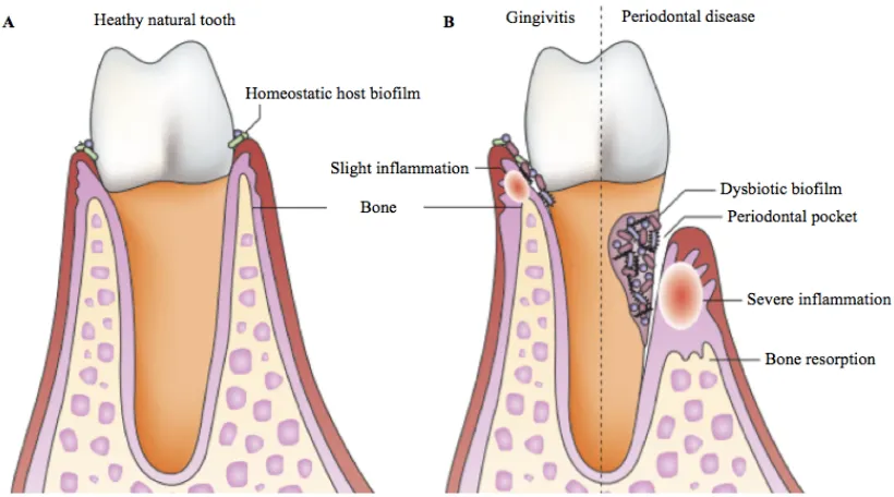
1.1 Progression from periodontal health to gingivitis and eventually periodontitis or periodontal disease……….……….. 4 1.2 Photographs of examples of distinct titanium dental implants………...…. 6
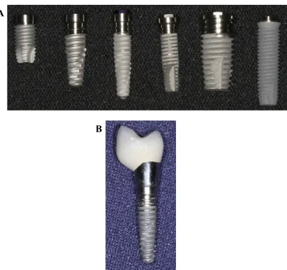
1.3 Diagram showing the genetic organisation of SCCmec types I-XII……….... 10
1.4 Photographs showing Candida species growing on CHROMagar Candida™ plate……….………. 18 3.1 Schematic diagram of microbial identification by matrix assisted laser desorption
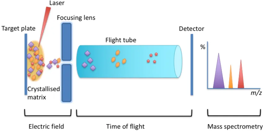
ionisation-time of flight mass spectrometry (MALDI-TOF
MS)………... 37
3.2 Photographs showing the appearance of staphylococcal species frequently recovered from clinical samples on chromogenic SaSelect™ agar after 48 h incubation………... 41 3.3 Photographs showing Candida species recovered from clinical samples on
chromogenic CHROMagar CandidaTM agar following 48 h incubation………….. 42 4.1 Prevalence of antimicrobial resistance (AR) and virulence factor (VF) genes
detected in S. aureus isolates by DNA microarray profiling………... 82 4.2 Prevalence of antimicrobial resistance (AR) and virulence factor (VF) genes
detected in S. epidermidis isolates by DNA microarray profiling………... 87 5.1 Schematic representation of the structural organisation of ACME type I... 101 5.2 Schematic of the Illumina Miseq sequencing technology………...… 105
5.3 Schematic representation of ACME type I elements harboured by two distinct S. epidermidis isolates characterised during the present study……… 126 5.4 Schematic representation of ACME II and ACME II composite elements
characterised in the present study………...…. 129 5.5 Schematic representation of ACME type III composite elements characterised in
the present study……….……. 135 6.1 UPGMA dendrogram based on concatenated MLST sequences of C. albicans
isolates recovered in the present study……….…………... 163 6.2 eBurst analysis of 3164 unique MLST profiles in the MLST database………...… 166 6.3 eBurst analyses of four distinct clonal complexes in the MLST database... 167
Abbreviations
aa Amino acid
ACME Arginine catabolic mobile element
AIDS Acquired immunodeficiency syndrome
agr Accessory gene regulator
ATP Adenosine triphosphate
BHIA Brain Heart Infusion Agar
BLAST Basic local alignment search tool
bp Base pair
BSI Blood stream infection
BURST Based upon related sequence types
ca. Approximately
CA Community associated
CA-MRSA Community associated methicillin resistant Staphylococcus aureus
CBA Columbia blood agar
CC Clonal complex
CDC Centers for disease control and prevention
CFU Colony forming units
CFU/ml Colony forming units per milliliter
CHIP Chemotaxis inhibitory protein
DDUH Dublin Dental University Hospital
DNA Deoxyribonucleic acid
dNTPs Deoxynucleotide triphosphate
DST Diploid sequence type
e.g. Exempli graita; for example
EARS-net Eruopean Antimicrobial resistance surveillance network
EDTA Ethylenediaminetetraacetic acid
et. al. Et alia; and others
etc. Et cetera; and the rest
EUCAST European committee on antimicrobial susceptibility testing
g Gravitational force
h Hours
HA Hospital acquired
HA-MRSA Hospital acquired methicillin resistant Staphylococcal aureus
HCL Hydrochloric acid
HGAP Hierarchical Genome Asssembly Process
HIV Human immunodeficiency virus
HRP Horseradish pyruvate
i.e. Id est; that is
IEC Immune evasion complex
IgG Immunoglobulin G
IS Insertion sequence
environment outside of a living organism.
kb Kilobase
L Litre
LA Livestock associated
M Molar
mg Milligram
MGE Mobile genetic element
min Minute
ml Millilitre
MLST Multi locus sequence type
mM Milli molar
MRSA Methicillin resistant Staphylococcus aureus MRSE Methicillin resistant Staphylococcus epidermidis
MSA Manitol salt agar
MS-CoNS Methicillin susceptible coagulase negative staphylococci
MSCRAMMs Microbial surface components recognizing adhesive matrix molecules
MSSA Methicillin susceptible Staphylococcus aureus MSSE Methicillin susceptible Staphylococcus epidermidis
N/A Not applicable
ORF Open reading frame
PBP Penicillin binding protein
PBS Phosphate buffer saline
PCR Polymerase chain reaction
PFGE Pulsed-field gel electrophoresis
pH potential hydrogen
PVL Panton-Valentine Leukocidin
RNA Ribonucleic acid
rRNA Ribosomal ribonucleic acid
rpm Revolutions per minute
RT-PCR Real-time polymerase chain reaction
s Seconds
SaPI Staphylococcal Pathogenicity Island
SCC Staphylococcal chromosomal cassette
SCCmec Staphylococcal chromosomal cassette mec
SCIN Staphylococcal complement inhibitor
SMRT Single molecule real time
SNV Single nucleotide variant
SSTI Skin and Soft Tissue Infection
ST Sequence Type
TBE Tris/Borate/EDTA
TE Tris/EDTA
TSA Trypticase soy agar
UK United Kingdom
UPGMA Unweighted pair group method according to arithmetic averages
USA United States of America
w/v Weight/volume
wgMLST Whole genome sequencing multi-locus sequence typing
WGS Whole genome sequencing
WHO World Health Organisation
°C Degrees celcius
µg Microgram
µl Microliter
% Percentage
> Greater than or equal to
< Less than or equal to
> Greater than
< Less than
Δ Deletion (of part of a gene)
n Number
α Alpha
β Beta
Publications
Some of the original work presented in this thesis has been published in a refereed international publication as listed below. Offprints of the proofs of the manuscripts are included at the end of the thesis (Appendix 1).
Chapter 1
1.1 The oral cavity and its microenvironments
The oral cavity consists of a wide variety of heterogeneous microenvironments composed of hard and soft tissue types as well as different shedding and non-shedding surfaces such as teeth, mucosa and the tongue (1, 2). Each of these distinct tissue types and surfaces give rise to numerous intraoral niches, which harbour distinct microbial communities. For example, each of the tongue’s different surfaces (e.g. dorsum, lateral border) are anatomically different to each other and harbour distinct microbial communities (1).
Oral microbial communities commonly exist as biofilms; highly structured and organised communities of microorganisms encased within a matrix of secreted extracellular polymer substances (3). The complex biofilms that form on the hard non-shedding surface of teeth are commonly known as dental plaque. Streptococcus, Actinomyces, Porphyromonas species and spirochetes are key pathogens in dental plaque. Saliva coats the teeth and provides the microbial residents of plaque with a constant source of peptides and proteins (4).
The formation and build-up of dental plaque is associated with oral diseases such as gingivitis, caries, halitosis and periodontal diseases due to the metabolites produced by the microbial residents of plaque biofilm. For example, the metabolic products formed by dental plaque microbial communities in subgingival plaque can induce host immune responses resulting in gingival inflammation and bleeding (gingivitis) (4). Microbes residing in supragingival plaque can metabolise carbohydrates present in saliva to organic acids such as lactic and acetic acid and prolonged exposure of the tooth to the acidic biofilm can result in dental caries (4). Good oral hygiene practices including regular teeth brushing, flossing, and periodic mechanical descaling are the most effective methods of controlling dental plaque in the oral cavity.
1.2 Periodontal disease
The periodontium exists as a support structure to the teeth and is composed of the gingival crevice, periodontal ligament, root cementum and alveolar bone (10). Periodontal disease is an inflammatory disease that is preceded by gingivitis, a less invasive reversible form of periodontal disease that causes expansion of the gingival margin and gingival swelling and bleeding (11). Visible dental plaque will start to develop on teeth 24 h following oral hygiene practices and an absence of oral hygiene practices for 10-21 days results in gingivitis. However, gingivitis can be reversed within one week if effective oral hygiene practices are reintroduced (12). If left untreated, the eventual detachment of the gingiva from the tooth results in the formation of a periodontal pocket (10). Periodontal pockets allow the build-up of dental plaque deep within the periodontal tissue, eventually leading to loss of supporting tissue, periodontal ligaments and the reabsorption of the alveolar bone around the tooth (Fig. 1.1) (7, 10). Porphyromonasgingivalis, Prevotella intermedia, Treponemadenticola and Tannerella forsythia were previously thought to be the causative pathogens of periodontal disease but research on staphylococcal and Candida populations in association with this disease are sparse. However, it has recently been suggested that the development of periodontal disease can be attributed to disruption of polymicrobial synergy between the resident microbes of dental plaque due to increased microbial diversity (7, 13, 14). This dysbiosis triggers the expression of virulence factors by resident pathogenic bacteria leading to the secretion of toxins which cause gum irritation (6, 7). The consequent host inflammatory response leads to an influx of innate immune cells such as neutrophils causing further destruction of the periodontal tissue (7).
The diagnosis of periodontal disease is based on signs of inflammation, probing depths, bleeding on probing and periodontal ligament attachment loss (clinical attachment loss >3 mm) (17, 18). The diagnoses of periodontal disease is subjective leading to difficulties calculating the global prevalence of this disease (16, 18). It is estimated that 50-90% of adults worldwide have gingivitis and 80% of adults worldwide will have a minimum one tooth affected by periodontal disease at some stage during their lifetime (6, 11).
1.3 Peri-implantitis
Dental implants are indwelling titanium medical devices that are inserted into the patients mandibular or maxillary bone and by a process known as osseointegration, integrates into the jawbone to form a structural and functional connection (Fig. 1.2). This process anchors the prosthetic restoration to the jaw where there has been tooth loss due to trauma, periodontal disease or other diseases (19, 20). Healthy dental implants have homoeostatic dental plaque at their gingival crevice and are fully integrated into bone. There are no periodontal ligaments surrounding an implant and therefore the gingival crevice is deeper around an implant than around a natural tooth. Healthy implants generally have a probing depth of 3 mm (20, 21). Implant failure can occur in the early stages of placement due to osseointegration failure, or in later stages of placement most commonly due to dysbiosis of the implant biofilm and microbes causing infection in periodontal tissue (20).
Figure 1.2 Photographs of examples of distinct titanium dental implants. A) Six examples of titanium dental implants which are used to replace teeth lost through trauma or oral disease. B) A titanium dental implant with the crown attached. Photographs reproduced with kind permission from Dr. I. Polyzois, Division of Restorative Dentistry and Periodontology, DDUH.
A
[image:29.595.109.520.64.450.2]are also associated with implant disease, however Staphylococcus aureus, Staphylococcus epidermidis, coliforms, Pseudomonas spp. and Candida spp. have also been isolated from peri-implantitis biofilms (20, 24).
Due to the slow progression from peri-implant mucositis to peri-implantitis, early diagnoses and treatment is possible. Treatment of peri-implant mucositis usually involves mechanical debridement of the dental plaque and regular maintenance of the implant until reduced sign of inflammation are achieved (21, 22).
Whilst the predisposing factors for the development of periodontal disease are comparable to those of peri-implantitis, smoking is also a risk factor for the development of peri-implantitis (25). A patient with a previous history of periodontal disease and tooth loss due to periodontal disease prior to implant placement is considered severely at risk for the future development of peri-implantitis (25, 26).
1.4 Staphylococcal species
aureus metabolises mannitol, altering the pH and causing a colour change in the agar surrounding the colony from red to yellow. Coagulase negative staphylococci can also grow on MSA as pale pink colonies that do not cause a colour change. Baird parker agar is a selective agar for CoPS that contains lithium chloride and potassium tellurite, S. aureus produce black colonies with a halo caused by tellurite reduction (28).
More recently chromogenic media is used for the presumptive identification of S. aureus and CoNS. The most widely used medium is SaSelect™, this medium is selective for staphylococcal species, S. aureus along with CoNS such as S. epidermidis, S. haemolyticus, and Staphylococcus saprophyticus can be presumptively identified based on colony colour and morphology on this medium (29).
1.4.2 Genetic transfer among staphylococci
Both CoPS and CoNS species can harbour a diverse range of antimicrobial resistance genes and virulence factor genes encoded on mobile genetic elements (MGEs) and frequently these MGEs are exchanged between different species (30). Mobile genetic elements can exist outside of the bacterial chromosome as plasmids, or integrate into genomic DNA such as insertion sequences, transposons, chromosomal cassettes, lysogenic bacteriophages and pathogenicity islands (31). Based on accumulating evidence it has been postulated that CoNS possibly act as a reservoir of MGEs that can transmit useful genetic material such as genes encoding antimicrobial resistance into S. aureus, enhancing the ability of this species to invade, cause tissue damage, and persist during infection of human and animal hosts (32, 33).
1.4.2.1Methicillin-resistance and the staphylococcal chromosomal cassette (SCC) Staphylococcal chromosomal cassettes (SCC), particularly those encoding the methicillin resistance gene mecA (SCCmec), are among the best characterised staphylococcal MGEs to date. These are large elements ranging from 20 to 67 kb in size that integrate into the chromosomal orfX locus in staphylococci and often harbour genes encoding antimicrobial resistance and virulence factors (34–36). These elements also encode regulatory genes and chromosome cassette recombinases (ccr) gene complexes, the latter of which regulate the insertion and excision of the MGE (37–39) into the bacterial chromosome.
worldwide. To date 12 different SCCmec types (I-XII) have been described in MRSA, with each type harbouring a different combination of the mec gene and ccr gene complexes (34–36, 40) (Fig. 1.3). The mec gene complex is composed of varying combinations of the mec gene (either mecA or mecC) and the mec regulatory genes (mecI, mecRI, and mecR2). Currently there are five classes (A-E) of the mec gene complex that have been detected in MRSA. The mec gene classes A-D all contain the mecA and various regulatory genes, whereas class E contains the recently described mecC gene and its accompanying regulatory genes (35). The ccr gene complex is composed of varying combinations of ccr genes: ccrA, ccrB, ccrC and ccrAA (37, 41). To date, eight different types of ccr genes complexes have been described in MRSA (42). The joining (J) regions are the genomic regions surrounding the ccr and mec gene complexes that can harbour MGEs containing additional antimicrobial resistance determinants (27, 43). Based on genomic differences such as the presence or absence of genes or non-coding sections in the J-region, SCCmec elements can be further subtyped. (42).
Several distinct SCCmec types that have been identified in MRSA have also been identified in S. epidermidis and other CoNS in addition to many SCCmec types yet to be identified in MRSA (27, 44–46). This has led to the hypothesis that SCCmec elements originated in CoNS (44, 47).
Pseudo-SCCmec elements which encode mecA but lack any ccr gene complexes, have been detected in MRSA and some CoNS such as S. hominis (27, 48). Other SCC elements lacking mecA have also been characterised that encode antibiotic resistance determinants such as fusidic acid resistance, or encode resistance to metalloids and transition metals such as cadmium or mercury which have bactericidal properties (27, 49–51).
1.4.3 Staphylococcus aureus
associated pathogen, and is known to be a significant cause of wound and device related infections (53). These infections can prove difficult to treat due to the emergence of antibiotic resistant strains of this species such as MRSA, a major cause of nosocomial infections worldwide (54, 55).
Compared to other staphylococci, the greatest impact of SCCmec has been observed in MRSA, although the prevalence and diversity of SCCmec is higher among the CoNS population.
Historically, three main lineages of MRSA evolved independently in hospital, community and agricultural settings; these were typically separated according to SCCmec types present and isolate origin. The typical features of healthcare-associated MRSA (HCA-MRSA) were the presence of SCCmec I, II or III and the acquisition of these strains in a healthcare environment, especially hospitals (41). Community-associated MRSA (CA-MRSA) emerged independently of HA-MRSA, and were considered more genetically diverse than HCA-MRSA populations, and tended to harbour smaller SCCmec elements such as SCCmec types IV and V (56). Community- associated MRSA emerged as a significant cause of infections in the community and was predominantly associated with SSTIs, however it has also been associated with life threatening infections (57–61). Most recently, MRSA has emerged in the agricultural sector and has been associated with infection in livestock (LA-MRSA). Livestock-associated MRSA clones are genetically distinct from HCA-MRSA and CA-MRSA as they do not harbour genes associated with the immune evasion cluster (IEC) which are required by MRSA to successfully infect a human host (62). Heathcare-assocaited MRSA and CA-MRSA typically harbour lysogenic prophages encoding the IEC integrated into the chromosomal beta-toxin gene hlb, a characteristic feature of human strains. As the global population of MRSA continues to evolve, the genotypic features associated with each population become less distinct, as evidenced in recent reports of MRSA outbreaks within hospitals caused by strains originally considered to be CA-MRSA (63–65).
1.4.3.1Adhesion, immune evasion and virulence
shock syndrome toxin (TSST-1), staphylococcal enterotoxins (SEs) and SE-like superantigens have been identified in S. aureus (66). These are small exoproteins that can bind to both MHC class II molecules and T-cell receptors, which can lead to a large cytokine release which causes toxic shock (66). Immune modulating proteins, such as the chemotaxis inhibitory protein of staphylococci (CHIPs) or the staphylococcus complement inhibitor (SCIN) target elements of the hosts innate immune response preventing neutrophil migration to the infection site and preventing opsonophagocytosis (67). Opsonophagocytosis is the immune process whereby an opsonin such as an antibody is bound to a pathogen marking if for phagocytosis by immune cells such as neutrophils. Panton-Valentine leucocidin (PVL) is a bi-component leukocidal toxin harboured primarily by CA-MRSA strains that can form pores in leukocytes (68).
Surface proteins expressed by S. aureus enable it to anchor to host tissues consisting of extracellular matrix proteins such as collagen, fibronectin, and fibrinogen. These S. aureus surface proteins are known as microbial surface components recognising adhesive matrix molecules (MSCRAMMs) (69). Some of the most well characterised MSCRAMMs are proteins FnbA and FnbB which bind to fibronectin, ClfA and ClfB which bind to fibrinogen, and Cna which is a collagen-binding protein (69, 70). These molecules can also enable S. aureus to evade the host’s immune response, causing further tissue damage or toxic shock (69).
1.4.3.2Antimicrobial Resistance
1.4.4 Staphylococcus epidermidis
Staphylococcus epidermidis is a commensal organism of the skin and nares, and like all CoNS, it is less virulent than S. aureus. However, based on its classification as a commensal organism, S. epidermidis was often regarded as a contaminant when isolated from clinical infections (73). More recently S. epidermidis has been associated with nosocomial infections, particularly with foreign body related infections such as catheter- related septicaemia, or prosthetic joint infections (27, 74, 75).
1.4.4.1Adhesion, immune evasion and virulence
The primary method by which S. epidermidis causes infection is by producing MSCRAMMs both for surface adherence and intercellular adhesion that are involved in the formation of an extracellular polysaccharide matrix also referred to as biofilm (76). This opportunistic pathogen is adept at building biofilms in vivo, which offer protection from antibiotics and the host’s immune response, while also being difficult to eradicate once established around an infected prosthesis (75, 77). Staphylococcus epidermidis can readily colonise metals such as titanium, which is a lightweight metal from which many prosthetic joint components are made, along with the majority of commercially available dental implants (78, 79). The MSCRAMMs known as serine-aspartate repeat (Sdr) proteins are expressed by S. epidermidis and play a critical role in the adherence of this species to fibrinogen-coated surfaces and binding to collagen I (80). The SesC protein is usually detected in biofilm-associated S. epidermidis isolates and is comparable to clumping factor A protein in S. aureus. Progression from the adherence phase of biofilm formation to the accumulation phase involves increased intercellular adhesion. The most well characterised protein involved in this stage progression is polysaccharide intercellular adhesin (PIA) which is encoded by icaA/D/B/C and is critical for the formation of the extracellular matrix (76, 80, 81).
source, and regulate the internal pH of bacteria in adverse environments. The precise role of opp3 is unknown, it encodes an oligopeptide permease ABC transporter, and it is hypothesised it could play a role in nutrient uptake. To date ACME has been detected in S. aureus, S. epidermidis and S. haemolyticus (54, 85).
1.4.4.2Antimicrobial resistance
Due to the diverse range of antibiotic resistance genes harboured by CoNS such as S. epidermidis, together with the propensity of these species to form biofilms, treating infections caused by CoNS can be difficult.
As mentioned previously the prevalence and diversity of SCC and SCCmec elements among CoNS is much greater compared to S. aureus. Species such as S. epidermidis and S. haemolyticus are thought to act as reservoirs for these MGEs, and CoNS often harbour non-typable SCCmec elements encoding novel mec and ccr gene complexes (45, 46, 86, 87). A higher prevalence of oxacillin resistance has been detected in clinical S. epidermidis and S. haemolyticus isolates (>80%) compared to other clinically important CoNS such as S. lugdunensis (7.9%) (27). Resistance to glycopeptides such as vancomycin was detected in S. epidermidis and S. haemolyticus prior to the first detection of reduced susceptibility in S. aureus, however the prevalence of glycopeptide resistance CoNS remains low despite the widespread use of vancomycin (27, 88). To date several distinct cfr-plasmids and multiple chromosomal cfr segments have been detected in CoNS (such as S. sciuri, S. simulans, and S. warneri) recovered from animals (89). The cfr gene can mediate PhLOPSA resistance and has also been detected in clinical CoNS such as S. epidermidis and S. haemolyticus, however, the overall prevalence of linezolid resistance is low among CoNS (90).
1.5 Candida species
1.5.1 Candida infection
Most Candida species are relatively harmless eukaryotic yeasts that are members of the Ascomycota phylum, and have been recovered from both mammalian and environmental sources. Candida species are commonly identified as part of the healthy microflora on mucosal surfaces including the oral cavity, gastrointestinal tract, and vaginal tract (92, 93). Certain Candida species are opportunistic pathogens that can cause infection when the hosts immunity is compromised, and to date over 17 different species have been associated with disease in humans (91, 92). There are many factors associated with increased risk of invasive Candida infections for example the use of broad-spectrum antibiotics, damage to the skin barrier associated with indwelling medical devices such as vascular catheters, the use of prosthetic devices such as dentures or dental implants, and burns (91–93). Individuals with underlying conditions such as cystic fibrosis, diabetes, or human immune deficiency virus (HIV) infection are also prone to Candida related infections (95–97).
Candida infections in humans are typically attributed to Candida albicans, Candida glabrata, Candida parapsilosis, Candida krusei, and Candida dubliniensis (91–93). Despite increasing rates of infection caused by non-C. albicans Candida species, C. albicans remains the most prevalent species and the predominant cause of systemic and oral candidiasis, as well as nosocomial Candida infections (91–93, 98). Interestingly, the overall prevalence of C. albicans infections has gradually declined over recent decades while cases of non-C. albicans Candida infections have increased (99). A major contributory factor to this change has been the increased use of the azole antifungal drug fluconazole, to which species such as C. krusei, amongst others, are inherently resistant (99). Another contributory factor is possibly the increase in life expectancy, as species like C. glabrata are more commonly associated with infections in the elderly. Candida glabrata and C. tropicalis are more commonly associated with patients with solid organ or haematological malignancies, whereas C. krusei is more common in patients undergoing fluconazole prophylaxis, or in haematopoietic stem cell recipients (99).
1.5.2 Identification of Candida species
Candida species and several distinct Candida species can be presumptively identified based on colony colour and morphology developed on this medium (Fig. 1.4).
Prior to the identification of C. dubliniensis, the ability to produce germ-tubes in serum was considered a definitive characteristic of C. albicans and was utilised to differentiate this dimorphic Candida species from other Candida species. However, this protocol has been adapted since the discovery of C. dubliniensis, a species that is very closely related to C. albicans and can also form germ-tubes in serum. Candida albicans can form germ-tubes following incubation in medium containing N-acetylglucosamine, whereas C. dubliniensis cannot (101), which enabled phenotypic differentiation between these species.
Both C. albicans and C. dubliniensis can both produce chlamydospores on Rice Agar Tween medium (RAT agar), differentiating them from other medically relevant Candida species. Chlamydospores are spore-like structures that usually are produced at the termini of pseudohyphae. Candida albicans typically produces a single chlamydospore at the end of a pseudohyphae when grown on RAT agar, whereas C. dubliniensis can produce multiple chlamydospores at pseudohyphal termini when grown on the same medium (102). However, not all C. dubliniensis isolates show this multiple chlamydospore characteristic (103).
Figure 1.4 Photographs showing Candida species growing on CHROMagar Candida™ agar following 48 h incubation at 37°C. Panels A and B show plates with C. albicans (larger light blue/green colonies) and C. dubliniensis (smaller dark blue/green colonies) colonies. Panel C show a plate containing C. albicans and C. dubliniensis colonies and one C. glabrata colony (dark pink with a pale pink edge) among
A
B
human, animal and plant populations. Investigations into microbial populations can also provide information on the complexities between commensal and infectious organisms, possible origins of infections, facilitate the tracking of the emergence of antimicrobial/antifungal resistance with microbial populations, and finally provide detailed information on the genetic relatedness between isolates within the same species (93). Systems developed to investigate microbial populations should be able to (i) accurately discriminate between isolates belonging to the same species that are highly related but non-identical; (ii) recognise identical strains among a collection of isolates and generate reproducible data; (iii) be unaffected by high-frequency genome reorganisation and evolutionary pressures so that genetic changes are reasonable stable over time and reflect evolutionary change only by mutating with a medium frequency; (iv) determine the genetic distances between isolates that vary in genetic relatedness; (v) be amenable to computer-based analysis to enable data analysis, normalisation and storage, and (vi) be directly comparable between different laboratories worldwide (93).
1.6.1 Molecular typing of staphylococci
sequence types (STs) and clonal complexes (CCs). This technique involves the amplification of seven highly conserved genes (often referred to as housekeeping genes) by PCR and sequencing of the PCR product (108). Sequence variations at each gene are identified as distinct alleles and assigned a specific integer, with the combination of the seven integers yielding an allelic profile. The allelic profile is used to assign a ST to the isolate using the international MLST database for the specific species. The STs can be assigned to distinct CCs based on their genetic relationship using the based upon related sequence types (BURST) algorithm, and this software can also deduce the probable founding genotype for each CC (109). Additionally, computer based analysis of sequence data or allelic profiles generated from MLST analysis can be used to create phylogenetic trees based on unweighted pair group method with arithmetic advantages (UPGMA) or neighbouring joining methods to visually display the genetic relatedness of the isolates being investigated (93).
MLST is used worldwide, the results are unambiguous as they are based on sequences, and is supported by an international nomenclature that enables consistent defining of isolates based on the ST or CC they belong to. MLST schemes have been developed for many bacterial and fungal species such as S. aureus, S. epidermidis, and C. albicans (110–112). Limitations of this typing method are that it only compares a small section of the core genome, is laborious, and expensive.
1.6.1.2Staphylococcus aureus protein A (spa) typing
Sequencing of the polymorphic X region of the protein A gene (spa) has been utilised as an effective typing technique suitable for both outbreak and long term investigations (113). The X region is highly diverse, variations in this region are caused by duplication and deletion of repetitive units, as well as point mutations within the repeat units (113). A spa type number is assigned to an isolate based on the sequence uploaded to a web-based database (http://spa.ridom.de/). This typing method is rapid and is supported by international nomenclature enabling inter-laboratory comparisons, and can detect greater population diversity compared to MLST (113).
1.6.1.3Staphylococcal cassette chromosome mec typing
been developed for MRSA, and typically involve numerous multiplex PCRs (114, 115). However, none of the current SCCmec PCR based typing schemes detect all SCCmec types and subtypes identified in MRSA to date. Combining SCCmec typing with MLST and spa type enables greater discrimination between MRSA isolates that belong to the same clone. A consensus international SCCmec typing nomenclature based on mec and ccr gene types has been established to streamline the naming of newly identified SCCmec types, subtypes and variants (42). To date, no SCCmec typing scheme has been developed for CoNS; SCCmec elements detected in CoNS tend to be typed based on the MRSA tying scheme (44). However, due to the higher variation in SCCmec elements harboured by CoNS, many of the elements detected are non-typeable SCCmec variants.
1.6.1.4DNA microarray profiling
The availability of the whole genome sequence of S. aureus lead to the development of the high-throughput DNA microarray platform, the S. aureus Genotyping Kit 2.0 (Alere, Jena, Germany). This microarray platform can be utilised to rapidly screen large numbers of S. aureus isolates for the presence of important virulence factor and antimicrobial resistance genes and typing markers. This technology can provide detailed genetic information and accurately assign isolates to an ST and/or CC, and identify SCCmec types (116). The S. aureus Genotyping Kit 2.0 contains oligonucleotide probes specific for 336 S. aureus gene sequences and alleles, these include antimicrobial resistance such as those encoding resistance to macrolides, tetracycline or beta-lactams and virulence factor genes such as MSCRAMMs, biofilm associated genes and ACME.
The S. aureus DNA microarray can also be used to investigate the prevalence of antimicrobial resistance gene, SCCmec types and some virulence factors such as ACME in S. epidermidis as both these species can harbour the same genes (46).
1.6.1.5Whole genome sequencing (WGS)
variation, in contrast to conventional MLST scheme based on seven loci (65). Core-genome MLST (cgMLST) excludes loci located on the accessory Core-genome and also offers a much higher discriminatory power than conventional MLST.
1.6.2 Molecular typing of Candida
1.6.2.1DNA fingerprinting using complex species-specific probes
Prior to the advent of direct sequencing methods and MLST, the most successful DNA-based typing technique for Candida isolates incorporated Southern hybridisation of restriction endonucleases-digested chromosomal DNA to a complex species-specific DNA fingerprinting probes that identified repetitive DNA sequences dispersed throughout the species genome (117, 118). This method has been used extensively due to its high discriminatory power, however the methodology is technically demanding and inter-laboratory comparisons are difficult (93). During the last two decades, the increased availability of sequencing technologies enabled the development of typing schemes based on direct DNA sequence comparisons.
1.6.2.2ABC Genotyping
Candida albicans ABC genotyping can be used to separate C. albicans isolates into three distinct genotypes based on the presence, absence or heterozygous presence of a transposable intron in the 25S rDNA gene (93, 119). ABC genotyping involves the PCR amplification of the internal transcribed spacer region of the 25S rDNA gene, and PCR product separation by agarose gel electrophoresis (119). Candida albicans genotypes A and B are identified by a single 450 bp or 840 bp product, respectively, whereas genotype C is identified by two products 450 bp and 840 bp (119). This typing technique is quick, cheap, and the results are easily interrupted, however ABC genotyping cannot detect the overall genetic diversity of the C. albicans population and is typically use in conjunction with other more specific typing methods to investigate C. albicans populations (93).
1.6.2.3Multilocus sequencing typing (MLST) of diploid species
albicans and most other Candida species, an increased potential for sequence variation exists due to heterozygous nucleotide sites, which provide additional genotypes. For this reason, for the majority of diploid organisms the ST is referred to as a diploid sequence type (DST). Using MLST for Candida population analysis has been highly successful due to its high reproducibility, the speed to which it can be carried out and it has a good discriminatory power. A major advantage of MLST, particularly for analysis of C. albicans populations, is that the data generated is directly comparable between different laboratories worldwide and usually made available online via the curated C. albicans MLST database (https://pubmlst.org/calbicans/).
1.6.2.4DNA microarray technology
More recently DNA microarray technology has been combined with MLST to develop an array for high-throughput SNP detection in C. albicans (122). This array detects SNPs previously detected by MLST, in conjunction with other SNPs on chromosomes that are not included in the MLST scheme (122). To date this MLST-biased SNP microarray has not be applied to large numbers of C. albicans isolates for population analysis.
1.6.2.5Whole genome sequence based typing systems
As WGS technologies develop and become more cost efficient, it is likely a whole genome-based MLST scheme will be established for Candida species, revolutionising epidemiological studies of Candida species, similar to what has already been developed for some bacterial species. The development of a wgMLST scheme for Candida species will be more difficult to create compared to a bacterial wgMLST schemes, due to the larger size of the Candida genomes, the presence of multiple chromosomes, and additionally, the diploid nature of the majority of these species which means heterozygous states need to be accommodated for in genomic analyses and associated software packages.
extensive range of antimicrobial resistance and virulence factor genes they can harbour that can enable them to persist in biofilm and cause persistent infection. To date, a comprehensive molecular characterisation of such species recovered from periodontal pockets, subgingival sites, and oral rinse samples of individuals during states of oral health and disease is lacking. Furthermore, the co-isolation of staphylococci and Candida, an important oral commensal and opportunistic yeast pathogen, in the oral cavities of healthy patients and those with plaque-related disease has not yet been determined.
The aims of the present study are:
• To determine if there is a significant difference in the prevalence and/or abundance of staphylococcal and Candida species in the oral rinse samples, periodontal pockets and subgingival sites of patients with periodontal disease, healthy patients with implants, and orally healthy participants.
• To undertake a comparative molecular characterisation and typing of S. aureus and S. epidermidis isolates recovered from the three distinct anatomical sites investigated of each participant group using DNA microarray technology. This technology was utilised to identify genes encoding antimicrobial resistance or virulence factors that may be particularly associated with distinct environments such as healthy or diseased subgingival sites and periodontal pockets.
• The molecular characterisation of recovered staphylococci indicated a high prevalence of the virulence factor ACME among S. epidermidis. The prevalence and types of ACME was further determined by PCR and WGS to investigate the genetic diversity of ACME and its association with particular environments such as periodontal pockets, subgingival sites and oral rinses samples in the participant groups sampled.
Chapter 2
2.1 General microbiology methods
2.1.1 Culture and storage
Staphylococcal species were routinely cultured on MSA, SaSelect™ (Bio-Rad, Hercules, CA, USA) or trypticase soy agar (TSA), at 37oC for 48 h (both supplied by Oxoid Ltd., Hampshire, UK) or on Columbia blood agar (CBA) (Fannin Ltd., Dublin, Ireland) for DNA extraction prior to DNA microarray analysis.
Candida species were routinely cultured on CHROMagar Candida™ medium (CHROMagar, Paris, France), or on yeast extract peptone dextrose agar (YEPD) (Oxoid Ltd.) at 37oC for 48 h. For liquid culture, Candida species were grown at 37oC in YEPD broth in an orbital incubator (Gallenkamp, Leicester, UK) set at 200 RPM for 24 h.
Staphylococcal and Candida species were maintained in Microbank cryogenic vials (Pro-lab Diagnostics, Cheshire, UK) at -80oC. Isolates were reactivated by removing a single bead from the vial using sterile tweezers and using this bead to inoculate an appropriate agar plate followed by incubation in a static incubator (Gallenkamp) for 48 h at 37oC.
2.1.2 Chemicals, enzymes, and oligonucleotides
All chemicals used, unless otherwise specified, were of analytical grade or molecular biology grade and were purchased from Sigma-Aldrich Ltd. (Co. Wicklow, Ireland). Enzymes and other reagents for molecular biology procedures such as dNTPS, 5X green GoTaq® flexi buffer, DNA loading dye, 1 kb and 100 bp DNA molecular size reference markers were purchased from the Promega Corporation (Madison, WI, USA). Lysozyme and lysostaphin were purchased from Sigma-Aldrich Ltd. GoTaq® DNA polymerase (Promega) was used in all polymerase chain reactions (PCRs). All custom synthesised oligonucleotides were purchased from Sigma-Aldrich Ltd. at a concentration of 100 µM and diluted to a working concentration of 10 µM using molecular biology reagent water (Sigma-Aldrich Ltd.) and stored at -20oC. GelRed™ (Biotium, Fremont, CA, USA) was used to fluorescently stain DNA for agarose gel electrophoresis.
using the QIAquick 96-well PCR purification kit (Qiagen, West Sussex, UK) according to the manufacturer’s instructions.
2.1.3 Buffers and solutions
A 5 × concentration of TBE buffer was prepared using 0.45 M Trizma base, 0.45 M boric acid, 0.01 M EDTA, pH 8. This was diluted to 0.5 × in purified water (Milli-Q® Direct 8 water purification system, Millipore Ireland, Cork, Ireland) for use as the buffer solution for agarose gel electrophoresis.
For extraction of nucleic acid from Candida isolates a cell lysis buffer consisting of 2% (v/v) Triton X-100, 1 mM EDTA, 1% (w/v) sodium dodecyl sulphate (SDS), 100 mM NaCl, and 10 mM Tris-HCl, pH 8 was prepared and stored at 4oC.
The solution phenol:chloroform:isoamyl alcohol (24:24:1) was prepared using 24 ml of liquefied phenol washed in Tris-buffer (Sigma-Aldrich Ltd.), 24 ml of chloroform, and 1 ml of isoamyl alcohol. This s