Full Terms & Conditions of access and use can be found at
http://www.tandfonline.com/action/journalInformation?journalCode=gcoo20
Journal of Coordination Chemistry
ISSN: 0095-8972 (Print) 1029-0389 (Online) Journal homepage: http://www.tandfonline.com/loi/gcoo20
Structural conversion of an oxazolidine ligand
upon treatment with copper(I) and (II) halides;
Structural, spectral, theoretical and docking
studies
Zahra Mardani, Vali Golsanamlou, Zahra Jabbarzadeh, Keyvan Moeini, Saba Khodavandegar, Cameron Carpenter-Warren, Alexandra M. Z. Slawin & J. Derek Woollins
To cite this article: Zahra Mardani, Vali Golsanamlou, Zahra Jabbarzadeh, Keyvan Moeini, Saba Khodavandegar, Cameron Carpenter-Warren, Alexandra M. Z. Slawin & J. Derek Woollins (2018): Structural conversion of an oxazolidine ligand upon treatment with copper(I) and (II) halides; Structural, spectral, theoretical and docking studies, Journal of Coordination Chemistry, DOI: 10.1080/00958972.2018.1536268
To link to this article: https://doi.org/10.1080/00958972.2018.1536268
Accepted author version posted online: 22 Oct 2018.
Submit your article to this journal
Article views: 2
Structural conversion of an oxazolidine ligand upon treatment with copper(I)
and (II) halides; Structural, spectral, theoretical and docking studies
ZAHRA MARDANI*†, VALI GOLSANAMLOU†, ZAHRA JABBARZADEH†, KEYVAN MOEINI‡, SABA KHODAVANDEGAR†, CAMERON CARPENTER-WARREN§, ALEXANDRA M. Z. SLAWIN§ and
J. DEREK WOOLLINS§
†Inorganic Chemistry Department, Faculty of Chemistry, Urmia University, 57561-51818 Urmia, I. R. Iran ‡ChemistryDepartment, Payame Noor University, 19395-4697 Tehran, I. R. Iran
§EaStCHEM School of Chemistry, University of St Andrews, St Andrews Fife UK, KY16 9ST
In this work, the 2-(2-(pyridin-2-yl)oxazolidin-3-yl)ethanol (AEPC) ligand was prepared under
solvent free conditions using ultrasonic irradiation, before reaction with a Cu(NO3)2/KSCN
mixture, CuCl2 and CuI, the products of which were characterized by elemental analysis,
UV-Vis, FT-IR spectroscopy and single-crystal X-ray diffraction. The X-ray analyses results
revealed that AEPC, after reactions with the three copper(I/II) halides, gave structures
([Cu(DEA)Cl2] (2), DEA = diethanolamine, [Cu(BHEG)2] (3), BHEG =
bis(2-hydroxyethyl)glycinato), however it retains its structure on treatment with
Cu(NO3)2/KSCN mixture ([Cu(AEPC)(NCS)2] (1)). The geometrical parameters for the
complexes were compared with the Cambridge Structural Database (CSD) and coordination
modes for thiocyanate ion were extracted. In the crystal structure of 1, the copper ion has a
distorted square-pyramidal geometry and a CuNpyN2NCSNtertOalc environment in which the AEPC
acts as NN'O-donor in a facial coordination mode. In the crystal structure of 2,the copper ion has
a Cu(Nsec)(Oalc)2Cl2 environment and distorted square-pyramidal geometry in which the DEA
ligand is coordinated as a mer-NO2-donor. The copper ion in 3 has a CuN2O4 environment and
distorted octahedral geometry. The ability of these compounds to interact with the nine
biomacromolecules (BRAF kinase, CatB, DNA gyrase, HDAC7, rHA, RNR, TrxR, TS and
Top II) was investigated by Docking calculations and compared with that of doxorubicin. The
thermodynamic stability of 1 and its isomer and also charge distribution patterns were studied by
DFT and NBO analysis, respectively.
Keywords: Oxazolidine; Copper halides; DFT Calculations; CSD Studies; Docking studies
*Corresponding author. Email: z.mardani@urmia.ac.ir
1. Introduction
The oxazolidine moiety is an important building block for pharmacologically active compounds
such as anti-diabetic [1], anti-tubercular [2], anti-convulsant [3] and aldose reductase inhibitors
[4]. It is called pseudo-proline to mimic the proline skeleton for investigation of peptide
biological activity [5]. The oxazolidine-based compounds are pseudo-irreversible inhibitors of
serine proteases [6] and are used as elastase inhibitors [7]. Some of these derivatives exhibited
very high binding affinities for both NK1 and NK2 receptors. There is speculation that a
combined NK1 and NK2 receptor antagonist might be an effective drug for the treatment of
asthma and chronic airway obstruction [8]. Oxazolidines have been studied extensively as
crosslinking agents [9] and for their anti-proliferative activity against cancer cell lines [10].
In order to extend the chemistry of the oxazolidines, we have recently reported two
complexes of cadmium and mercury with 2-(2-(pyridin-2-yl)oxazolidin-3-yl)ethanol (AEPC,
scheme 1) [11] and in this work, coordination of this ligand to both copper halides and
thiocyanate are described. Based on the X-ray analysis, the AEPC ligand converts to the other
structures during complexation to the copper(I/II) halides (Cl, I) while keeping its base in
treatment with Cu(NO3)2/KSCN mixture (scheme 2) to produce complexes of
[Cu(AEPC)(NCS)2] (1), [Cu(DEA)Cl2] (2), DEA = diethanolamine and [Cu(BHEG)2] (3),
BHEG = bis(2-hydroxyethyl)glycinato. In addition to this, synthesis of AEPC by the new
optimized method (ultrasonic irradiation) with a higher yield is described, along with the
characterization of the compounds and theoretical study of some complexes.
+ → + H2O
AEPC
Scheme 1. The synthetic route of 2-(2-(pyridin-2-yl)oxazolidin-3-yl)ethanol (AEPC).
Scheme 2. The structural conversion of the AEPC ligand during complexation process.
In addition to the expected biological properties of AEPC, binding the copper(II) ion to
this unit makes these complexes a good choice for biologically active compounds. The copper(II)
provides a rapid anti-microbial action without the risk of resistance development [12] and, at the
same time, has the ability to modulate angiogenesis, a crucial challenge of current tissue
engineering technologies. Moreover, copper(II) is naturally present in the human body, contrary,
for instance, to silver [13]. This ion is biocompatible and exhibits many significant roles in
biological systems. A further advantage of the copper is that its cost is significantly lower than
the therapeutic metals (Pt, Ru, Rh and Au) currently used in the preparation of metal-based
cancer agents. A large number of the copper(II) complexes have been reported as potential
anti-tumor agents and they have been found to be active both in vitro and in vivo [14-17].
For predicting the biological activities of the ligand and complexes, docking calculations
were run to investigate the possibility of an interaction between these compounds and nine
protein targets [18-20], including BRAF kinase, Cathepsin B (CatB), DNA gyrase, Histone
deacetylase (HDAC7), recombinant Human albumin (rHA), Ribonucleotide reductases (RNR),
Thioredoxin reductase (TrxR), Thymidylate synthase (TS) and Topoisomerase II (Top II). These
proteins were selected either due to their reported roles in the cancer growth or as transport
agents that affect drug pharmacokinetic properties (e.g., rHA). Also, DNA gyrase was included
to study the possibility of anticancer properties of the compounds, also acting as antimalarial
agents [21].
2. Experimental
2.1. Materials and instrumentation
All starting chemicals and solvents were from Merck and used as received. Infrared spectra (as
KBr pellets) from 4000–400 cm–1 were recorded with a FT-IR 8400-Shimadzu
spectrophotometer. The carbon, hydrogen and nitrogen contents were determined using a
Thermo Finnigan Flash Elemental Analyzer 1112 EA. The melting points were measured with a
Barnsted Electrothermal 9200 electrically heated apparatus. The ultrasonic-assisted reaction was
carried out using an ultrasonic bath Sonica 2200ETH S3-Soltec. The electronic spectra were
recorded in H2O using a Shimadzu model 2550 UV-Vis spectrophotometer (190–900 nm).
2.1.1. Synthesis of 2-(2-(pyridin-2-yl)oxazolidin-3-yl)ethanol, (AEPC). A mixture of 0.21 g
(2 mmol) of 2,2'-azanediylbis(ethan-1-ol) and 0.21 g (2 mmol) of picolinaldehyde was irradiated
under reflux condition inside an ultrasonic bath for one hour at 60 °C under solvent-free
conditions. After ultrasonic irradiation, a thick brown oil was obtained and the redundant
precursors removed by rotary evaporation. Yield: 0.37 g, 95%. Anal. Calcd for C10H14N2O2 (%):
C, 61.84; H, 7.27; N, 14.42. Found: C, 62.13; H, 7.11; N, 14.30. IR (KBr disk): 3363 (ν OH),
3010 (ν CH)ar, 2942 (ν CH), 1603 (ν C=N), 1400 (δas CH2 and/or ν C=C), 1387 (δs CH2), 1250
(ν C–O), 1040 (ν C–N), 753 and 702 (γ py) cm–1. 1H NMR (300 MHz, [D6]DMSO): δ = 8.48
(d, 1 H, C10H), 7.80 (t, 1 H, C8H), 7.52–7.55 (d, 1 H, C7H), 7.32 (m, 1H, C9H), 4.84 (s, 1H,
C5H), 4.50 (s, 1H, OH), 2.73−3.97 (m, 8H, C1H2−C4H2) ppm.
2.1.2. Synthesis of (2-(2-(pyridin-2-yl)oxazolidin-3-yl)ethanoldi(thiocyanato)copper(II),
[Cu(AEPC)(NCS)2] (1). A solution of 0.19 g (1 mmol) of AEPC, dissolved in ethanol (10 mL),
was added to a stirring solution of 0.24 g (1 mmol) of Cu(NO3)2·3H2Oand 0.29 g (3 mmol) of
KSCN in ethanol (10 mL). The reaction mixture was stirred for five days at 60 °C and then
filtered. Suitable green crystal prisms for X-ray diffraction studies were obtained by slow
evaporation of the solution for four days and collected by filtration. Yield: 0.23 g, 63%; m.p.
3.75; N, 14.90. IR (KBr disk): 3433 w (ν OH), 2173 m and 2066 w (CNNCS), 1608 s (ν C=N)py,
1475 w (δas CH2), 1384 s (δs CH2), 1293 m (ν C−O), 1054 w (ν C−N), 825 w (ν CS), 767 w and
695 w (γ py) 481 w (δ NCS) cm−1. UV-Vis (H2O, λmax (nm)/ε): 298/137 (d→d).
2.1.3. Synthesis of dichlorodiethanolaminecopper(II), [Cu(DEA)Cl2] (2). A solution of 0.19 g
(1 mmol) of AEPC, dissolved in ethanol (15 mL), was added to a stirring solution containing
0.17 g (1 mmol) of CuCl2·2H2Oin the same solvent (5 mL). The reaction mixture was stirred for
six hours at 50 °C and then filtered. Suitable blue crystal prisms for X-ray diffraction studies
were obtained by slow evaporation of the solution for four days and collected by filtration. Yield:
0.1 g, 30%; m.p. 133 °C. Anal. Calcd for C4H11Cl2CuNO2 (%): C, 20.05; H, 4.63; N, 5.85.
Found: C, 20.18; H, 4.62; N, 5.76. IR (KBr disk): 3389 (ν OH), 3259 (ν NH), 2965 (ν CH), 1463
(δas CH2), 1373 (δs CH2), 1235 (ν CO), 1090 (ν CN) cm−1. UV-Vis (H2O, λmax (nm)/ε): 338/881
(d→d).
2.1.4. Synthesis of bis(bis(2-hydroxyethyl)glycinato)copper(II), [Cu(BHEG)2](3). A solution
of 0.51 g (2.63 mmol) of AEPC, dissolved in ethanol (15 mL), was added to a stirring solution of
0.50 g (2.63 mmol) of CuIin ethanol (5 mL). The reaction mixture was stirred for eight hours at
room temperature and then filtered. After a week, the solvent evaporated and an oily compound
was obtained. Acetone (15 mL) was added to the resultant oil and stirred for a day before
filtration. After evaporation of the acetone, an oily compound formed. By adding distilled water
(15 mL) and stirring for one hour and then filtering, the final solution was obtained. The suitable
blue crystal prisms for X-ray diffraction studies were obtained by slow evaporation of the
solution and collected by filtration. Yield: 0.03 g, 6%; m.p. 203 °C decomposed. Anal. Calcd for
C12H24CuN2O8 (%): C, 37.16; H, 6.24; N, 7.22. Found: C, 37.32; H, 6.31; N, 7.08. IR (KBr
disk): 3310 (ν OH), 2946 (ν CH), 1610 (νas COO), 1405 (νs COO), 1463 (δas CH2), 1372 (δs
CH2), 1242 (ν C−O), 1052 (ν C−N), 665 (δ OCO) cm−1. UV-Vis (H2O, λmax (nm)/ε): 379/114
(d→d).
2.2. Crystal structure determination
X-ray diffraction data for 1 were collected at 93 K using a Rigaku FR-X Ultrahigh Brilliance
Microfocus RA generator/confocal optics with XtaLAB P200 diffractometer. Compound 2 was
analyzed at 173 K using a Rigaku SCXmini CCD diffractometer with a SHINE monochromator.
Mo Kα radiation (λ = 0.71075 Å) was used and intensity data were collected using ω steps
accumulating area detector images spanning at least a hemisphere of reciprocal space. All data
were corrected for Lorentz polarization effects. A multiscan absorption correction was applied
using CrystalClear [22] or CrysAlisPro [23]. Structures were solved by dual space methods
(SHELXT [24]) and refined by full-matrix least-squares against F2 (SHELXL-2013 [25]).
Non-hydrogen atoms were refined anisotropically, and N-H and O-H Non-hydrogen atoms were refined
with DFIX restraints, while all other hydrogen atoms were placed geometrically using a riding
model. All calculations were performed using the CrystalStructure interface [26]. Selected
crystallographic data are presented in table 1. Diagrams of the molecular structure and unit cell
were created using Ortep-III [27, 28] and Diamond [29]. Selected bond lengths and angles are
displayed in table 2 and hydrogen bond geometries in table 3.
2.3. Computational details
All structures were optimized with the Gaussian 09 software [30] and calculated for an isolated
molecule using Density Functional Theory (DFT) [31] at the B3LYP/LanL2DZ and
B3LYP/6-31+G level of theory for complexes and AEPC, respectively, as well as for NBO analysis. The
cif file of 1 and also a similar complex containing an O-donor oxazolidine ligand [32] were used
as input files for the theoretical calculations.
2.4. Docking details
The pdb files 4r5y, 3ai8, 5cdn, 3c0z, 2bx8, 1peo, 3qfa, 1njb, 4gfh for the nine receptors, BRAF
kinase, Cathepsin B (CatB), DNA gyrase, Histone deacetylase (HDAC7), recombinant Human
albumin (rHA), Ribonucleotide reductases (RNR), Thioredoxin reductase (TrxR), Thymidylate
synthase (TS), and Topoisomerase II (Top II), respectively, used in this research were obtained
from the Protein Data Bank (pdb) [33]. The full version of Genetic Optimisation for Ligand
Docking (GOLD) 5.5 [34] was used for the docking. The Hermes visualizer in the GOLD Suite
was used to further prepare the metal complexes and the receptors for docking. The optimized
AEPC ligand and 1 were used for docking calculations. The region of interest used for Gold
docking was defined as all the protein residues within the 6 Å of the reference ligand “A” that
accompanied the downloaded protein. All free water molecules in the structure of the proteins
were deleted before docking. Default values of all other parameters were used and the complexes
were submitted to 10 genetic algorithm runs using the GOLDScore fitness function.
3. Results and discussion
2,2'-Azanediylbis(ethan-1-ol) in the reaction with picolinaldehyde under solvent free conditions
using ultrasonic irradiation gave AEPC via an oxazolidination reaction (scheme 1). Reaction of
AEPC with an ethanolic solution of Cu(NO3)2/KSCN (1:2) mixture, CuCl2 and CuI in a molar
ratio of 1:1 resulted in the formation of 1-3. The complexes are air-stable and soluble in DMSO.
Study of the literature revealed that a similar structure to 3 [35-37] has been reported previously
from different precursors than those we used. In all CSD searches which have been presented,
for more precise results, the structures containing any error or disorder have been omitted.
3.1. Spectroscopic characterization
The frequencies of IR bands for the free ligand are different from those of the corresponding
complexes providing significant indications of bonding sites of the AEPC. In the IR spectrum of
AEPC, a broad peak at 3363 cm−1 can be assigned to the ν (OH) which shifted to higher
frequencies in the spectrum of 1 by 70 cm−1, confirming the coordination of an alcohol group to
the copper ion. A slight shift (5 cm−1) to higher frequency was observed for the ν(C=N) of the
pyridine ring.
The most interesting part of the spectrum of 1 is the region above 2000 cm–1, where the
absorptions due to pseudohalides are observed [38]. Presence of the thiocyanate groups in 1
affects its IR spectrum in three regions: 2000–2200 cm–1 for CN stretches, 700–900 cm–1 for CS
stretches and 400–500 cm–1 for SCN bending vibrations [39]. These vibrations can be used to
determine the coordination modes of thiocyanato ligands in complexes [38]. In 1, the peak
corresponding to ν(CN) is split into two peaks, indicating the existence of two non-equivalent
coordinated thiocyanate groups and hence mutual cis coordination [38, 39]. These peaks appear
at 2173 and 2066 cm–1 which are higher than those of typical N-bonded thiocyanato (in
N-bonded thiocyanato, ν(CN) appears below 2100 cm–1) [38] which can be attributed to
participation of the sulfur atom of the thiocyanato ligand in the hydrogen bonding. The ν(CS)
and δ(NCS) in 1 are observed at 825 and 481 cm–1, respectively, which are characteristic
frequencies for an N-bonded thiocyanato ligand [38].
In the FT-IR spectrum of 2, the region above 3000 cm–1, corresponding to the alcohol and
amine groups, can be useful for predicting the coordination mode of the DEA ligand. In
comparison to the FT-IR spectrum of DEA [39], the ν(OH) and ν(NH) are shifted about 89 and
59 cm–1, respectively, to higher frequencies than in the free ligand, confirming NO2-donation of
DEA to the copper ion.
In the 1H NMR spectrum of the ligand (see scheme 1 for numbering), the aromatic
hydrogen atoms of the pyridine ring are observed at the lowest magnetic field. Among these
protons, C10H, which is the closest to the nitrogen atom of the pyridine ring, has the highest ppm
value. In the aliphatic region and with increasing the magnetic field, two singlet peaks belonging
to the hydrogen atom of the chiral carbon and alcoholic proton are revealed, respectively. The
peaks belonging to the other hydrogen atoms of the aliphatic moieties appear in range of
2.7−4.0 ppm.
UV–Vis spectra of the complexes in aqueous solution exhibited a broad absorption
attributed to d–d transitions of the copper(II) complexes. The order of energy for the d–d
transition is 1 > 2, showing that the ligand field strength in 1 is higher than in 2.
3.2. Description of the crystal structures
3.2.1. Crystal structure of (2-(2-(pyridin-2-yl)oxazolidin-3-yl)ethanol
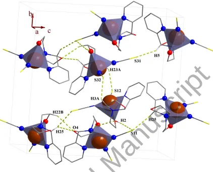
di(thiocyanato)copper(II), [Cu(AEPC)(NCS)2] (1). In the crystal structure of 1 (figure 1), there
are two independent molecules of [Cu(AEPC)(NCS)2] in the asymmetric unit with slightly
different geometrical parameters. Each AEPC ligand acts as a tridentate NN'O-donor, with a
pyridyl nitrogen donor atom, tertiary amine nitrogen donor atom and alcohol oxygen donor atom,
forming two five-membered non-planar chelate rings. The angle between the mean planes
through the two chelate rings of the AEPC ligand is 77.12(13) and 73.26(14)°, respectively, for
Cu1 and Cu21, showing that the ligand binds to the copper ion in the fac formation. A similar
coordination manner has been observed for AEPC in reaction with CdCl2 [40]. The C5 carbon
atom on the AEPC ligand (scheme 1) has four different substituents and is chiral. In addition to
this, a new chiral center is formed (nitrogen atom of oxazolidine ring) upon coordination. Thus
each of the two molecules in the asymmetric unit has both a C- and N-chiral centers. The two
molecules in the asymmetric unit are different enantiomeric forms of one another, one R,S and
the other S,R.
The 2-(pyridin-2-yl)oxazolidine unit is potentially a tridentate ligand that can bind to
metal ions through one O- and two N- atoms. A survey of the CSD reveals that this unit has two
different coordination modes including NpyNoxa- and NpyOoxa-donor of which the NpyNoxa mode is
common. There is only one example for NpyOoxa-donor mode [32]. The angle between the two
mean planes through the pyridine and oxazolidine rings in 1 is 75.94(16) and 74.03(17)°,
respectively, for Cu1 and Cu21 proving these rings are almost perpendicular to each other as
“face to face” form and the bond angles for chiral carbon atoms in the chelate rings are 111.1(2)
and 110.6(2)° for Cu1 and Cu21, respectively (for “face to side” form the bond angle is larger)
[11]. The pyridine and oxazolidine rings of the AEPC in the reported cadmium complex have
“face to face” direction while in a mercury complex they have “face to side” form [11].
In this structure, the copper ion has a coordination number of five, by coordination of the
one oxygen and two nitrogen atoms of one AEPC ligand and two nitrogen atoms of two
thiocyanate ions. A five-coordinate geometry of 1 may adopt either a square pyramidal or a
trigonal bipyramidal structure which is determined by applying the formula of Addison et al.
[41, 42]. The angular structural parameters, τ (τ = (β – α)/60, where α and β are the two largest
angles at the copper ion with β ≥ α), were calculated to be 0.19 and 0.24, respectively, for Cu1
and Cu21, indicating a distorted square-pyramidal geometry for both (figures 1 and 2). In this
geometry, the oxygen atom occupies the axial position and four nitrogen atoms lie on the
equatorial plane. Among the four copper-nitrogen bond lengths around Cu1, Cu–NNCS
(1.944(3) Å, average of two bond lengths) and Cu–Noxa (2.052(3) Å) are the shortest and longest
ones. The bond distance of the Cu–O is about 0.169 Å longer than the longest Cu–N, showing
the elongated distance along the z-axis. Similar results were observed for the other molecule in
the asymmetric unit. A search of the Cambridge Structural Database (CSD) [43] revealed that
there are so far no examples of a CuNpyN2NCSNtertOalc environment, complex 1 is the first one.
The thiocyanato ligands in 1 coordinate through their nitrogen atom. For studying the
different coordination modes of thiocyanate to copper ions, a structural survey using CSD data
was carried out and the results are presented in table 4. Based on these data, the terminally
N-bonded mode, observed in 1, is common. The percentage of terminally bonded modes (53%)
is slightly higher than the bridging (47%). Interestingly, the thiocyanate ion is also capable of
forming a four-membered chelate ring with a copper ion, however this is a very rare mode [44].
Among the bridging modes, bridging NS-donor thiocyanate between two copper ions is more
common than any other mode.
For comparing the geometrical parameters of the terminal N-donor coordinated
thiocyanato ligands of 1 with the CSD analogues, the bond lengths and angles average for all
reported complexes were calculated and presented in scheme 3(a). The results revealed that the
N-bonded thiocyanate ligand and copper ion do not form a linear structure (scheme 3(a)). The
C−N−Cu angles average and N−Cu bond lengths average in 1 are 172.1° and 1.950 Å, which are
higher and lower than the CSD average (scheme 3(a)), respectively. To compare the geometry of
the thiocyanate ion in its coordinated and uncoordinated states, a CSD search was performed for
free thiocyanate ions (scheme 3(b)). Based on these data, after coordination of thiocyanate the
CN and SC bond lengths are slightly increased and decreased, respectively, which is in
agreement with the literature for N-bonded thiocyanato ligands [45, 46].
(a) (b)
Scheme 3. (a) The CSD average for geometrical parameters in complexes containing the terminal
N-donor thiocyanato ligand. (b) The CSD average for bond lengths and angles for all
non-bonded thiocyanate ions.
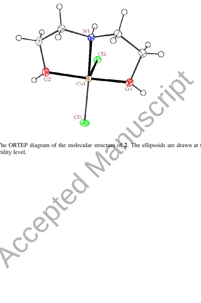
3.2.2. Crystal structure of dichlorodiethanolaminecopper(II), [Cu(DEA)Cl2] (2). In the
crystal structure of 2 (figure 3), the copper ion is coordinated to one nitrogen and two oxygen
atoms of a DEA ligand and two chloride ions, giving a total coordination number of five. The
angular structural parameter, τ, was calculated to be 0.40 for the copper ion, indicating a
distorted square-pyramidal geometry (figures 3 and 4). In this geometry, the Cl2 ligand occupies
the axial position and three donor sites of the DEA along with the Cl1 ligand lie on the equatorial
plane. Studying the CSD database revealed that there are no examples of Cu(Nsec)(Oalc)2Cl2
environments that would allow us to compare the geometric parameters with 2. In another study,
chloride ions) and τ values in the range of 0.00−0.50 (similar as to 2) were extracted (observed
range, 0.04−0.31, three complexes without the equatorial plane were omitted [47-49]). These
complexes can be classified in three types (table 5). The (a) type, in which one chloro ligand is
located in the axial position, has the largest difference (∆, table 5) between the bond lengths of
the two chloro ligands (the distance of axial Cu−Cl is larger than the equatorial). The mean
deviation of the atoms from their equatorial plane is greater in type (a) structures than any of the
others. Complex 2 belongs to the (a) type complexes (table 5) with ∆ = 0.34 Å and 0.31 Å for the
distance of the copper ion from coordinated plane (d).
The DEA ligand acts as tridentate NO2-donor through a secondary amine nitrogen and
two alcohol oxygen atoms and forms two five-membered non-planar chelate rings. Each
tridentate ligand can coordinate to the metal in facial or meridional forms. In the mer form there
are two angles of 90° and one at 180°; in the fac form there are three angles of 90°. In 2, two
angles of coordinated DEA are deviating from 90° due the chelating bite angle, while the third
one is about 149°, confirming mer form (135°, exactly half way between fac and mer) [50, 51].
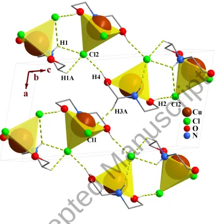
3.2.3. Crystal network interactions. In the crystal networks of 1 and 2 (figures 2 and 4,
respectively) intermolecular O–H···S (1) and O–H···Cl (2) hydrogen bonds appear between
different moieties. In this way the sulfur atom and chloride ion act as proton acceptors and
oxygen atoms participate in hydrogen bonding as proton donors and acceptors, simultaneously.
In addition to hydrogen bonds, there are short contact interactions between the sulfur atoms of
the adjacent thiocyanate ions (1).
In the crystal packing of the complexes, the O–H···Cl (2) hydrogen bonds participate in
the formation of very different hydrogen bond motifs such as R22(8), R64(20), R65(22) and
R66(24) [50, 52] between adjacent complexes.
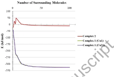
Total intermolecular interaction energies for single molecules of each complex were
calculated using Mercury [53] and its CSD-materials tool [33, 54]. For this, the sum of the
intermolecular interactions energies in a molecular packing shell containing 100 molecules
around the one complexes 1 and 2 were calculated to be −325.60 (complex 1 containing Cu1),
−326.07 (complex 1 containing Cu21) and –10.34 kJ/mol (figure 5), respectively, confirming
that one molecule of 1 is more stabilized in the solid state by its network interactions than +2.
Also the interactions of the enantiomer containing Cu21, in 1, were slightly stronger than its Cu1
enantiomer. In 1, 90% (Cu1 enantiomer) and 88% (Cu21 enantiomer) of the total energy is
corresponding to the interactions with its 14 closest neighboring molecules (figure 5). The
interactions between one molecule of 2 with three molecules in the distance range of 6−7 Å
increase the energy component of the molecule by +90.63 kJ/mol. Other interactions decrease
the energy level of the studied unit in 2.
3.3. Theoretical studies of AEPC and 1
Study of the literature revealed that of the two possible NpyNoxa- and NpyOoxa-donor modes for
2-(pyridin-2-yl)oxazolidine-based ligands, the N2-donor mode is common while other mode is
rare [11]. Based on this observation, the energy level for the optimized complex 1 (1opt) in which
2-(pyridin-2-yl)oxazolidine unit acts as a NpyNoxa-donor was compared with a possible isomer
containing NpyOoxa-donor AEPC, [Cu(AEPC)(NCS)2] (1'opt, figure 6). Owing to the spatial
restriction effects when the oxazolidine ring is coordinated through the oxygen atom, the
alcoholic group on the AEPC ligand cannot be coordinated. Thus in 1'opt, the AEPC acts as a
NO-donor while in 1opt as N2O-donor. The DFT calculation revealed that the isolated complex
1opt is about −6.57 Kcal/mol more thermodynamically stable than the 1'opt, which is in agreement
with the solid state result.
For studying the charge distribution before and after complexation, an NBO analysis was
done on the free AEPC and 1 (table 6). The results reveal that the calculated charge on the
copper ion is about +0.91 and lower than the formal charge (+2), owing to the electron donation
of the ligand during complexation. Based on the calculated total charge values, the total charge
of the nitrogen, carbon and oxygen atoms in 1 is more negative than that of the free ligand, while
the total charge of hydrogen atoms is more positive than in the free ligand. This observation
reveals that the hydrogen atoms play an important role in electron donation toward the metal ion,
thus decreasing the charge of the copper ion. The nitrogen atoms on the thiocyanato ligands are
more negative than those on the AEPC, showing the nitrogen atoms of the thiocyanate ions are
more electronegative than the AEPC nitrogen atoms.
In the optimized AEPC, the HOMO is delocalized on the oxazolidine ring and partially
on the ethanolic side arm while the LUMO is delocalized on the pyridine ring (table 7). In 1opt,
the HOMO is almost delocalized on the thiocyanato ligands while the LUMO is delocalized on
the pyridine ring of the coordinated AEPC. The metal ion does not have any significant
participation in the frontier molecular orbitals (table 7).
Similar to the solid phase results, in the isolated molecule of 1opt, the copper ion has a τ
value of 0.04 and square-pyramidal geometry. In this structure, the AEPC ligand is coordinated
in fac form (the angle between two mean planes through the chelate rings of AEPC is 86.25).
3.4. Docking studies of AEPC and 1-3
For predicting the biological activities of AEPC and 1-3, interactions of these compounds with
nine macromolecule receptors using Gold [34] docking software were studied. The Gold docking
results are reported in terms of the values of fitness which means that the higher the fitness, the
better the docked interaction of the compounds [18-21]. The results of the docking presented in
this work are the best binding results out of the ten favorites predicted by Gold. Also for
evaluation of the calculated fitness values, these scores were compared with those of the famous
anti-cancer drug, doxorubicin (a cancer medication that interferes with the growth and spread of
cancer cells in the body [55]).
The general features from the Gold docking prediction (table 8) show that all studied
structures can be considered as biologically active compounds [18-20]. The best predicted targets
for AEPC is HDAC7, while for the studied complexes TrxR is the target. A comparison of the
GOLDScore fitness values for the ligand and 1 showed that 1 had a better interaction with the
biomacromolecules (except for CatB which exhibited comparable fitness values). A fitness value
comparison between 1-3 showed the general trend 1 > 3 > 2 in their binding ability towards
proteins. The docking results of the interaction between the ligand and the complexes with
BRAF kinase protein are shown in figures 7-10, respectively. In addition to the alcohol and
amino moieties in the structures of the complexes, the thiocyanato (1) and chloro (2) ligands
participate in hydrogen bonding with proteins (table 9). Data of table 8 revealed that the ligand
or complexes or both of them, in some cases (HDAC7, CatB), have comparable fitness values
with the doxorubicin in binding toward the studied biomacromolecules, thus we suggest that the
anticancer activities of these compounds will be studied.
4. Conclusion
In this work, four complexes of copper(I/II), [Cu(AEPC)(NCS)2] (1), AEPC =
2-(2-(pyridin-2-yl)oxazolidin-3-yl)ethanol, [Cu(DEA)Cl2] (2), DEA = diethanolamine, [Cu(BHEG)2] (3), BHEG
= bis(2-hydroxyethyl)glycinato, were synthesized in a reaction between AEPC with
Cu(NO3)2/KSCN mixture (1) and copper(I/II) halides (2, 3). Their spectral (IR, UV-Vis,
1
H NMR) and structural (single crystal X-ray diffraction) properties were investigated. These
structural analyses revealed that the AEPC ligand can convert to the other structures during the
complexation process. In the crystal structure of 1, the copper ion has a distorted
square-pyramidal geometry and CuNpyN2NCSNtertOalc environment, in which AEPC acts as NN'O-donor
in the fac form and thiocyanto ligands adopts a terminally N-bonded mode, which is the most
common mode of coordination among the CSD selected analogues (53%). In the structure of 2,
the AEPC converts to the DEA ligand and coordinates as a mer-NO2-donor to form a distorted
square-pyramidal geometry around the copper ion. In the crystal networks of the complexes, the
N–H···Cl (2) and O–H···Cl (2) hydrogen bonds form very different hydrogen bond motifs. The
theoretical studies revealed that the optimized copper(II) complex (1opt), which has a similar
structure to 1, is thermodynamically more stable than its isomer containing a NpyOoxa-donor
AEPC, [Cu(AEPC)(NCS)2] (1'opt). The docking studies revealed that AEPC and 1, 2 and 3 can
interact with the nine biomacromolecules (BRAF kinase, CatB, DNA gyrase, HDAC7, rHA,
RNR, TrxR, TS and Top II). Also the best predicted target for the AEPC is HDAC7, while for
the other complexes it is TrxR. The order of the binding affinity of the compounds towards
studied proteins is determined as 1 > 3 > 2.
Supplementary data
CCDC 1814322 and 1814321, respectively, for 1 and 3 contain the supplementary
crystallographic data for this paper. These data can be obtained free of charge via
http://www.ccdc.cam.ac.uk/conts/retrieving.html, or from the Cambridge Crystallographic Data
Centre, 12 Union Road, Cambridge CB2 1EZ, UK; Fax: (+44) 1223-336-033; or E-mail:
deposit@ccdc.cam.ac.uk.
References
[1] R.L. Dow, B.M. Bechle, T.T. Chou, D.A. Clark, B. Hulin, R.W. Stevenson. J. Med.
Chem., 34, 1538 (1991).
[2] R.S. Goncalves, C.R. Kaiser, M.C. Lourenco, M.V. De Souza, J.L. Wardell, S.M.
Wardell, A.D. Da Silva. Eur. J. Med. Chem., 45, 6095 (2010).
[3] S.L. Shapiro, I.M. Rose, F.C. Testa, E. Roskin, L. Freedman. J. Am. Chem. Soc., 81, 6498
(1959).
[4] R.C. Schnur, R. Sarges, M.J. Peterson. J. Med. Chem., 25, 1451 (1982).
[5] G. Tuchscherer, D. Grell, Y. Tatsu, P. Durieux, J. Fernandez-Carneado, B. Hengst, C.
Kardinal, S. Feller. Angew. Chem. Int. Ed., 40, 2844 (2001).
[6] A.B. Santana, S.D. Lucas, L.M. Gonçalves, H.F. Correia, T.A.F. Cardote, R.C. Guedes, J.
Iley, R. Moreira. Bioorg. Med. Chem. Lett., 22, 3993 (2012).
[7] R. Moreira, A.B. Santana, J. Iley, J. Neres, K.T. Douglas, P.N. Horton, M.B. Hursthouse.
J. Med. Chem., 48, 4861 (2005).
[8] T. Nishi, T. Fukazawa, K. Ishibashi, K. Nakajima, Y. Sugioka, Y. Iio, H. Kurata, K. Itoh,
O. Mukaiyama, Y. Satoh, T. Yamaguchi. Bioorg. Med. Chem. Lett., 9, 875 (1999).
[9] H. Chen, Z.-H. Shana. Int. J. Biol. Macromol., 46, 535 (2010).
[10] S.F. Andrade, B.G. Oliveira, L.C. Pereira, J.P. Ramos, A.R. Joaquim, M. Steppe, E.M.
Souza-Fagundes, R.J. Alves. Eur. J. Med. Chem., 138, 13 (2017).
[11] Z. Mardani, V. Golsanamlou, S. Khodavandegar, K. Moeini, A.M.Z. Slawin, J.D.
Woollins. J. Coord. Chem., 71, 120 (2018).
[12] M. Vincent, P. Hartemann, M. Engels-Deutsch. Int. J. Hyg. Environ. Health, 219, 585
(2016).
[13] H. Xie, Y. Kang. Curr. Med. Chem., 16, 1304 (2009).
[14] S. Ramakrishnan, V. Rajendiran, M. Palaniandavar, V.S. Periasamy, B.S. Srinag, H.
Krishnamurthy, M.A. Akbarsha. Inorg. Chem., 48, 1309 (2009).
[15] Q. Wang, M. Zhu, L. Lu, C. Yuan, S. Xing, X. Fu. Dalton Trans., 40, 12926 (2011).
[16] S. J. Tan, Y. K. Yan, P. P. Lee, K. H. Lim. Future Med. Chem., 2, 1591 (2010).
[17] M. Alagesan, N.S.P. Bhuvanesh, N. Dharmaraj. Eur. J. Med. Chem., 78, 281 (2014).
[18] F. Marandi, K. Moeini, F. Alizadeh, Z. Mardani, C.K. Quah, W.-S. Loh, J.D. Woollins.
Inorg. Chim. Acta, 482, 717 (2018).
[19] F. Marandi, K. Moeini, F. Alizadeh, Z. Mardani, C.K. Quah, W.-S. Loh. Z. Naturforsch.,
73b, 369 (2018).
[20] Z. Mardani, R. Kazemshoar-Duzduzani, K. Moeini, A. Hajabbas-Farshchi, C.
Carpenter-Warren, A.M.Z. Slawin, J.D. Woollins. RSC Adv., 8, 28810 (2018).
[21] A.A. Adeniyi, P.A. Ajibade. Molecules, 18, 3760 (2013).
[22] CrystalClear-SM Expert, v3.1b27. Rigaku Americas, The Woodlands, Texas, USA, and
Rigaku Corporation, Tokyo, Japan (2013).
[23] CrysAlisPro v1.171.38.41. Rigaku Oxford Diffraction, Rigaku Corporation, Oxford, U.K.
(2015).
[24] G. Sheldrick. Acta Crystallogr., A71, 3 (2015).
[25] G. Sheldrick. Acta Crystallogr., C71, 3 (2015).
[26] CrystalStructure v4.2. Rigaku Americas, The Woodlands, Texas, USA, and Rigaku
Corporation, Tokyo, Japan (2015).
[27] L.J. Farrugia. J. Appl. Crystallogr., 30, 565 (1997).
[28] M.N. Burnett, C.K. Johnson, Ortep-III, Report ORNL-6895. Oak Ridge National
Laboratory, Oak Ridge, Tennessee, USA (1996).
[29] G. Bergerhof, M. Berndt, K. Brandenburg. J. Res. Natl. Stand. Technol., 101, 221 (1996).
[30] M.J. Frisch, G.W. Trucks, H.B. Schlegel, G.E. Scuseria, M.A. Robb, J.R. Cheeseman, G.
Scalmani, V. Barone, B. Mennucci, G.A. Petersson, H. Nakatsuji, M. Caricato, X. Li,
H.P. Hratchian, A. F. Izmaylov, J. Bloino, G. Zheng, J.L. Sonnenberg, M. Hada, M.
Ehara, K. Toyota, R. Fukuda, J. Hasegawa, M. Ishida, T. Nakajima, Y. Honda, O. Kitao,
H. Nakai, T. Vreven, J.A. Montgomery Jr., J.E. Peralta, F. Ogliaro, M.J. Bearpark, J.
Heyd, E.N. Brothers, K.N. Kudin, V.N. Staroverov, R. Kobayashi, J. Normand, K.
Raghavachari, A.P. Rendell, J.C. Burant, S.S. Iyengar, J. Tomasi, M. Cossi, N. Rega, N.J.
Millam, M. Klene, J.E. Knox, J.B. Cross, V. Bakken, C. Adamo, J. Jaramillo, R.
Gomperts, R.E. Stratmann, O. Yazyev, A.J. Austin, R. Cammi, C. Pomelli, J.W.
Ochterski, R.L. Martin, K. Morokuma, V.G. Zakrzewski, G.A. Voth, P. Salvador, J.J.
Dannenberg, S. Dapprich, A.D. Daniels, Ö. Farkas, J.B. Foresman, J.V. Ortiz, J.
Cioslowski, D.J. Fox, Gaussian 09, in, Gaussian, Inc., Wallingford, CT, USA (2009).
[31] J.P. Perdew. Phys. Rev., B33, 8822 (1986).
[32] G.A. Ardizzoia, S. Brenna, B. Therrien. Dalton Trans., 41, 783 (2012).
[33] A. Gavezzotti. Acc. Chem. Res., 27, 309 (1994).
[34] G. Jones, P. Willett, R.C. Glen, A.R. Leach, R. Taylor. J. Mol. Biol., 267, 727 (1997).
[35] N.N. Anan'eva, I.N. Polyakova, T.N. Polynova, M.A. Porai-Koshits, N.D. Mitrofanova.
J. Struct. Chem., 16, 448 (1975).
[36] H. Thakuria, G. Das. Polyhedron, 26, 149 (2007).
[37] M.K. Ammar, F.B. Amor, A. Driss, T. Jouini. Z. Kristallogr. - New Cryst. Struct., 216,
665 (2001).
[38] K. Nakamoto, 6th Edn., Infrared and Raman Spectra of Inorganic and Coordination
Compounds, John Wiley, Hoboken (2009), pp. 232.
[39] F. Marandi, K. Moeini, A. Rudbari Hadi. Z. Naturforsch., 71b, 959 (2016).
[40] Z. Mardani, V. Golsanamlou, S. Khodavandegar, K. Moeini, A.M.Z. Slawin, J.D.
Woollins. J. Coord. Chem., 71, 120 (2018).
[41] A.W. Addison, T. Rao, J. Reedjik, J.V. Rijn, G. Verschoor. Dalton Trans., 1349 (1984).
[42] M. Hakimi, Z. Mardani, K. Moeini, F. Mohr. Polyhedron, 102, 569 (2015).
[43] F.H. Allen. Acta Crystallogr., B58, 380 (2002).
[44] M. Kabesova, J. Soldanova, M. Dunaj-Jurco. Proc. Conf. Coord. Chem., 10, 183 (1985).
[45] A. Lalegani, M. Khaledi Sardashti, R. Gajda, K. Woźniak. J. Mol. Struct., 1149, 777
(2017).
[46] G.L. Miessler, D.A. Tarr, Inorganic Chemistry, Upper Saddle River, N.J., Pearson
Education (2004), pp. 54.
[47] P.-D. Mao, L.-L. Yan, W.-N. Wu, B.-X. Yao, M.-Q. Liu, Y. Wang. Wuji Huaxue Xuebao,
32, 1476 (2016).
[48] L. Lin, X.-H. Li, B. Zhang, Z.-Y. Zhang, W.-N. Wu, Y. Wang. Wuji Huaxue Xuebao, 32,
1653 (2016).
[49] Z.-L. You, H.-L. Zhu. Acta Crystallogr., C60, m445 (2004).
[50] M. Hakimi, Z. Mardani, K. Moeini, F. Mohr, M.A. Fernandes. Polyhedron, 67, 27
(2014).
[51] M. Hakimi, Z. Mardani, K. Moeini, N. Feizi, F. Mohr. J. Coord. Chem., 70, 1247 (2017).
[52] M. Hakimi, K. Moeini, Z. Mardani, F. Mohr. Polyhedron, 70, 92 (2014).
[53] C.F. Macrae, I.J. Bruno, J.A. Chisholm, P.R. Edgington, P. Mccabe, E. Pidcock, L.
Rodriguez-Monge, R. Taylor, J. Van De Streek, P.A. Wood. J. Appl. Crystallogr., 41,
466 (2008).
[54] A. Gavezzotti, G. Filippini. J. Phys. Chem., 98, 4831 (1994).
[image:19.612.73.517.209.522.2][55] https://www.drugs.com/mtm/doxorubicin.html, in, (7/19/2018).
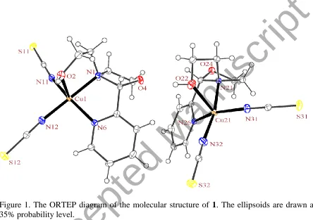
Figure 1. The ORTEP diagram of the molecular structure of 1. The ellipsoids are drawn at the 35% probability level.
Figure 2. Packing of 1 showing the hydrogen bonds. Only the hydrogen atoms involved in hydrogen bonding are shown. Each CuN4O unit is shown as square pyramid.
[image:20.612.96.518.76.418.2]Figure 3. The ORTEP diagram of the molecular structure of 2. The ellipsoids are drawn at the 35% probability level.
Figure 4. Packing of 2 showing the hydrogen bonds. Only the hydrogen atoms involved in hydrogen bonding are shown. Each CuNO2Cl2 unit is shown as square pyramid.
[image:22.612.97.522.69.508.2]Figure 5. Variation diagram of total intermolecular interactions energy (E) for 1 and 2 with increasing the number of surrounding molecules.
[image:23.612.130.511.73.332.2]Figure 6. Optimized structure for the complex containing NpyOoxa-donor AEPC, [Cu(AEPC)(NCS)2] (1'opt), possible isomer for 1.
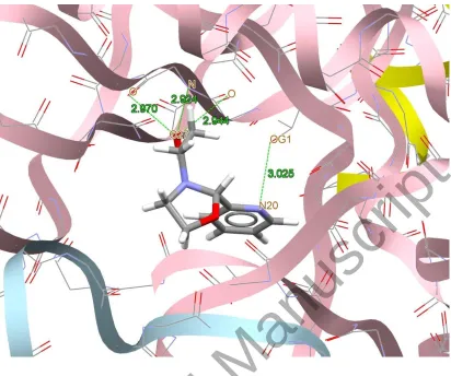
[image:24.612.101.524.72.679.2]Figure 7. Docking study results showing the interaction between AEPC ligand and BRAF kinase protein.
Figure 8. Docking study results showing the interaction between 1 and BRAF kinase protein.
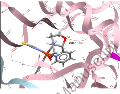
Figure 9. Docking study results showing the interaction between 2 and BRAF kinase protein.
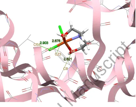
Figure 10. Docking study results showing the interaction between 3 and BRAF kinase protein.
Table 1. Crystal data and structure refinement for 1 and 2.
1 2
Empirical formula C24H28Cu2N8O4S4 C4H11Cl2CuNO2
Formula weight, g mol−1 747.87 239.59
Crystal size, mm3 0.12 × 0.12 × 0.09 0.15 × 0.15 × 0.06
Temperature, K 93 173
Crystal system Orthorhombic Monoclinic Space group P212121 P21/n
Unit cell dimensions (Å, °)
a 9.7908(3) 7.7998(6)
b 17.3016(4) 7.8465(5)
c 18.1819(4) 14.4389(11)
α
β 90.00 103.817(8)
γ
Volume, Å3 3079.95(14) 858.11(11)
Z 4 4
Calculated density, g cm−3 1.613 1.854 Absorption coefficient, mm−1 1.70 3.11
F(000), e 1528 484
2θ range for data collection (°) 4.6–56.4 5.0–63.2
h, k, l ranges −11 ≤ h ≤ 13, −16 ≤ k ≤ 21, −23 ≤ l ≤ 19
−11 ≤ h ≤ 11, −11 ≤ k ≤ 11, −21 ≤ l ≤ 20
Reflections collected / independent / Rint 18136 / 6420 / 0.035 10395 / 2827 / 0.032
Data / ref. parameters 6420 / 383 2827 / 103 Goodness-of-fit on F2 1.07 1.07
Final R indexes [I>=2σ (I)] R1 = 0.026, wR2 = 0.0650 R1 = 0.028, wR2 = 0.0541
Final R indexes [all data] R1 = 0.0299, wR2 = 0.0659 R1 = 0.0501, wR2 = 0.0600
Largest diff. peak / hole, e Å–3 0.39 / −0.35 0.46 / –0.38
Table 2. Selected bond length (Å) and angles (°) for 1 and 2 with estimated standard deviations in parentheses.
1 2
Dis
ta
nces
Cu1−O2 2.277(2) Cu1−O2 2.0165(13) Cu1−N1 2.058(3) Cu1−O4 2.0189(13) Cu1−N6 1.989(3) Cu1−N1 2.0078(14) Cu1−N11 1.962(3) Cu1−Cl1 2.2122(5) Cu1−N12 1.944(3) Cu1−Cl2 2.5476(5) Cu21−O22 2.293(2)
Cu21−N21 2.052(3) Cu21−N26 1.994(3) Cu21−N31 1.949(3) Cu21−N32 1.946(3)
Ang
les
N1−Cu1−O2 77.83(9) O2−Cu1−N1 82.54(6) O2−Cu1−N11 96.13(10) N1−Cu1−O4 82.73(6) N11−Cu1−N12 94.23(11) O4−Cu1−Cl1 96.33(4) N12−Cu1−N6 93.79(11) Cl1−Cu1−Cl2 97.068(18) N6−Cu1−N1 83.06(10) Cl2−Cu1−O2 103.37(4) N6−Cu1−O2 104.87(10) N1−Cu1−Cl1 173.06(5) N1−Cu1−N12 168.60(12) O2−Cu1−O4 148.87(6) N6−Cu1−N11 157.12(11)
N21−Cu21−O22 78.77(9) O22−Cu21−N31 95.24(10) N31−Cu21−N32 94.16(12) N32−Cu21−N26 94.30(11) N21−Cu21−N32 168.60(12) N26−Cu21−N31 154.14(11) N26−Cu21−O22 108.88(10) N26−Cu21−N21 83.12(10)
[image:30.612.69.546.98.630.2]Table 3. Hydrogen bond and short contact interactions dimensions (Å and °) in 1
and 2.
D–H···A d(D–H) d(H···A) <(DHA) d(D···A)
1
O(2)−H(2)∙∙∙S(11) 0.90(3) 2.29(3) 172(3) 3.186(3) O(22)−H(22)∙∙∙S(31) 0.93(3) 2.38(3) 169(3) 3.298(3)
2
O(2)−H(2)∙∙∙Cl(2) 0.95(2) 2.09(2) 170(2) 3.0290(14) O(4)−H(4)∙∙∙Cl(2) 0.931(15) 2.171(17) 165.2(18) 3.0801(13)
Table 4. All coordination modes of the thiocyanato ligand with copper ion. T er m in ally co o rd in atio n m o d es (5 3 %)
Percent 3.1% 48.1% 0.6% 1.4% 0.1%
B rid g in g co o rd in atio n m o d es (4 7 %)
Percent 36.8% 9.4% 0.3% 0.1%
Table 5. All complexes with CuNO2Cl2 environment (any types of O- and N-donor ligand with terminal chloride
ions) and τ value in range of 0.00−0.50. The ∆ value refers to the difference between bond lengths of two chloro ligands and d refers to the distance of the copper ion from coordinated plane.
Stru
ctu
res
∆ 0.30 Å 0.06 Å 0.03 Å
d 0.30 Å 0.16 Å 0.20 Å
(a) (b) (c)
[image:33.612.71.546.108.673.2]Table 6. The NBO analysis results for AEPC ligand and 1opt isolated complex. The values are the total of charge on the similar atoms. The ∆ show the variation of charge on the atoms after coordination.
Carbon Hydrogen Nitrogen Oxygen SNCS CNCS NNCS Metal AEPC –0.52 2.97 –1.09 –1.36 – – – –
1opt –0.57 3.49 –1.15 –1.39 –0.24 0.35 –1.41 0.91
∆ –0.05 +0.52 –0.06 –0.03 – – – –
Table 7. HOHO and LUMO orbitals for the optimized structures of AEPC and 1opt.
HOMO LUMO
Total energy (Kcal/mol)
AE
PC
−4
07
62
0
[C
u
(AE
PC
)(
NC
S)
2
]
Opt
(
1
Opt
)
−6
59
76
6
Table 8. The calculated fitness values for AEPC and 1-3 along with the doxorubicin. Top II TS TrxR RNR rHA HDAC7 DNA-Gyrase CatB BRAF-Kinase 35.49 35.41 38.50 31.76 34.96 41.43 32.68 26.71 33.71 AEPC 45.46 48.41 51.90 42.55 43.08 50.25 38.09 26.65 41.79 [Cu(AEPC)(NCS)2] (1)
25.04 27.02 30.65 25.95 26.89 30.53 22.59 15.66 25.70 [Cu(DEA)Cl2] (2)
40.72 34.10 46.05 36.78 41.01 33.77 34.58 24.36 40.72 [Cu(BHEG)2](3)
Table 9. Hydrogen bonds dimensions (Å and °) between proteins and 1-3.
Proteins D–H···A d(D···A) Compounds Proteins D–H···A d(D···A) Compounds
BRAF-Kinase
O–HAEPC···O 2.944
AEPC
RNR
N–H···OAEPC 2.937
AEPC O–HAEPC···O 2.970 O–H···NAEPC 2.797
N–H···OAEPC 2.924 N–H···NAEPC 2.770 O–H···NAEPC 3.025 N–H···OAEPC 2.647 O–H···O1 2.377 1 O–H1···O 2.569
1
C–H···O2 2.621
2
N–H···O1 2.839
C–H···Cl2 2.903 N–H2···O 3.052 2
C–H···O2 2.676 N–H···O4 2.512
3
O–H4···O 3.027
3
O–H···O4 2.584 N–H···O4 3.062 O–H4···O 2.979 O–H4···O 2.455 N–H···O4 2.660 O–H4···O 3.029 N–H···O4 3.056
CatB
C–H···πAEPC 2.865 AEPC
TrxR
O–H···OAEPC 2.974
AEPC N–H···O1 2.773 1 N–H···NAEPC 2.898
N–H···Cl2 2.739 2 N–H···O1 2.172
1
N–H···O4 2.426
3
N–H···O1 2.258 O–H4···O 2.722 O–H1···O 2.731 O–H···O4 2.610 O–H2···O 2.927
2
O–H···O4 2.426 O–H2···O 2.952 O–H2···O 3.038
DNA-Gyrase
N–H···NAEPC 2.681 AEPC N–H···Cl2 2.825 O–H1···N 2.819 1 O–H4···O 2.702
3
N–H···O1 2.705
2
N–H···O4 2.610 N–H···Cl2 2.916
N–H···Cl2 2.584
TS
N–H···NAEPC 2.492
AEPC N–H···O4 2.757
3
S–H···OAEPC 3.107
O–H···O4 2.506 O–H···S1 2.752 1
O–H4···O 2.842 O–H···O2 2.458 2
N–H···O4 2.797
3
HDAC7
O–H···OAEPC 2.769 AEPC O–H4···O 2.952 C–H···S1 2.759 1 O–H4···O 2.918 N–H···Cl2 3.200
2
N–H···Cl2 3.063
Top II
N–H···OAEPC 2.690
AEPC N–H···O4 2.324
3
O–HAEPC···O 2.585 N–H···O4 2.407 O–HAEPC···O 2.552 O–H4···N 2.826 N–H···O1 2.922
1
N–H···O1 2.935
rHA
O–HAEPC···O 2.850 AEPC O–H1···O 3.026 O–H···O1 2.588
1 O–H
2···O 2.724
2
N–H···O1 2.603 N–H···Cl2 3.280 N–H···O1 2.591 2 N–H···Cl2 3.174 N–H···O4 2.804
3
N–H···Cl2 3.137 O–H···O4 2.548 N–H···O4 3.049
3
O–H4···N 2.963 O–H···O4 2.336 O–H4···O 2.753 O–H4···O 2.927 N–H···O4 2.530
![Figure 6. Optimized structure for the complex containing N pyOoxa-donor AEPC, [Cu(AEPC)(NCS)Accepted Manuscript2] (1'opt), possible isomer for 1](https://thumb-us.123doks.com/thumbv2/123dok_us/9018957.398494/24.612.101.524.72.679/figure-optimized-structure-complex-containing-accepted-manuscript-possible.webp)