ISSN Print: 2325-7458
DOI: 10.4236/jacen.2019.83010 Aug. 8, 2019 115 Journal of Agricultural Chemistry and Environment
A Phytotoxic and Antifungal Metabolite
(Pyrichalasin H) from a Fungus Infecting
Brachiaria eruciformis
(Signal Grass)
Kumudini M. Meepagala
1*, Brandon M. Clausen
1,2, Robert D. Johnson, David E. Wedge
1,
Stephen O. Duke
11USDA-ARS, Natural Products Utilization Research Unit, University, MS, USA
2Sally McDonnell Barksdale Honors College, University of Mississippi, University, MS, USA
Abstract
Brachiaria eruciformis (sm.) Griseb, locally known as “signal grass”, is a common weed in lawns and turfs in Mississippi, USA. During late spring and early summer months, leaves of B. eruciformis are infected with a fungus causing necrosis. The infected leaves ultimately turn brown and wither. As part of our search for potential new natural product-based agrochemicals, we studied this plant pathogen in order to investigate phytotoxic and fungitoxic metabolites produced by the fungus. The causative fungus was isolated from an infected leaf of B. eruciformis, cultured in potato dextrose agar plates and identified via molecular techniques as Pyricularia grisea. A phytotoxic com-pound was isolated from Czapek-Dox broth liquid culture medium and iden-tified as pyrichalasin H by spectroscopic techniques. Pyrichalasin H was toxic to the fungal plant pathogen Colletotrichum fragariae in a TLC bioautogra-phy assay and bioautogra-phytotoxic to two monocot and one dicot plants. This is the first report of antifungal activity of pyrichalasin H against phytopathogens. Pyrichalasin H isolated from Pyricularia grisea, a pathogen infecting B. eru-ciformis (signal grass) was shown to be phytotoxic and fungicidal to Colleto-trichum fragariae.
Keywords
Mycotoxin, Brachiaria eruciformis,Phytotoxicity, Pyricularia grisea,
Pyrichalasin H
1. Introduction
Microbes are good sources of bioactive compounds [1]. Of particular interest in
How to cite this paper: Meepagala, K.M., Clausen, B.M., Johnson, R.D., Wedge, D.E. and Duke, S.O. (2019) A Phytotoxic and Antifungal Metabolite (Pyrichalasin H) from a Fungus Infecting Brachiaria eruciformis (Signal Grass). Journal of Agricultural Chemistry and Environment, 8, 115-128. https://doi.org/10.4236/jacen.2019.83010
Received: June 15, 2019 Accepted: August 5, 2019 Published: August 8, 2019
Copyright © 2019 by author(s) and Scientific Research Publishing Inc. This work is licensed under the Creative Commons Attribution International License (CC BY 4.0).
http://creativecommons.org/licenses/by/4.0/
DOI: 10.4236/jacen.2019.83010 116 Journal of Agricultural Chemistry and Environment our research are phytopathogenic fungi and their metabolites. Phytopathogenic fungi use the host plant as a source of nutrients for its growth and development. In this process, the fungi often produce toxins that are lethal to the host plant and often toxic to other plant species, and these toxins may also have insecticid-al, antibacterial and antifungal activities as a result of coevolution and competi-tion among species to survive in the biosphere [2] [3] [4].
Weeds are a major threat to crop production worldwide. Modern agricul-tural practices primarily rely on synthetic herbicides, due to their high efficacy, selectivity and low cost [5] [6]. Repeated use of synthetic herbicides for pro-longed periods of time has facilitated the widespread evolution of herbicide re-sistance among weed species exposed to them [7]. Herbicide-resistant weeds have caused an increased need for new herbicides with new modes of action. Natural products offer broad chemical diversity with a wide range of bioactivi-ties, including phytotoxicity. These compounds may have varying and multiple molecular target sites and thus can be used as novel compounds or templates for development of pest control agents [8]. This study focused on isolation of bioactive compounds from a pathogenic fungus infecting B. eruciformis (sig-nal grass).
2. Materials and Methods
2.1. General Procedures
Potato dextrose agar (PDA) plates were prepared by dissolving 19.6 g of DifcoTM (Detroit, MI) potato dextrose agar and 8.0 g of BD BactoTM agar in 1 L of deio-nized water. The solution was autoclaved for 30 min at 120˚C. PDA solution was poured into sterile plastic petri dishes (BD FalconTM) inside a class II-A biologi-cal safety cabinet. Czapek-Dox broth media was made by dissolving 35.0 g of Czapek-Dox broth culture medium (FlukaTM), 1.5 g of malt extract (BD Bac-toTM), and 1.5 g of yeast extract (BD BactoTM) in each 1 L of deionized water in thirty 2 L Erlenmeyer flasks. All media were sterilized by autoclave for 30 min at 120˚C. Fungal broth extracts were analyzed on 250 µm silica gel TLC plates with
fluorescent indicator (Analtech, Newark, DE). UV light (254 and 365 nm).
p-Anisaldehyde spray reagent and iodine vapor were used for visualization of compounds. Isolation and purification of metabolites were performed with a Biotage IsoleraTM Flash Chromatography system (Charlotte, NC) using hexane, ethyl acetate, dichloromethane and methanol in various percentages. 1H and 13C NMR spectra were recorded on a Bruker AMX spectrometer (Billerica, MA) op-erating at 400 MHz for 1H and at 125 MHz for 13C NMR. High-resolution mass spectra were obtained using JEOL ACCU TOF JMS-T1000 LC mass spectrome-ter (Peabody, MA).
2.2. Fungal Material
DOI: 10.4236/jacen.2019.83010 117 Journal of Agricultural Chemistry and Environment
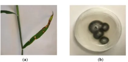
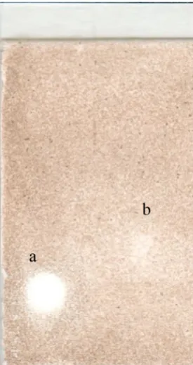
Figure 1. (a) Infected Brachiaria eruciformis leaves showing necrosis; (b): Pyricularia
grisea growing on PDB plate.
was surface sterilized by immersing it in aqueous sodium hypochlorite (5%) so-lution for 30 s, followed by thoroughly rinsing with sterile deionized water. A PDA plate (half strength potato-dextrose-agar, DifcoTM) was inoculated by plac-ing a small piece of leaf tissue (approx. 2 × 2 mm) cut by a sterile scalpel from an infection site. This plate was allowed to grow in a growth chamber at 24˚C under 12-h light cycle for one week. A single colony from this plate was sub-cultured on another PDA plate under similar conditions as above by placing a 0.5 cm diameter plug of the fungal colony (Figure 1(b)). This plate was incubated and fungal culture was allowed to grow for 7 days, and this fungal colony was used to inoculate a 500 mL Erlenmeyer flask containing 250 mL Czapek-Dox broth. The liquid culture was allowed to grow for 7 days in an orbital shaker rotating at 90 rpm at 24˚C. Aliquots of 5 mL of liquid culture broth was added to thirty 2-L flasks each containing 1 L of Czapek-Dox broth, and these culture broths were allowed to grow in an orbital shaker (80 rpm) under the same conditions as above.
2.3. Molecular Identification of Fungus
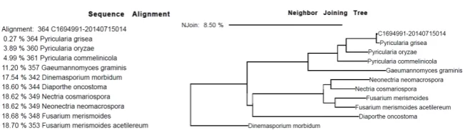
DNA sequencing and alignment of ITS region was performed by Accugenix® (Newark, DE). Consensus C169499120140715014 was aligned, using the Neigh-bor Joining Tree method with Dinemasporium morbidum as the out-group (Figure 2).
2.4. Extraction and Isolation of Phytotoxins in the Fungal Broth
DOI: 10.4236/jacen.2019.83010 118 Journal of Agricultural Chemistry and Environment
Figure 2. Sequence alignment and neighbor joining tree for the identification of
Pyricu-laria grisea.
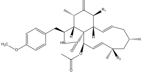
which was identical to the white solid precipitated from the crude ethyl acetate ex-tract and was identified as pyrichalasin H (Figure 3) by NMR and high-resolution mass spectroscopy.
Pyrichalasin H (1): ESI-HRMS m/z 524.30136 [M + H] (calcd for 524.30124), [α]D-18.9 (CHCl3 c = 0.5)mp 208˚C - 209˚C. 1H NMR (400 MHz, CDCl3) δ 0.98 (d, J = 6.7 HZ, 3H, CH3-11), 1.04 (d, J = 6.2 Hz, CH3-22), 1.34 (s, CH3-23), 1.54 (dd, J = 14, 2 Hz, Ha-17), 1.79 (m, H-16), 1.8 (m, Ha-15), 1.88 (dd, J = 14, 2 Hz, Hb-17), 2.03 (m, Hb-15), 2.11 (t, J = 4.4, H-4), 2.24 (s, OAc), 2.59 (dd, J = 14, 10, Ha-10), 2.77 (m, H-5), 2.81 (dd, J = 14, 10, Hb-10), 2.93 (dd, J = 8, 8 Hz, H-8), 3.21 (m, H-3), 3.79 (s, OMe), 3.83 (d, J = 12 Hz, H-7), 5.10 (br s, Ha-12), 5.34 (br s, Hb-12), 5.36 - 5.44 (m, H-14), 5.51 (d, J = 2 Hz), 5.54 (dd, J = 15, 2 Hz, H-20), 5.6 (s, NH), 5.74 (dd, J = 16, 9 Hz, H-13), 5.88 (d, J = 2 Hz H-19), 6.85 (d, J = 8Hz), 7.06(d, J = 8 Hz); 13C NMR (100 MHz, CDCl
3) δ 13.88, 14.30, 20.99, 21.14, 21.24, 26.52, 28.47, 31.17, 33.18, 42.78, 44.57, 44.61, 49.84, 51.86, 53.80, 53.83, 55.39, 60.51, 72.36, 74.35, 114.36, 114.94, 125.98, 126.60, 129.35, 130.26, 136.60, 137.98, 145.95, 158.71, 170.24, 174.18.
Acetylation of pyrichalasin H: To a solution of pyrichalasin H (200 mg, 0.382 mmol) in pyridine (4 mL), 4 eq of acetic anhydride (72 µL) was added and the mixture was stirred for 12 h at room temperature. The reaction mixture was cooled in ice, acidified with 0.5 M cold HCl solution and the product was ex-tracted with ethyl acetate (100 mL × 2). The ethyl acetate extract was dried over anhydrous Na2SO4 and the solvent was evaporated to obtain a semi-solid. The product 2 was purified by (SNAP 50 g Biotage column) using 5% - 100% ethyl acetate in hexane to afford white crystalline solid (157 mg). ESI-HRMS m/z 566.30648 [M + H] (calcd for 566.31178), [α]D-56.9 (CHCl3 c = 0.5)mp 135˚C - 137˚C. 1H NMR (400 MHz, CDCl
3) δ 0.91 (d, J = 6.7 Hz, 3H),1.02 (d, J = 6.1 Hz, 3H), 1.26 (t, J = 7.1 Hz, 1H), 1.32 (s, 3H), 1.53 (dd, J = 14.2, 2.4 Hz, 1H), 1.66 - 1.8 (m, 3H), 1.85 (dd, J = 14.2, 2.7 Hz, 1H),1.94 (s, 3H), 2.12 (dd, J = 5.0, 3.5 Hz, 1H), 2.17 (s, 3H), 2.26 (s, 3H), 2.64 (dd, J = 13.6, 8.9 Hz, 1H), 2.86 - 2.72 (m, 2H), 3.15 (dd, J = 11.1, 9.6 Hz, 1H), 3.26 - 3.18 (m, 1H), 3.79 (s, 3H), 5.04 (t, J = 1.7 Hz, 1H), 5.34 - 5.15 (m, 3H), 5.66 - 5.43 (m, 3H), 5.89 - 5.73 (m, 2H), 6.90 - 6.78 (m, 2H), 7.12 - 7.02 (m, 2H); 13C NMR (100 MHz, CDCl
DOI: 10.4236/jacen.2019.83010 119 Journal of Agricultural Chemistry and Environment
Figure 3. Structures of pyrichalasin H (1) and its acetate analog (2): R1 = R2 = OH; (2):
R1 = OAc, R2 = OH.
2.5. Phytotoxicity Bioassay on Seed Germination
Fungal extract, column chromatography fractions and isolated compounds and their analogs were tested with the phytotoxicity bioassay described by Dayan et al. [9]. Phytotoxic effects of the compounds and the extract, column fractions and pure compounds were evaluated by the germination and growth effects on
Lactuca sativa (lettuce; dicot, Crisphead cultivar from Burpee seeds, Warmister, PA) and Agrostis stolonifera (bentgrass; monocot, Penncross variety from Turf-Seed, Inc of Hubbard, OR) seeds in 24-well plates. The negative control was deionized sterile water and a solvent control consisted of 10% acetone in deionized water. Each 24-well plate was placed in an incubator under 16/8 h light/dark condition at 26˚C and 120 μmol∙s−1∙m−2 average photosynthetically ac-tive radiation (PAR). Phytotoxic activity was ranked qualitaac-tively on a scale of 0 to 5 after 7 days for L. sativa and after 10 days for A. stolonifera where a ranking of 0 means no difference between the control and treated seeds and a ranking of 5 indicates complete inhibition of germination, with intermediate effects (2, 3 and 4) indicating increasing effect on seedling growth and development.
2.6. Phytotoxicity Evaluation on
Lemna pausicostata
The method described by Michel et al. [10] using duckweed (Lemna pausicosta-ta) was used to quantitatively evaluate (e.g., accurate IC50 values) phytotoxicity. Two duckweed plants with three fronds each were placed in a six-well plate with 4950 µL of Hoagland’s media and 50 µL of water, or the solvent, or the com-pound dissolved in the appropriate solvent (at a concentration of 100×). The concentration of acetone in the wells was therefore 1% by volume. The plates were incubated in the Percival incubator as described above. Duckweed plant areas were measured at day 0 and day 7 using a Lemnatec Scanalyzer PL with Lemna Launcher and Lemna Miner software (Lemna Tec GmbH, Schumanstr 19, 52, 146 Würselen, Germany). The image analysis software was used to meas-ure and monitor frond number, total frond area as well as color classes (healthy, chlorotic and necrotic tissue). Replicate tests at varying concentrations of test compounds allowed for determination of IC50 values using R Studio software (Version 0.99.491).
O H3C
H
R1
R2
O O
O
DOI: 10.4236/jacen.2019.83010 120 Journal of Agricultural Chemistry and Environment
2.7. Cellular Leakage Test on
Cucumis sativus
(Cucumber) Leaf
Disks
To determine if the phytotoxic compounds cause membrane leakage, a mod-ified method developed by Duke and Kenyon [11] was used. Cucumis sativus
(cucumber, from Burpee seeds, Warmister, PA) plants were grown from seeds in a Conviron growth chamber (Model E7/2; Winnipeg, Canada) at 26˚C un-der 173 μmol∙s−1∙m−2 PAR for 6 days. Fifty 4-mm disks were cut using a cork borer from C. sativus leaves and placed in Petri dishes along with 5 mL of 1 mM 2-(4-morpholino) ethane sulfonic acid (MES) buffer solution with 2% w/v su-crose. The pH of the solution was adjusted to 6.5 with 1 M NaOH. The test compounds were dissolved in acetone and were added to MES buffer solution such that the final concentration of acetone was 1%. Aciflurofen, a herbicide that causes rapid plasma membrane destruction in the light [12] was used as the pos-itive control in the experiment. Electrical conductivities of the solutions in the dishes were measured using a dip cell at various time intervals (0, 1, 2, 4, 6 and 8) after exposure to the chemical. All dishes were covered with aluminum foil and kept in the dark for 18 h. The dishes were then exposed to 200 μmol∙s−1∙m−2 photosynthetically active radiation (PAR) light and electrical conductivity mea-surements were taken at the same time intervals. Experiments were done in trip-licate, and the averages were graphed as % conductivity change after treatment. The maximum leakage reading for the leaf disks was taken by measuring the conductivity of the boiled solution with leaf disks.
2.8. Effect of Pyrichalasin H on the Growth and Chlorophyll
Content of Lettuce (
Lactuca sativa
) and Barley
(
Hordeum vulgare
L.) Seedlings
Pyrichalasin H was also tested at varying concentrations on growth of lettuce and barley seeds.
DOI: 10.4236/jacen.2019.83010 121 Journal of Agricultural Chemistry and Environment DMSO. The tubes were incubated at 65˚C - 68˚C for 2h. The DMSO was trans-ferred to another tube and the process was repeated with another 2 mL of DMSO for 30 min. DMSO extracts were combined and the concentration of chlorophyll was determined using Shimadzu UV-3101 UV-VIS NIR spectro-photometer. The absorbance values at 663 and 645 nm were taken using 1-cm plastic cuvettes. The instrument was zeroed with DMSO. The total chlorophyll concentration was determined by the following equation:
Total Chlorophyll Concentration = 0.0202 A663 + 0.00802 A645
where A663 and A645 are the absorbance readings at 663 and 645 nm. Using this value, the amount of chlorophyll per fresh weight of shoots was calculated and graphed against the concentration of pyrichalasin H.
2.9. Bioautography
Silica gel TLC plate-based bioautography was carried out to identify the anti-fungal activity of compounds and extracts against Colletotrichum fragariae ac-cording to a previously published method [14]. Column fractions and pure compounds were eluted on silica gel TLC plates with appropriate solvent and were air-dried. The plates were sprayed with a spore suspension of C. fragariae
(105 spores/mL) and were incubated in a moisture chamber for 3 days at 26˚C with a 12-h photoperiod. Clear zones on the TLC plates indicate the presence of antifungal constituents.
2.10. Micro-Bioassay for Quantitative Fungicide Activity
To evaluate the quantitative fungicide activity, the purified compounds were evaluated in a dose response manner (0.3, 3, and 30 µM) in a 96-well mi-cro-bioassay against Colletotrichum fragariae, C. gloeosporioides, C. acutatum,
Botrytis cinerea, Fusarium oxysporium and Phomopsis obscurans with compar-ison to commercial fungicides (captan and azoxystrobin) according to published methods [15] [16].
3. Results and Discussion
The fungus infecting B. eruciformis was identified by molecular analysis (by Accugenix®, Newark, DE USA) as Pyricularia grisea. ITS sequence alignment of consensus C169499120140715014 (Figure 2), using Dinemasporium morbidum
as the out-group, showed a 99.73% match.
DOI: 10.4236/jacen.2019.83010 122 Journal of Agricultural Chemistry and Environment
Table 1. Phytotoxicity bioassay results of crude ethyl acetate extract of the culture broth
and isolated pyrichalasin H and acetate analogs (at varying concentrations).
Sample Concentration Lettuce Agrostis Culture broth EtOAc extract 1 mg/mL 3 3
Fraction 7 1 mg/mL 2 4
Fraction 8 1 mg/mL 2 4
Pyrichalasin H
0 μM 0 0
10 μM 0 0
33 μM 2 3
100 μM 2 3
330 μM 2 4
1000 μM 2 4
Pyrichalasin Hacetate 1 mg/mL 0 0
activity and gain insight into the mode of action of pyrichalasin H, further bio-assays were conducted. The cellular leakage test on cucumber cotyledon disks using pyrichalasin H as the test compound was conducted in order to see if the phytotoxic activity was due to cellular leakage caused by the test compound. The plot (Figure 4(a)) showed minimal to no change in conductivity even at the highest concentration of pyrichalasin H (1000 µM) that was tested. Acifluorfen, a herbicide that causes massive cellular leakage in the light was used as the posi-tive control (Figure 4(b)) This observation of lack of leakage or no change in conductivity of cucumber cotyledons due to pyrichalasin H suggests that the mechanism of action of phytotoxicity does not involve disruption of the plasma membrane.
To gain more quantitative phytotoxic activity, the effect of pyrichalasin H was examined at varying concentrations on the monocot duckweed (L. pausicostata) (Figure 5). Growth of duckweed plants decreased with increasing concentration of pyrichalasin H, with an IC50 value of 150 µm. At higher concentrations (greater than 330 µm) pyrichalasin H started to separate from the solution mak-ing a layer of white powder in the test solution. It was also observed that duck-weed plants started to show chlorosis at the concentrations 100 µm and higher, indicating that pyrichalasin H may inhibit synthesis of chlorophyll.
DOI: 10.4236/jacen.2019.83010 123 Journal of Agricultural Chemistry and Environment
Figure 4. (a) Changes of the conductivity of solutions of the cucumber leaf disks treated
[image:9.595.287.462.298.465.2]at varying concentrations (1, 10, 100 and 1000 µM) of pyrichalasin H in the dark and after exposure to light (arrow) at 18 h. The dotted line shows the maximum leakage value ob-tained by using boiled leaf disks in MES buffer. (b) The same measurements at 1 µM concentration of acifluorifen.
Figure 5. Effect of pyrichalasin H on Lemna pausicostata growth. Dotted line indicates
IC50. Concentrations greater than 300 µm could not be used as pyrichalasin H
precipi-tated from the aqueous medium.
Figure 6. Root length of lettuce (a) and barley (b) plants at varying concentration of
[image:9.595.218.530.522.691.2]DOI: 10.4236/jacen.2019.83010 124 Journal of Agricultural Chemistry and Environment
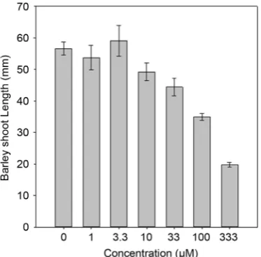
Figure 7. Shoot length of barley plants at varying concentration of pyrichalasin H
meas-ured at 7 days after treatment. Shoot length measurements were not taken for lettuce plants.
Figure 8. Chlorophyll per fresh weight of shoots lettuce (a) and barley (b) at varying
concentration of pyrichalasin H measured at 7 days after treatment.
An acetate analog of pyrichalasin H was synthesized. Acetylation of pyrichala-sin H upyrichala-sing acetic anhydride in pyridine afforded only the mono acetate analog, where the secondary OH group was acetylated. The acetate analog was not phy-totoxic (Table 1) in lettuce and bentgrass bioassays at 1 mg/mL concentration. Therefore, the presence of OH groups is needed for the phytotoxic activity.
[image:10.595.214.537.322.495.2]DOI: 10.4236/jacen.2019.83010 125 Journal of Agricultural Chemistry and Environment
Figure 9. TLC bioautography of a: pyrichalasin H (1); b: pyrichalasin H monoacetate (2)
The plate was eluted in 40% ethylacetate in hexane and sprayed with spores of C. fraga-riae.
activity in comparison to two commercial fungicides captan and azoxystrobin. The compounds were evaluated at 0.3, 3.0, 30 µm concentrations at 24 and 72 h for Colletotrichum fragariae, C. gloeosporioides, C. acutatum, Botrytis cinerea,
and Fusarium oxysporium and for 120 and 144 h for Phomopsis obscurans. C. acutatum, F. oxysporium and P. obscurans were not inhibited by pyrichalasin H (data not shown). Botrytis cinerea and C. gloeosporioides were marginally inhi-bitory to pyrichalasin H at 48 h after treatment and the activity diminished at 72 h (Figure 10(a) and Figure 10(b)). C. fragariae treated at 30 µm concentration of pyrichalasin H was inhibited about 60% and 30% at 48 h and 72 h after treat-ment, respectively (Figure 10(c)). At the same concentration of azoxystrobin and captan C. fragariae growth was inhibited 85% and 80% - 70% at 48 h and 72 h after treatment, respectively. C. fragariae was the most sensitive fungal species to pyrichalasin H among the fungi that we tested.
From the phytotoxicity bioassays, it is evident that even at the highest con-centration of pyrichalasin H (1000 µm) germination inhibition was not com-plete. Root and shoot growth were reduced by pyrichalasin H concentrations as low as 33 µM.
DOI: 10.4236/jacen.2019.83010 126 Journal of Agricultural Chemistry and Environment
Figure 10. Microbioassay results showing % inhibition of fungal growth in comparison to
that caused by commercial fungicides captan and azoxystrobin. ((a) & (b)) Inhibition caused by C. gloeosporioides ((c) & (d)) Inhibition caused to C. fragariae ((a) & (c)) re-sults at 48 h ((b) & (d)) rere-sults at 72 h.
phytotocity of pyrichalasijn H. The amount of pyrichalain H produced by dif-ferent isolates of Pyricularia grisea correlates positively with their virulence against Digitaria plants [20]. Pyrichalasin H was previously found to inhibit growth and cause curling of the shoots of rice seedlings, but produced no symp-toms in the leaf [21]. Finger millet (Eleusine coracana L.) seeing growth is inhi-bited by pyrichalasin H [22]. It causes effects on plant cell morphology similar to those caused by actin polymerization inhibitors [23]. Cytochalasin H, an analog of pyrichalasin H, where the OMe group is replaced with H, is a potent inhibitor for elongation of actin fibers [24]. Cytochalasin H is also a plant growth inhibi-tor [25]. In our experiments, we observed that the growth of plants was not completely inhibited by pyrichalasin H at concentrations as high as 1 mM, but the elongation of shoots and roots were significantly inhibited, and chlorophyll levels were decreased at concentrations as low as 33 µM. We hypothesize that the mode of action of pyrichalasin H is similar to the mode of action of cytochalasin H, and that the effects that we have documents are secondary and/or tertiary ef-fects of this primary effect. Experiments with pyrichalasin H on actin fiber po-lymerization should be conducted to confirm this hypothesis.
4. Conclusion
The causative fungus infecting Brachiaria eruciformis (signal grass) was isolated and identified as Pyricularia grisea. From the culture filtrate of Pyricularia grisea
DOI: 10.4236/jacen.2019.83010 127 Journal of Agricultural Chemistry and Environment
Acknowledgements
We thank Linda Robertson, Ramona Pace and William Eric Briscoe for their technical assistance.
Conflicts of Interest
The authors declare no conflicts of interest regarding the publication of this pa-per.
References
[1] Monciardini, P., Iorio, M., Maffioli, S., Sosio, M. and Donadio, S. (2014) Discover-ing New Bioactive Molecules from Microbial Sources. Microbial Biotechnology, 7, 209-220.https://doi.org/10.1111/1751-7915.12123
[2] Macheleidt, J., Mattern, D.J., Fischer, J., Netzker, T., Weber, J., Schroeckh, V., Va-liante, V. and Brakhage, A.A. (2016) Regulation and Role of Fungal Secondary Me-tabolites. Annual Review of Genetics, 50, 371-392.
https://doi.org/10.1146/annurev-genet-120215-035203
[3] Ogorek, R. (2016) Enzymatic Activity of Potential Fungal Plant Pathogens and the Effect of Their Culture Filtrates on Seed Germination and Seedling Growth of Gar-den Cress (Lepidium sativum L.). European Journal of Plant Pathology, 145, 469-481. https://doi.org/10.1007/s10658-016-0860-7
[4] Kim, W., Park, J.-J., Dugan, F.M., Peever, T.L., Gang, D.R., Vandemark, G. and Chen, W. (2017) Production of the Antibiotic Secondary Metabolite Solanapyrone A by the Fungal Plant Pathogen Ascochytarabiei during Fruiting Body Formation in Saprobic Growth. Environmental Microbiology, 19, 1822-1835.
https://doi.org/10.1111/1462-2920.13673
[5] Duke, S.O. and Dayan, F.E. (2018) Herbicides. John Wiley & Sons, Chichester. https://doi.org/10.1002/9780470015902.a0025264
[6] Dayan, F.E., Cantrell, C.L. and Duke, S.O. (2009) Natural Products in Crop Protec-tion. Bioorganic & Medicinal Chemistry, 17, 4022-4034.
https://doi.org/10.1016/j.bmc.2009.01.046
[7] Heap, I. and Duke, S.O. (2018) Overview of Glyphosate-Resistant Weeds World-wide. Pest Management Science, 74, 1040-1049. https://doi.org/10.1002/ps.4760
[8] Sparks, T.C., Hahn, D.R. and Garizi, N.V. (2017) Natural Products, Their Deriva-tives, Mimics and Synthetic Equivalents: Role in Agrochemical Discovery. Pest Management Science, 74, 700-715.https://doi.org/10.1002/ps.4458
[9] Dayan, F.E., Romagni, J.G. and Duke, S.O. (2000) Investigating the Mode of Action of Natural Phytotoxins. Journal of Chemical Ecology, 2, 2079-2094.
https://doi.org/10.1023/A:1005512331061
[10] Michel, A., Johnson, R.D., Duke, S.O. and Scheffler, B.E. (2004) Dose-Response Re-lationships between Herbicides with Different Modes of Action and Growth of Lemnapaucicostata—An Improved Ecotoxicological Method. Environmental Toxi-cology and Chemistry, 23, 1074-1079. https://doi.org/10.1897/03-256
[11] Duke, S.O. and Kenyon, W.H. (1993) Peroxidizing Activity Determined by Cellular Leakage. In: Boger, P. and Sandman, G., Eds., Target Assays for Modern Herbicides and Related Phytotoxic Compounds, Lewis, Boca Raton, 61-66.
DOI: 10.4236/jacen.2019.83010 128 Journal of Agricultural Chemistry and Environment
Slikker, W.J. and Van Hemmon, J., Eds., Haye’s Handbook of Pesticide Toxicology, 3rd Edition, Vol. 2, Academic Press, Elsevier, San Diego, 1733-1751.
https://doi.org/10.1016/B978-0-12-374367-1.00081-1
[13] Hiscox, J.D. and Israelstam, G.F. (1979) A Method for the Extraction of Chlorophyll from Leaf Tissue without Maceration. Canadian Journal of Botany, 57, 1332-1334. https://doi.org/10.1139/b79-163
[14] Wedge, D.E. and Nagle, D.G. (2000) A New 2D TLC Bioautography Method for the Discovery of Novel Antifungal Agents to Control Plant Pathogens. Journal of Nat-ural Products, 63, 1050-1054. https://doi.org/10.1021/np990628r
[15] Oliva, A., Meepagala, K.M., Wedge, D.E., Harries, D., Hale, A.L., Aliotta, G. and Duke, S.O. (2003) Natural Fungicides from Ruta graveolens L. Leaves, Including a New Quinolone Alkaloid. Journal of Agricultural and Food Chemistry, 51, 890-896. https://doi.org/10.1021/jf0259361
[16] Meepagala, K.M., Schrader, K.K., Burandt, C.L., Wedge, D.E. and Duke, S.O. (2010) New Class of Algicidal Compounds and Fungicidal Activities Derived from a Chromene Amide of Amyris texana. Journal of Agricultural and Food Chemistry, 58, 9476-9482.https://doi.org/10.1021/jf101626g
[17] Scherlach, K., Boettger, D., Remme, N. and Hertweck, C. (2010) The Chemistry and Biology of Cytochalasans. Natural Product Reports, 27, 869-886.
https://doi.org/10.1039/b903913a
[18] Ismaiel, A.A. and Papenbrock, J. (2015) Fungal Phytotoxins with Potential Herbi-cidal Activity: Chemical and Biological Characterization. Agriculture, 5, 492-537. https://doi.org/10.3390/agriculture5030492
[19] Cimmino, A., Masi, M., Evidente, M., Superchi, S. and Evidente, A. (2015) Fungal Phytotoxins with Potential Herbicidal Activity: Chemical and Biological Characte-rization. Natural Product Reports, 32, 1629-1653.
https://doi.org/10.1039/C5NP00081E
[20] Tsurushima, T., Don, L.D., Kawashima, K., Murakami, J., Nakayashiki, H., Tosa, Y. and Mayama, S. (2005) Pyrichalasin H Production and Pathogenicity of Digita-ria-Specific Isolates of Pyriculariagrisea. Molecular Plant Pathology, 6, 605-613. https://doi.org/10.1111/j.1364-3703.2005.00309.x
[21] Nukina, M. (1987) Pyrichalasin H, a New Phytotoxic Metabolite Belonging to the Cytochalasansfrom Pyriculariagrisea (Cooke) Saccardo. Agricultural and Biological Chemistry, 51, 2625-2628. https://doi.org/10.1271/bbb1961.51.2625
[22] Sanmathi, K.R.P., Shanthala, L., Anikumar, T.B. and Sudharshana, L. (2006) Phyto-toxins from Pyriculariagrisea and Their Effect on Finger Millet. Journal of Plant Bi-ochemistry and Biotechnology, 15, 63-66. https://doi.org/10.1007/BF03321905
[23] Hirose, T., Izawa,Y., Koyama, K., Natori, S., Iida, K., Yahara, I., Shimaoka, S. and Maruyama (1990) The Effects of New Cytochalasins from Phomopsis sp. and the Derivatives on Cellular Structure and Actin Polymerization. Chemical and Phar-maceutical Bulletin, 38, 971-974.https://doi.org/10.1248/cpb.38.971
[24] Yahara, I., Harada, F., Sekita, S., et al. (1982) Correlation between Effects of 24 Dif-ferent Cytochalas Ins on Cellular Structures and Cellular Events and Those on Actin
in Vitro. The Journal of Cell Biology, 92, 69-78. https://doi.org/10.1083/jcb.92.1.69
[25] Cutler, H.G., Cutler, S.J. and Matesic, D. (2004) Mode of Action of Phytotoxic Fun-gal Metabolites. In: Macias, F.A., Galindo, J.C.G., Molinillo, J.M.G. and Cutler, H.G., Eds., Allelopathy: Chemistry and Mode of Action of Allelochemicals, CRC Press, Boca Raton, 253-270.https://doi.org/10.1201/9780203492789.ch13