CUTANEOUS MALIGNANCY: OUR OBSERVATIONS AND REVIEW OF LITERATURE
1,*
Harish Kumar Hanumappa,
5
Diviya K Kariappa and
1
Assistant Professor, Dept. of Surgical Oncology
2
Associate Professor, Dept. of Surgical Oncology
3
Professor, Dept., of Plastic Surgery
4
Senior resident, Dept. of Surgical Oncology
5,6,
M.Ch Resident, SJMCH, St Johns Medical College Hospital, SJMCH,
7
Junior resident, Dept. of Surgical Oncology, SJMCH, St Johns Medical College Hospital, SJMCH,
ARTICLE INFO ABSTRACT
Background:
of the world. Rarity of its incidence could be due to protective effects of melanin.Though it is the largest organ by surface area, occurrence of cutaneous malignanc
Presentation is usually earlier due to the fact that it is easily noticeable.
sex incidence, common site of presentation, type of histology,optimal management and its outcome, als avoidable risk factors.
and managed in the department of surgical oncology at tertiary care hospital from January 2016 to December 2018.
were managed over period of 3 years.3 cases were melanoma and 9 cases were squamous cell carcinoma and basal cell carcinoma was the final diagnosis in another 3cases. Surgical excision with adequate margins were done in 13 cases and palliative chemotherapy was given in one case and no cancer directed treatment was given in a case which had extensive skin lesion on scalp with poor general condition of the patient. Conclusion:
regarding warning signs of skin cancer. Nonsurgical options needs to be considered especially in syndromic conditions and inoperable situations. Surgery with adequate margins offers the best option
Copyright © 2018, Harish Kumar Hanumappa et al. This unrestricted use, distribution, and reproduction in any medium,
INTRODUCTION
The Reported incidence of cutaneous malignancy is as follows. Basal cell carcinomacontributes to about 65%
cancers in whites and 20%-30% in Asian
Squamous cell carcinomarepresents 30%-65% of skin cancers in blacks and Indians and 15%-25% in whites
2006).Non melanoma skin cancerare relatively more common in India compared to melanoma.Approximately 87,110 individuals are predicted to be diagnosed with melanoma in the United States alone (Surveillance, 2015). Markovic
observed that males are approximately 1.5-times more likely to develop melanoma than females.Other studies have shown that the incidence rate of melanoma is grater in women than men until they reach the age of 40 years, however, by 75 years of age, the incidence is almost 3-times as high in men versus women (145.6 vs. 47.3 per 100,000) (Rigel
*Corresponding author: Harish Kumar Hanumappa,
Assistant Professor, Dept. of surgical oncology, SJMCH, St Johns Medical College Hospital, SJMCH, Bangalore.
ISSN: 0975-833X
Article History:
Received 17th September, 2018
Received in revised form 20th October, 2018
Accepted 10th November, 2018 Published online 31st December, 2018
Citation: Harish Kumar Hanumappa, Rakesh S Ramesh, Sunder Raj Ellur, Pradeep Hopkins, P., Diviya K Kariappa and malignancy: our observations and review of literature
Key Words:
Cutaneous Malignancy, Melanoma,
Non Melanoma Skin Cancer, Excision, Reconstruction. KeyWords:
Cutaneous Malignancy, Melanoma,
Non Melanoma Skin Cancer, Excision, Reconstruction.
RESEARCH ARTICLE
CUTANEOUS MALIGNANCY: OUR OBSERVATIONS AND REVIEW OF LITERATURE
Harish Kumar Hanumappa,
2Rakesh S Ramesh,
3Sunder Raj Ellur,
4Pradeep Hopkins, P.,
Diviya K Kariappa and
6Raxit sringeri and
7Sushil joseph rao
Surgical Oncology, SJMCH, St Johns Medical College Hospital, SJMCH,
Surgical Oncology, SJMCH, St Johns Medical College Hospital, SJMCH,
Plastic Surgery, SJMCH, St Johns Medical College Hospital, SJMCH,
Surgical Oncology, SJMCH, St Johns Medical College Hospital, SJMCH,
Resident, SJMCH, St Johns Medical College Hospital, SJMCH, Bangalore
Oncology, SJMCH, St Johns Medical College Hospital, SJMCH,
ABSTRACT
Background: Cutaneous malignancies are relatively rare in Indian subcontinent compared to western part of the world. Rarity of its incidence could be due to protective effects of melanin.Though it is the largest organ by surface area, occurrence of cutaneous malignancy is quite less compared to other organs. Presentation is usually earlier due to the fact that it is easily noticeable.
sex incidence, common site of presentation, type of histology,optimal management and its outcome, als avoidable risk factors. Methods: It is retrospective analysis of all the cutaneous malignancies diagnosed and managed in the department of surgical oncology at tertiary care hospital from January 2016 to December 2018. Results: With this background, we present 15 cases of cutaneous malignancies which were managed over period of 3 years.3 cases were melanoma and 9 cases were squamous cell carcinoma and basal cell carcinoma was the final diagnosis in another 3cases. Surgical excision with adequate margins ere done in 13 cases and palliative chemotherapy was given in one case and no cancer directed treatment was given in a case which had extensive skin lesion on scalp with poor general condition of the patient. Conclusion: Increasing incidence of cutaneous malignancy should alert the clinicians to create awareness regarding warning signs of skin cancer. Nonsurgical options needs to be considered especially in syndromic conditions and inoperable situations. Surgery with adequate margins offers the best option
This is an open access article distributed under the Creative Commons medium, provided the original work is properly cited.
The Reported incidence of cutaneous malignancy is as follows. Basal cell carcinomacontributes to about 65%-75% of skin 30% in Asian Indians and 65% of skin cancers 25% in whites (Gloster, .Non melanoma skin cancerare relatively more common Approximately 87,110 d to be diagnosed with melanoma in the Markovic et al. have times more likely to develop melanoma than females.Other studies have shown that the incidence rate of melanoma is grater in women than men until they reach the age of 40 years, however, by 75 years of times as high in men
Rigel, 2010; 4).
Assistant Professor, Dept. of surgical oncology, SJMCH, St Johns Medical
The most common areas for melanoma and the arms and legs for women
having greater incidence, the mortality of BCC and SCC is still low as compared to the alarmingly high mortality of malignant melanoma (Lewis et al., 2007).
Risk factors
Squamous cell carcinoma: Commonly proposed risk factors
are UVB (Ultraviolet B) radiation
Fitzpatrick skin types I and II, outdoor occupation, human papillomavirus (HPV) types 16, 18 and 31, and cutaneous genetically inherited skin diseases, like albinism, xerodermapigmentosum and epidermodysplasia verruciformis (Surveillance, 2015). However,
is represented by UV radiation and sunlight 2015). Other risk factors porposed are ARSENIC 2014), chronic leg ulcer (Patricia senet recipients have a 30-80-fold higher risk (Moloney et al., 2006).
International Journal of Current Research
Vol. 10, Issue, 12, pp.76613-76618, December, 2018
DOI: https://doi.org/10.24941/ijcr.33631.12.2018
Rakesh S Ramesh, Sunder Raj Ellur, Pradeep Hopkins, P., Diviya K Kariappa and malignancy: our observations and review of literature”, International Journal of Current Research, 10, (12), 76613-76618
CUTANEOUS MALIGNANCY: OUR OBSERVATIONS AND REVIEW OF LITERATURE
Pradeep Hopkins, P.,
Sushil joseph rao
, SJMCH, St Johns Medical College Hospital, SJMCH, Bangalore
Johns Medical College Hospital, SJMCH, Bangalore
, SJMCH, St Johns Medical College Hospital, SJMCH, Bangalore
, SJMCH, St Johns Medical College Hospital, SJMCH, Bangalore
Bangalore,
Oncology, SJMCH, St Johns Medical College Hospital, SJMCH, Bangalore
Cutaneous malignancies are relatively rare in Indian subcontinent compared to western part of the world. Rarity of its incidence could be due to protective effects of melanin.Though it is the largest y is quite less compared to other organs. Presentation is usually earlier due to the fact that it is easily noticeable. Objective:To determine the age and sex incidence, common site of presentation, type of histology,optimal management and its outcome, also It is retrospective analysis of all the cutaneous malignancies diagnosed and managed in the department of surgical oncology at tertiary care hospital from January 2016 to resent 15 cases of cutaneous malignancies which were managed over period of 3 years.3 cases were melanoma and 9 cases were squamous cell carcinoma and basal cell carcinoma was the final diagnosis in another 3cases. Surgical excision with adequate margins ere done in 13 cases and palliative chemotherapy was given in one case and no cancer directed treatment was given in a case which had extensive skin lesion on scalp with poor general condition of the patient. alignancy should alert the clinicians to create awareness regarding warning signs of skin cancer. Nonsurgical options needs to be considered especially in syndromic conditions and inoperable situations. Surgery with adequate margins offers the best option of cure.
Commons Attribution License, which permits
The most common areas for melanoma are the back for men and the arms and legs for women (Markovic, 2007). Although having greater incidence, the mortality of BCC and SCC is still low as compared to the alarmingly high mortality of malignant
.
Commonly proposed risk factors (Ultraviolet B) radiation (Armstrong, 2001), Fitzpatrick skin types I and II, outdoor occupation, human papillomavirus (HPV) types 16, 18 and 31, and cutaneous genetically inherited skin diseases, like albinism, xerodermapigmentosum and epidermodysplasia verruciformis . However, the most important risk factor is represented by UV radiation and sunlight (Calzavara-Pinton, Other risk factors porposed are ARSENIC (Hunt et al., Patricia senet, 2014), transplant fold higher risk of developing NMSC INTERNATIONAL JOURNAL OF CURRENT RESEARCH
AK is the most common precursor of cutaneous SCC, and it represents a disease continuum (Apalla et al., 2017) Between 0.025% and 16% of AKs evolve to SCC every year (Chen et al., 2013; Glogau, 2000). Cytological atypia at the basal layer of the AK can determine progression to SCC (Ratushny et al., 2012). Chronic discoid lupus erythematosus is also known risk factor for squamous cell carcinoma (Dawn et al., 1994).
Basal cell carcinoma: Individual risk factors for BCC include
gender, age, immunosuppression, genetic diseases (e.g., Gorlin–Goltz syndrome), and Fitzpatrick skin types I and II (Apalla et al., 2017). However, ultraviolet (UV) radiation plays the most important (Apalla et al., 2017), Lichter et al. reported that therapeutic ionizing radiations such as X-rays, lead to an increased risk of both BCC and SCC (Lichter et al., 2000). In particular, radiation therapy for acne has been reported to be associated with about a threefold risk of a new BCC (Karagas et al., 1996). HIV (Human immunodeficiency virus) infection has been proposed to be one of the risk factor (
Crum-Cianflone, 2009).
Melanoma: It is more common in fair skinned people
compared to dark. Exposure to Ultraviolet radiations is one of the common proposed etiological factor. A direct relationship between UVB and melanoma has been demonstrated, with a 10% increase in average annual UVB irradiation correlating with a 19% increased risk of melanoma (Fears et al., 2002), encouraging the use of sunscreen during outdoor activities, utilization of protective clothing, wide-brimmed hats, use of shaded areas, and being mindful of the daily UV index (American Academy of Pediatrics, 1999). Other risk factors proposed are Voriconazole (Lawrence et al., 2017) specially for squamous cell carcinoma.Some of the common premalignant skin lesions areActinic keratoses, Bowen’s disease, Bowenoid papulosis, and parapsoriasis (Berna Aksoy et al., 2017). Mast cells have been proposed to be the contributing factor for tumorigenesis of cutaneous malignancies (SydneyCh’ng, 2006).
MATERIALS AND METHODS
It is a retrospective analysis of cases admitted and treated under Department of surgical oncology at tertiary care hospital from January 2016 to December 2018.Literature review was conducted using database pubmed, google scholar with key words cutaneous malignancy, skin cancer .Patients survival data is till the latest follow up.Patients treated by both surgical and nonsurgical methods were included.Staging was done according 8th edition AJCC staging system. Penile cancer was staged according to staging system used for Carcinoma penis.
RESULTS
Cases in our series ranged from 24 years to 82 years, with 11 males and 4 females.9 cases were squamous cell carcinoma, 3 cases were basal cell carcinoma and 3 cases melanoma. Lesions were found in scalp in 4 cases,face in 5cases,leg in 3 cases,thumb was involved in 1 cases, penile skin was involved in 1 case, back was involved in 1 case.Size ranged from 1.2 cm to 15 cm.14 cases are under follow up till date,with evidence of recurrence in 1 case. Reconstruction was done using split skin graft in 3 cases,rotation flap in 2 cases,supratrochlear flap in 2 cases, advancement flap in 1 case, free flap in 2 cases, and cross finger flap in 1 case.No major flap related complications found.In one of the case, excision was done at other hospital, for which details of size was not available.
Above knee amputation was done in one case, and total penectomy was done in case of carcinoma penile skin destroying whole of the shaft with bilateral ilioinguinal lymph node dissection.
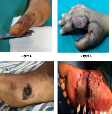
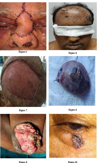
Clinical and histological images of representative cases:
Figure legend:
Subungual melanoma insitu of right thumb
Cross finger flap to cover the defect
Malignant melanoma of foot
Free anterolateral thigh flap covering the defect
Supratrochlear flap covering the defect over nose after excision of basal cell carcinoma
Multiple squamous cell carcinoma over scalp in a known case of epidermolysis verruciformis
Split skin graft to cover the large defect over scalp
Excised specimen of squamous cell carcinoma over scalp
Squamous cell carcinoma destroying whole of penis
Basal cell carcinoma over infraorbital region over face.
DISCUSSION
Protective effect of eumelanin against Ultraviolet rays induced skin damage has been proposed as possible explaination for relatively lesser incidence of cutaneous malignancy among indian population (Yamaguchi, 2008). Prognostically, Non melanoma skin cancer has better overall survival compared to melanoma. TNM (tumor, node, metastasis) Staging used are different for melanoma and non-melanoma (AJCC, 8th edition). Simple edge biopsy from skin lesion establishes the diagnosis in almost all cases. High risk lesions of cutaneous SCC are location (ear, lip, anogenital and scars),diameter more than 2 cm /depth more than 4 mm or beyond subcutaneous fat, perineural invasion, poor differentiation, infiltrative or desmoplastic typr of growth pattern (Jennings et al., 2010). Role of Sentinel lymph node biopsy is still a matter of debate for non melanoma skin cancer (Matthey-Giè et al., 2013). In case of cutaneous melanoma, sentinel lymph node biopsy is indicated for tumor thickness (Breslow depth) greater than equal to 0.76mm (Phan et al., 2009).
Table 1. Demographic, histomorphological data, treatment and its outcome
SL.NO. AGE SEX HISTOLOGY Size(cm) SITE Treatment outcome
1 60 M SCC 4x4 BACK A+SSG C
2 48 F SCC 1.5x1.5 FACE A+Rotation flap C
3 26 M SCC 4.5x4 SCALP A+SSG C
4 30 M SCC 3x3.5 SCALP A+SSG Recurred
5 26 M SCC 5x5 SCALP A+Rotation flap C
6 80 F BCC 1.2x1.4 FACE A+Advancement flap C 7 46 M BCC 1.5x1.5 FACE A+suprarochlear flap C 8 82 M BCC 1.2x1.3 NOSE A+supratrochlear flap C
9 75 M SCC 4x4 FACE A+MRND+RFFF C
10 50 M SCC 12x15 SCALP B
11 24 F Melanoma in Situ ** SUBUNGUAL (Right thumb)
A+CROSS FINGER FLAP C
12 56 M Melanoma 3.5x3.5 FOOT A+ALT flap C
13 58 M Melanoma Tx FOOT* Popliteal fossa LND C
14 60 F SCC 6x6 LEG AKA+IILND C
15 56 M SCC 7x4 PENILE SKIN TP+BilateralIILND+Adjuvan t RT+CT
C
SCC: Squamous cell carcinomaA- Wide excision BCC: Basal cell carcinoma SSG- Split skin grating
M –Male MRND- Modified radical lymph node dissection F – Female RFFF- Radial free forearm flap
C- On follow up with no evidence of recurrence ALT – Anterolateral thigh flap IILND- Ilioinguinal lymph node dissection AKA – Above knee amputation CT- chemotherapy
RT- Radiotherapy TP- Total penectomy
B- Extensive disease with poor general condition of patient ** - Due to previous biopsy, exact size could not be measured.
*- Excision was done in other hospital, and no details were available regarding the size of the lesion.
Figure 1. Figure 2.
[image:3.595.111.479.409.782.2]One study found that 52% of NAM cases had been misdiagnosed by the first clinician who saw the patient, and this misdiagnosis was responsible for an 18-month median delay in diagnosis (Metzger, 1998). In our series, we had one case of subungual melanoma in situ, who underwent wide excision rather than disarticulation and is on follow up till date without any evidence of recureence.
Cases described in our series were of age group which ranged from 24 to as 82years,with mean age of 51.8 years , with majority being males (73%), which matches with the studies proposed by Samaila and Jain et al. (2005; Jain, 2008). Squamous cell carcinoma was the most common histology in our series(60%),which is also supported by the study done by Samaila and Jain et al, but basal cell carcinoma was the most
Figure 5.
Figure 6.
Figure 7. Figure 8.
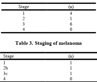
[image:4.595.118.507.49.703.2]Table 2. Staging of Non melanoma skin cancr
Stage (n)
1 4
2 1
3 6
[image:5.595.79.245.70.211.2]4 0
Table 3. Staging of melanoma
Stage (n)
1 1
2b 1
3c 1
4 0
common subtype seen in the study done by Faridehjowkar (Farideh Jowkar, 2015). Face and scalp was the most common subsite seen in our series, which is in agreement with the studies mentioned above (Samaila, 2005; Jain, 2018; Farideh Jowkar, 2015). Mean size of the lesion for squamous cell carcinoma, basal cell carcinoma were 5.2 cm and 1.3 cm respectively. Chronic sun exposure and bare foot walker with chronic skin irritation were some of the few possible risk factors that could be observed. In two of the cases, who were siblings, had epidermolysis verruciformis with transformation to invasive squamous cell carcinomas. In case of subungual melanoma in situ (case no.5), full thickness excision rather than disarticulation was done in view of very early stage of disease (melanoma in situ). Lymph node dissection was done when indicated. Adjuvant radiation and chemotherapy was given in case of carcinoma penis with multiple positive lymph nodes. Cases were observed till last follow up.one case of squamous cell carcinoma recurred, who is being treated with 5-fluorouracil cream in view syndromic nature of the disease.
Limitations of our study: Small sample size, short duration of
follow up, no data on HPV (Human papilloma virus) status were some of the limiting factors. Sentinel node biopsy was not done due to logistic issues.
Conclusion
Increasing incidence of cutaneous malignancy should alert the clinicians to create awareness regarding warning signs of skin cancer. Nonsurgical options needs to be considered specially in syndromic conditions and inoperable situations. Surgery with adequate margins offers the best option of cure.
Keypoints
Avoidance of too much of exposure to sun, use of sunscreens might have protective effect
In situ lesions involving nail apparatus needs to be excised without disarticulation
Sentinel node biopsy needs to be considered when appropriate
Regional nodal clearance with appropriate adjuvant treatment needs to be given when indicated
Appropriate reconstruction options needs to be considered based on size and depth of the defect.
Non surgical options needs to be given priority in syndromic situations.
Conflict of Interest: Nil
Funding Statement: Not Applicable
REFERENCES
Apalla, Z., Nashan, D., Weller, R.B., Castellsagué, X. 2017. Skin Cancer: Epidemiology, Disease Burden, Pathophysiology, Diagnosis, and Therapeutic Approaches. Dermatol. Ther. 7, 5–19
Armstrong BK., Kricker A. 2001. The epidemiology of UV induced skin cancer. J Photochem Photobiol B 63:8-18. Berna Aksoy, 1,2 Aslı Tatlıparmak,1,3 Funda Tamer,4 Can
Ergin, 5 ErolKoç. 2017. The Incidence of Precancerous and Cancerous Skin Lesions: A Retrospective Multicenter Study. South. Clin. Ist. Euras., 28(3):199-203.
Calzavara-Pinton, P., Ortel, B., Venturini, M. Non-melanoma skin cancer, sun exposure and sun protection. G. Ital. Dermatol. Venereol. 2015, 150, 369–378.
Chen, A.C., Halliday, G.M., Damian, D.L. 2013. Non-Melanoma Skin Cancer: Carcinogenesis and Chemoprevention. Pathology, 45, 331–341.
Chren, M.M., Torres, J. S. S. E. Stuart, D. Bertenthal, R. J. Labrador, and W. J. Boscardin, 2011. “Recurrence after treatment of nonmelanoma skin cancer: a prospective cohort study,” Archives of Dermatology, vol. 147, no. 5, pp. 540–546.
Crum-Cianflone, N., Hullsiek, K. H., Satter, E., Marconi, V., Weintrob, A., Ganesan, A., Barthel, R. V., Fraser, S., … Agan, B. K. 2009. Cutaneous malignancies among HIV-infected persons. Archives of internal medicine, 169(12), 1130-8.
Dawn G., Kanwar AJ., Dhar S., Nanda R. 1994. Squamous
cell carcinoma over disseminated discoid lupus
erythematosus on non-photoexposed skin. Indian J
Dermatol Venereol Leprol., 60:217-8
Doina Ivan and Victor G. Prieto 2011. An Update on Reporting Histopathologic Prognostic Factors in Melanoma. Archives of Pathology & Laboratory Medicine: July. 135, No. 7, pp. 825-829.
FaridehJowkar, Maryam Sadat Sadati Iman Ahrari, Fatemeh Sari Aslani. 2015. Analysis of Surgically Treated Cutaneous Malignancies in a Tertiary Dermatology Center during Six-Year Period.Middle east journal of cancer; July, 6(3):151-156.
Faries MB., Thompson JF., Cochran AJ. et al. 2017. Completion dissection or observation for sentinel-node metastasis in melanoma. N Engl J Med., 376: 2211-22 Fears T. R., Bird C. C., Guerry D., 4th, et al. 2002. Average
midrange ultraviolet radiation flux and time outdoors predict melanoma risk. Cancer Res., 62:3992–3996 Glogau, R.G. 2000. The risk of progression to invasive disease.
J. Am. Acad. Dermatol. 42, 23–24 .
Gloster HM Jr, Neal K. 2006. Skin cancer in skin of color. J Am Acad Dermatol., 55:741-64.
Hunt, K.M., Srivastava, R.K., Elmets, C.A., Athar, M. 2014. The mechanistic basis of arsenicosis: Pathogenesis of skin cancer. Cancer Lett. 354, 211–219.
J American Academy of Pediatrics, Committee on Environmental Health. Ultraviolet light: a hazard to children. Pediatrics. 1999;2:328–333..
Jain A et al. 2018. An analysis of surgically treated cutaneous malignancies in central India Int J Res Med Sci., Jun;6(6):2159-2164
Karagas, M.R., McDonald, J.A., Greenberg, E.R., Stukel, T.A., Weiss, J.E., Baron, J.A., Stevens, M.M. 1996. Risk of basal cell and squamous cell skin cancers after ionizing radiation therapy. For The Skin Cancer Prevention Study Group. J. Natl. Cancer Inst. 88, 1848–1853
Lasithiotakis K, Leiter U, KrügerMacKie RM, Hauschild A, E ggermont AM: Epidemiology of invasive cutaneous melanoma. Ann Oncol 20(Suppl 6): vi1-7, 2010.
Lawrence F. Kuklinski, Shufeng Li, Margaret R. Karagas, Wen-Kai Weng, and Bernice Y. 2017. KwongEffect of voriconazole on risk of nonmelanoma skin cancer after hematopoietic celltransplantation. J Am Acad Dermatol. October ; 77(4): 706–712
Lewis KG., Weinstock MA. 2007. Trends in Nonmelanoma Skin Cancer Mortality Rates in The United States, 1969 through 2000. J Invest Dermatol., 127(10):2323-7
Lichter, M.D., Karagas, M.R., Mott, L.A., Spencer, S.K., Stukel, T.A., Greenberg, E.R. 2000. Therapeutic ionizing radiation and the incidence of basal cell carcinoma and squamous cell carcinoma. The New Hampshire Skin Cancer Study Group. Arch. Dermatol. 136, 1007–1011. Markovic SNrickson LA, Rao RD, Weenig RH, Pockaj BA, B
ardia A et al. Epidemiology, risk factors, screening, prevention, and diagnosis. Mayo Clin Proc 3: 364-380, 2007.
Matthey-Giè, M. L., Boubaker, A., Letovanec, I., Demartines, N., & Matter, M. 2013. Sentinel lymph node biopsy in nonmelanoma skin cancer patients. Journal of skin cancer, 2013,267474.
Moloney, F.J., Comber, H., O’Lorcain, P., O’Kelly, P., Conlon, P.J., Murphy, G.M. 2006. A population-based study of skin cancer incidence and prevalence in renal transplant recipients. Br. J. Dermatol. 154, 498–504 Patricia senet et al. 2014. Cutaneous cancers and chronic leg
ulcers. Phlebolymphology. 21(2):75-80.
Phan GQ1, Messina JL, Sondak VK, Zager JS. 2009. Sentinel lymph node biopsy for melanoma: indications and rationale.Cancer Control. Jul;16(3):234-9.
Ratushny, V., Gober, M.D., Hick, R., Ridky, T.W., Seykora, J.T. 2012. From keratinocyte to cancer: The pathogenesis and modeling of cutaneous squamous cell carcinoma. J. Clin. Investig., 122, 464–472
Rigel DS. 2010. Epidemiology of melanoma. SeminCutan Med Surg 4: 204-209.
Samaila M.O.A and Adewuyi. 2005. A histopathological analysis of cutaneousmalignancies in a tropical African population. Nigerian journal of surgical Research Vol 7 No 3 – 4,300-304
Samarasinghe V. and Madan, V. 2012. “Nonmelanoma skin cancer,” Journal of Cutaneous and Aesthetic Surgery, vol. 5, no. 1, pp. 3– 10.
Surveillance, Epidemiology, and End Results (SEER). Program Cancer Statistics Review, 1975–2013, National Cancer Institute (Internet) Nov, 2015
SydneyCh’ng, Richard A Wallis, Lan Yuan, Paul F Davisand Swee T Tan. 2006. Mast cells and cutaneous malignancies. Modern Pathology 19, 149–15
Szpringer E., Lutnicki K., Marciniak A. 2004. Photodynamic
therapy - mechanism and employment. Ann UnivMariae
Curie Sklodowska Med., 59:498-502
Thai KE Young R Sinclair RD. 2001. Nail apparatus melanoma. Australas J Dermatol., 42 (2) 71- 81, quiz 82-8335.Metzger SEllwanger UStroebel WSchiebel URassner G Fierlbeck G Extent and consequences of physician delay in the diagnosis of acral melanoma. Melanoma Res 1998;8 (2) 181- 186
Yamaguchi Y, Beer JZ, Hearing VJ. Melanin mediated apoptosis of epidermal cells damaged by ultraviolet radiation: factors influencing the incidence of skin cancer. Arch Dermatol Res. 2008;300:S43-50.