PHARYNGEAL MOVEMENTS DURING FEEDING
SEQUENCES OF NAVANAX INERMIS (GASTROPODA:
OPISTHOBRANCHIA) IN SUCCESSIVE STAGES OF
DISSECTION
BY ABRAHAM J. SUSSWEIN, YAIR ACHITUV
Department of Life Sciences, Bar Ilan University, Ramat Gan 52 100, Israel MITCHELL S. CAPPELL AND MICHAEL V. L. BENNETT Department of Neuro science, Albert Einstein College of Medicine, Bronx,
NY 10461, USA
Accepted 3 November 1986
SUMMARY
Feeding in Navanax inermis Cooper was filmed and analysed after various dissections. In preparations with a cut through the body wall exposing the pharynx and buccal ganglia, completely normal feeding was observed. In addition to seven motor acts previously described in intact animals, an eighth act, peristalsis, was observed. In preparations with the pharynx excised from the animal but attached to the buccal ganglia, four motor acts were observed: flaring, expansion, contraction and peristalsis. In addition to increasing information about the nature of feeding movements in Navanax, these data indicate that preparations suitable for neuro-physiological studies are capable of producing a variety of feeding acts.
INTRODUCTION
Neurobiological studies combining behavioural and cellular observations must establish links between the two levels. Since dissected or anaesthetized preparations are often used in cellular studies, it is difficult to demonstrate that a cellular event is related to a specific behaviour. This problem is less acute when examining simple invertebrate behaviour, which can be performed even after radical dissections (Kandel, 1976). However, as more complex behavioural processes are examined, the problem of bridging levels of analysis arises in these animals as well.
This paper demonstrates a preparation that ties together behavioural and cellular levels: we observe a complex behavioural sequence in progressively more dissected preparations. In late stages of dissection the entire sequence no longer occurs, but individual acts from which the sequence is composed are still recognizable. In the future, it should be possible to investigate cellular events underlying specific behavioural components, and then reconstruct the entire sequence. A previous paper (Susswein et al. 1984) demonstrated seven distinct motor acts that make up feeding sequences in intact Navanax:
324 A. J. SUSSWEIN AND OTHERS
(1) Protraction of the pharynx, initiated by touch of food to the tentacles. (2) Lip flaring, occurring simultaneously with protraction; the lips are an anterior extension of the pharynx.
(3) Prey seizure: strike, initiated by contact of food to the lips. This is a fast lunge of the pharynx towards the prey.
(4) Prey seizure: lip closure, occurring simultaneously with the strike. This movement consists of the lips snapping shut around the prey.
(5) Expansion of the pharynx, initiated by food in the anterior pharynx. This movement sucks prey into the pharynx.
(6) Retraction of the pharynx, which may occur as an alternative movement to prey seizure if food is withdrawn; if prey seizure occurs, retraction follows both successful and unsuccessful attempts to capture prey.
(7) Contraction of the pharynx, leading to expulsion of the pharyngeal contents. This can occur instead of expansion or subsequent to it, and may represent prey rejection, or clearing of the pharynx in preparation for a new attempt at prey seizure.
Recently, Leonard & Lukowiak (1984) have published an ethogram of Navanax. In their terminology, 1-Coil, 2-Strike, 3-Hold and 4-Relax seem to correspond with (1) protraction and lip flaring, (2) prey seizure, (3) expansion and (4) contraction, respectively.
Data below confirm the picture of feeding outlined above, and indicate that feeding acts of known function occur in preparations suitable for cellular analysis. A similar preparation has been used to determine, by optical methods, activity during feeding of many units simultaneously (London, Zecevic & Cohen, 1984; Zecevic, London & Cohen, 1985).
MATERIALS AND METHODS
Subjects were Navanax inermis (Pacific Biomarine, Venice, California). Tech-niques for maintaining Navanax and for filming and analysing feeding behaviour have been described (Susswein et al. 1984).
In some preparations pharyngeal movements were filmed in the same individual while it was intact, after each of a number of enlargements of a hole through the skin above the pharynx had been made, and after the pharynx had been completely excised. Progressive dissection allowed observation of a movement in an individual at different stages of dissection. However, by the later stages animals had been exposed to very rough handling and had performed many feeding responses, which produced habituation of feeding (Susswein & Bennett, 1979). To overcome this problem, radical dissection was sometimes performed without previous feeding.
To make a 'pharyngeal window', a fold of skin was lifted with fine forceps, and a quick cut removed the fold. Animals contracted when cut, but quickly relaxed. Feeding movements were elicited and filmed within minutes of the cut.
occurred, feeding movements ceased. When the gut and pharynx were pushed back into the animal, feeding movements proceeded. If the gut and pharynx could not be returned to the interior, dissection to an isolated pharynx preparation was performed.
In experiments utilizing an excised pharynx, the gut and pharynx were pinned to a wax-covered dish. The buccal ganglia innervating the pharynx remained in place throughout. In some experiments the circumoesophageal ganglia remained attached to the buccal ganglia by way of the cerebrobuccal connectives, but all potential connections from the circumoesophageal ganglia to the buccal musculature were severed. In other experiments, the cerebrobuccal connectives were cut and the circumoesophageal ganglia removed.
RESULTS
Preparations with a window through the body wall
The first stage of dissection was cutting a window through the body wall, exposing the pharyngeal musculature.
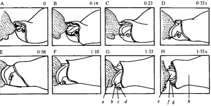
Figs 1 and 2 show feeding sequences in which the same individual sucked a large Bulla partially into the pharynx and then rejected it, before and after a ventral cut over the posterior pharynx. The sequences were strikingly similar: prey seizure was followed by pharyngeal expansion and contraction, leading to expulsion of the oversize prey. Time for prey seizure was almost identical (Fig. 1A-D and Fig. 2A—D), while expansion and contraction were somewhat slower following the
0 B 0-14 0-23 D 0-33 s
[image:3.451.46.411.391.573.2]0-58 110 G 1-33 l-55s
Fig. 1. Line drawing traced from film of a feeding sequence in an intact animal. The sequence consists of prey seizure, expansion and contraction of the pharynx leading to expulsion of the prey. At the start of the sequence (A), the pharynx is already protracted and the lips are flared. In this and following figures numbers in each frame represent seconds following the first frame, a, edge of the Bulla prey; b, pharynx; c, tentacle;
326
A. J. SUSSWEIN AND OTHERS [image:4.451.44.412.171.337.2]cut. The data indicate that strike, lip closure, expansion and contraction are minimally affected by the pharyngeal window. The first frames in Figs 1 and 2 show a pharynx that is already protracted and flared, indicating that the ability to perform these acts is unaffected.
Fig. 3 shows a preparation with a large cut exposing the entire ventral pharynx and the buccal ganglia. The cut was not preceded by smaller cuts, or by feeding
0 B 012 016 D 0-36s
1-83 F 2-64 2-80 H 2-97 s
Fig. 2. Line drawing traced from film of a feeding sequence from the same individual as in Fig. 1, following a cut which exposed the posterior pharynx. Events occurring before and after the cut are similar, a, the pharyngeal window exposing the interior of the animal.
0 B 0-36 0-84 D l-06s
[image:4.451.52.406.407.582.2]1-33 2-94 4-50 H 8-38s
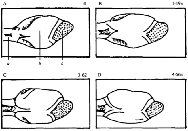
movements. Fig. 3A shows the rest position of the pharynx, which is somewhat expanded. Before prey seizure the posterior pharynx constricted (Fig. 3B). In this sequence the lips are not seen; when the lips are visible, protraction and flaring occur at this time (Susswein et al. 1984). Prey seizure began at Fig. 3C and ended at Fig. 3E. Prey seizure is characterized by rapid movement of the pharynx, and even at a speed of 64 frames s"1 the pharynx changes from frame to frame (Susswein et al. 1984). Fig. 3C-E shows that during prey seizure the pharynx lunged outwards, and prey entered the anterior pharynx; considerable pharyngeal expansion also occurred. Prey seizure was followed by a much slower expansion of the pharynx (Fig. 3E—G), most prominent posteriorly. The pharynx remained expanded for a considerable time (Fig. 3G-H). The prey was eventually released, and left the mouth. The data indicate that normal feeding occurs even when the pharyngeal window is very large, exposing the entire ventral pharynx and buccal ganglia.
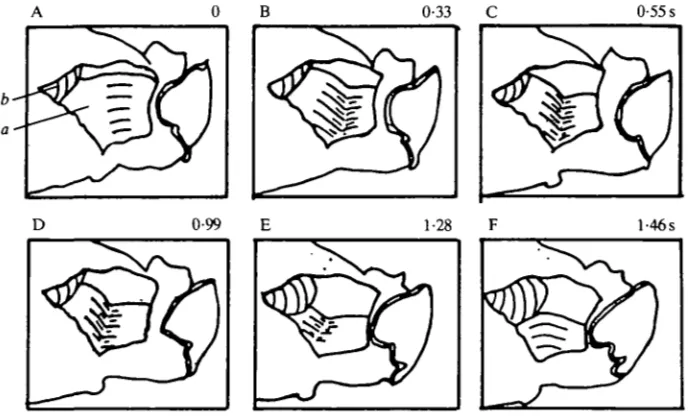
[image:5.451.55.400.393.603.2]Movements subsequent to prey seizure push food from the pharynx into the gut. Fig. 4 shows a sequence in which 3 s after prey seizure (not shown) peristalsis occurred. At the start (Fig. 4A), the pharynx was relatively expanded. The anterior pharynx then contracted, followed by a posterior contraction, while the anterior region remained contracted (Fig. 4A-E). The form of the contraction is consistent with two bands of innervation anteriorly and posteriorly. With small prey the movement pushes an expanded portion of the pharynx containing prey towards the gut. Peristalsis ended with a coordinated action of the gut and pharynx, the gut enlarging to accommodate the prey. Peristalsis was a relatively slow movement, taking close to 1 -5 s.
0-33 0-55 s
D 0-99 1-28 l-46s
328 A . J . SUSSWEIN AND OTHERS
The CNS-isolated pharynx preparation
The next stage of reduction examined was the excised pharynx and anterior gut attached to the buccal ganglia and, via the cerebrobuccal connective, to the circumoesophageal ganglia.
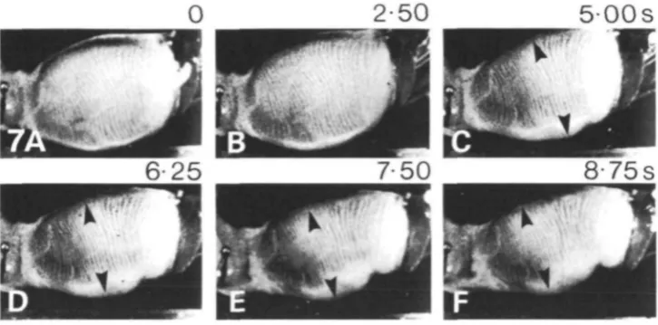
[image:6.451.63.390.367.595.2]Expansion, contraction and peristalsis were readily observed in this preparation. Fig. 5 shows a modest expansion that took over 4-5s. Fig. 6 shows a sequence in which expansion (not shown) was followed by contraction and prey expulsion. Contraction occurred as a constriction beginning posteriorly, pushing prey out of the pharynx. Contraction took over 3-8 s in the sequence, compared to about 3 s in intact animals (Susswein et al. 1984). At the end of the contraction the mouth was still open, as it is in intact animals (Susswein et al. 1984) and in animals with a window through the body wall (Fig. 2). Fig. 7 shows a peristaltic movement that pushed a bolus of material within an expanded region of the pharynx towards the gut. Peristalsis lasted over 6s (Fig. 7B-F), as compared to about 1-5s (Fig. 4) in preparations with a window. A similar result was also illustrated by Bennett (1974). Although well-organized feeding movements occurred in this preparation, sensory control was impaired. Peristalsis was difficult to elicit and was observed only occasionally. In contrast, expansion was easier to obtain than in more intact animals: placing food directly into the pharynx, or tactile stimuli to the lips or the posterior pharynx, occasionally elicited expansion in the CNS-isolated pharynx preparation, but never elicited expansion in intact animals (Susswein et al. 1984), or in animals with a window through the skin.
A OB 119s
•m
* >-
'3-P
3-62 4-56s
Protraction, strike and retraction were never observed in the CNS—isolated pharynx preparation, probably because these acts depend in part on extrapharyngeal musculature. More surprisingly, we never observed the lips to snap shut. This may mean that some muscles responsible for the movement are extrapharyngeal, or that activity of extrapharyngeal muscle receptors initiates the movement.
0-75
1-31S
1-50
1-94
3-81S
Fig. 6. Expulsion of prey, caused by contraction of the pharynx. (A) The pharynx is expanded, and a piece of Bulla meat is present inside the pharynx. (B-F) The piece of
Bulla is slowly pushed out of the pharynx as it constricts. Magnification, X1 • 1.
[image:7.451.49.412.409.588.2]2-50
500s
3 3 0 A . J . SUSSWEIN AND OTHERS
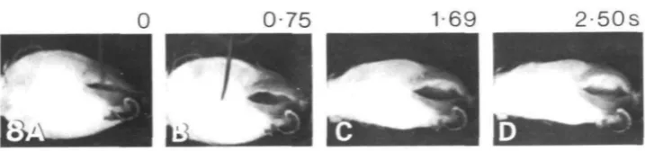
[image:8.451.46.405.63.147.2]0 0-75 1-69 2-50s
fci^^i | fl|
Fig. 8. Flaring of the anterior pharynx, with constriction of the posterior pharynx, in the buccal ganglia-isolated pharynx preparation. (A) The stimulus for the movement is rubbing the length of a pin on the lips. The initial state of the pharynx is partially expanded. (B-D) Constriction of the posterior pharynx, while the lips flare. Magnification, XO8.
The buccal ganglia—isolated pharynx preparation
The next stage of reduction examined was the pharynx and anterior gut innervated by the buccal ganglia alone. In this preparation, expansion and contraction move-ments virtually identical with those shown in Figs 5 and 6 were observed, indicating that neural circuitry entirely within the buccal ganglia is sufficient for these acts. Peristalsis was not observed, but since it occurred only rarely in the CNS-isolated pharynx preparation as well, the absence of this movement does not mean that the movement is due to circuitry present in the circumoesophageal ganglia. Tactile stimulation of the pharyngeal walls evoked motoneurone activity suggestive of peristalsis in more radically dissected preparations with only buccal ganglia present (Spira, Spray & Bennett, 1980).
Contraction movements were easily evoked after the circumoesophageal ganglia had been removed. Fig. 8 shows a sequence in which a tactile stimulus to the lips (touch of the side of a pin) produced a constriction of the body of the pharynx and lip flaring. This was a slow movement, occurring over 2-5 s, similar to the time course of flaring in intact animals (Sussweinef al. 1984).
DISCUSSION
are wholly suitable for detailed physiological analysis (Spira & Bennett, 1972; Woollacott, 1974; S p i r a e a / . 1980; Spray, Spira & Bennett, 1980a,6).
Our results conflict with those of Woollacott (1974), who found that noxious stimuli associated with dissection produced permanent withdrawal of the phalliform organs, sensory structures near the lips that initiate feeding (Murray, 1971). The difference is probably due to herniation of the gut and pharynx in Woollacott's experiments: the drawings by Woollacott (1974) indicate that substantial herniation had occurred. We found that dissected preparations fed only when the gut and pharynx were within the body cavity.
Our observations indicate that two previously identified acts, flaring and con-traction, are similar: in both, the posterior pharynx constricts while the anterior pharynx is expanded. Although further studies may demonstrate that different pattern generators, motoneurones or muscles are involved, on a behavioural level the movements are distinguished only by the stimuli eliciting them, and by their context. Since expulsion of pharyngeal contents and mouth opening should precede an expansion for maximal effectiveness, the movements may in fact be the same. A common pattern generator may underlie both acts, perhaps by being driven by two kinds of sensory inputs.
It is not clear what muscles act during the strike. Activity of the protractor muscles, an increase in coelomic pressure by body wall circumferential muscles, contraction of pharyngeal circumferential muscles or 'inverse jet propulsion' through expansion may all contribute in intact animals. In dissected preparations, coelomic pressure could not contribute.
Radial muscles causing expansion are active in the resting state (Fig. 3). During feeding, these muscles are active in flaring, prey seizure and expansion. In flaring, the anterior pharynx is expanded, whereas in later movements more posterior regions also expand (Fig. 3). Previous studies (Hall, Spray & Bennett, 1983; Levitan, Tauc & Segundo, 1970; Spira & Bennett, 1972; Spira et al. 1980) have demonstrated electrical coupling between radial muscle motoneurones. Coupling is modulated by synaptic input, presumably allowing the motoneurones to function both indepen-dently and as a unit (Spira & Bennett, 1972; Spira et al. 1980). The present data confirm that both local and global expansion movements occur.
332 A. J. SUSSWEIN AND OTHERS
Reversal of coupling probably occurs during peristalsis and lip closure, but is unlikely to be functional in contraction and flaring.
Studies on other gastropods (for reviews see Benjamin, 1983; Kohn, 1983) have shown that feeding movements are driven by central pattern generators. Although many sensory stimuli can initiate or modulate feeding, the motor sequences that make up feeding are relatively fixed. In contrast, this and a previous paper {Susswein et al. 1984) indicate that feeding in Navanax seems to consist of a chain of discrete acts which can combine in different ways to form sequences appropriate to a specific prey stimulus.
Cellular studies on feeding in Navanax to date have utilized a number of reduced preparations. These studies have produced information on properties of receptor fields and synaptic interactions between primary sensory neurones (Spray et al. 1980a,b) as well as on effects on muscles of identified motoneurones (Cappell et al. 1979, 1980; Spira & Bennett, 1972; Spira et al. 1976, 1980; Woollacott, 1974; Zimering et al. 1981). Electrical and chemical synaptic interactions between moto-neurones and between motomoto-neurones and intermoto-neurones have also been described (Bennett et al. 1985; H a l l o a / . 1983; Levitan et al. 1970; Murray, 1971; Spira & Bennett, 1972; Spira et al. 1976, 1980; Woollacott, 1974). The present study explores the behavioural abilities of a number of preparations allowing considerable latitude for application of electrophysiological techniques. By using these prep-arations, it should be possible to determine the role of specific cells in producing a behavioural sequence. An entire feeding sequence can then be built by tying together a number of specific acts.
We would like to thank D. C. Spray for valuable suggestions enabling us to begin the project and for advice, J. H. Byrne for comments on the manuscript, S. Markovich for technical assistance, and M. Achituv for help with the figures. The research was supported by grant no. 19120 of the National Science Foundation, grants nos HD-02428, NS-07512 and NS-12627 of the National Institutes of Health and NIH Post-Doctoral Fellowship 5 F32 NS 05D11.
REFERENCES
BENJAMIN, P. R. (1983). Gastropod feeding: behavioural and neural analysis of a complex multicomponent system. In Neural Origin of Rhythmic Movements (ed. A. Roberts & B. Roberts), pp. 159—193. Cambridge: Cambridge University Press.
BENNETT, M. V. L. (1974). Flexibility and rigidity in electrotonically coupled systems. In
Synaptic Transmission and Neuronal Interaction (ed. M. V. L. Bennett), pp. 153-178. New
York: Raven Press.
BENNETT, M. V. L., ZIMERING, M. B., SPIRA, M. E. & SPRAY, D. C. (1985). Interactions of
electrical and chemical synapses. In Gap Junctions (ed. M. V. L. Bennett & D. C. Spray), pp. 355—366. Cold Spring Harbor: Cold Spring Harbor Laboratory.
CAPPELL, M. S., HALL, D. H., SUSSWEIN, A. J., SPRAY, D. C. & BENNETT, M. V. L. (1979).
Identified motoneurons innervate identified pharyngeal muscle bands and regions in Navanax, an opisthobranch mollusc. Soc. Neurosd. Abstr. 5, 243.
CAPPELL, M. S., SPRAY, D . C , HALL, D. H. & BENNETT, M. V. L. (1980). Peristaltic swallowing in Navanax: Functional organization of circumferential muscle and motor units. Soc. Neurosd.
HALL, D . H., SPRAY, D. C. & BENNETT, M. V. L. (1983). Gap junctions and septate-like junctions
between neurons of the opisthobranch mollusc Navanax inermis.J. Neurocytol. 12, 831-846. KANDEL, E. R. (1976). Cellular Basis of Behavior: An Introduction to Behavioral Neurobiology.
San Francisco: Freeman.
KOHN, A. J. (1983). Feeding and digestion in gastropods. In The Mollusca, vol. 5, Physiology, part 2 (ed. K. Wilbur), pp. 1-64. New York: Academic Press.
LEONARD, J. L. & LUKOWIAX, K. (1984). An ethogram of the sea slug, Navanax inermis (Gastropoda, Opisthobranchia). Z. Tierpsychol. 65, 327-345.
LEVTTAN, H., TAUC, L. & SEGUNDO, J. P. (1970). Electrical transmission among neurons in the buccal ganglion of a mollusc, Navanax inermis.J. gen. Physiol. 55, 484—496.
LONDON, J., ZECEVIC, D. & COHEN, L. B. (1984). Optical monitoring of activity from buccal ganglia during pharyngeal expansion (feeding) in a minimally dissected Navanax. Soc.
Neuwsci. Abstr. 10, 508.
MURRAY, M. J. (1971). The Biology of a Carnivorous Mollusc: Anatomical, Behavioral and
Electrophysiological Observations on Navanax inermis. Ph.D. dissertation, University of
California, Berkeley, 189 pp.
SPIRA, M. E. & BENNETT, M. V. L. (1972). Synaptic control of electrotonic coupling between neurons. Brain Res. 37, 294-300.
SPIRA, M. E., SPRAY, D . C. & BENNETT, M. V. L. (1976). Electrotonic coupling: effective sign reversal by inhibitory neurons. Science 194, 1065-1067.
SPIRA, M. E., SPRAY, D. C. & BENNETT, M. V. L. (1980). Synaptic organization of expansion
motoneurons of Navanax inermis. Brain Res. 195, 241-270.
SPRAY, D. C , SPIRA, M. E. & BENNETT, M. V. L. (1980a). Peripheral fields and branching
patterns of buccal mechanosensitive neurons in the opisthobranch mollusc, Navanax inermis.
Brain Res. 182, 253-270.
SPRAY, D . C , SPIRA, M. E. & BENNETT, M. V. L. (19806). Synaptic connections of buccal
mechanosensory neurons in the opisthobranch mollusc, Navanax inermis. Brain Res. 182, 271-286.
SUSSWEIN, A. J., ACHITUV, Y., CAPPELL, M. S., SPRAY, D. C. & BENNETT, M. V. L. (1984).
Pharyngeal movements during feeding sequences in Navanax inermis: A cinematographic analysis. J . comp. Physiol. 155, 209-218.
SUSSWEIN, A. J. & BENNETT, M. V. L. (1979). Plasticity of feeding behavior in the opisthobranch mollusc Navanax. J. Neurobiol. 10, 521-534.
WEISS, K. R., CHIEL, H. J., KOCH, U. & KUPFERMANN, I. (1986). Activity of an identified histaminergic neuron, and its possible role in arousal of feeding behavior in semi-intact Aplysia.
J. Neuwsci. 6, 2403-2415.
WOOLLACOTT, M. (1974). Patterned neural activity associated with prey capture in Navanax (Gastropoda, Aplysiacea). J. comp. Physiol. 94, 69-84.
ZECEVIC, D., LONDON, J. A. & COHEN, L. B. (1985). Optical measurements, using absorption, of activity from buccal ganglia during spontaneous pharyngeal expansion and during feeding. Soc.
Neuroci. Abstr. 11, 641.
ZIMERING, M. B., SPRAY, D. C. & BENNETT, M. V. L. (1981). Synaptic relations underlying