ADAPTIVE MODIFICATIONS IN THE FLIGHT SYSTEM OF
THE LOCUST AFTER THE REMOVAL OF WING
PROPRIOCEPTORS
BY ANSGAR BUSCHGES AND KEIR G. PEARSON
Department of Physiology, Faculty of Medicine, University of Alberta, Edmonton T6G 2H7, Alberta, Canada
Accepted 25 January 1991
Summary
Previous investigations on the flight system of the locust have found that removal of the wing tegulae in mature locusts (Locusta migratoria) results in an immediate change in the flight motor pattern: the wingbeat frequency (WBF) decreases, the interval between the activity of the depressor and the elevator muscles (the D - E interval) increases, and the phase of the elevator activity in the depressor cycle increases. Here we report the results of a detailed quantitative analysis of these changes. We also examined the flight motor pattern for up to 14 days after removal of the tegulae and found that the changes caused by this operation were not permanent. Beginning on the first day after the operation there was a time-dependent recovery of the WBF, the D - E interval and the phase towards their normal values. In about 80 % of the experimental animals the flight motor pattern recovered almost completely. Intracellular recordings from elev-ator motoneurones showed that this recovery was associated with changes in the pattern of excitatory input to these motoneurones. The modification of activity in elevator motoneurones was dependent on afferent input since complete deaffer-entation of recovered animals resulted in a motor pattern similar to that following deafferentation of normal animals.
Introduction
an initial phase of misorientation in response to wind puffs the orientation response returns towards normal over a period of 4 weeks. Similarly, the phonotactic behaviour of female crickets adapts to the loss of an ear (Huber et al. 1984; Huber, 1987; Schildberger and Huber, 1988; Schildberger and Kleindienst, 1989; Schmitz, 1989). After recovery, the female cricket can maintain a stable course with respect to a sound source emitting the conspecific calling song of a male.
The present study focuses on the adaptive capabilities of the flight system in the mature locust. Two studies have already indicated that this system has adaptive properties: Kutsch (1974) showed that the wingbeat frequency (WBF) in adult mature locusts increases after its initial decrease following the removal of all wing stretch receptors; and Mohl (1988) showed that the steering system of the locust adapts to forced preset timing relationships between different steering muscles. In this study we investigated whether the fully developed pattern-generating system for flight is able to adapt to the removal of proprioceptive input, i.e. whether slow changes occur that return the motor pattern to normal. We chose to examine the effects of ablating the wing tegulae, because the function of these wing propriocep-tors in the generation of the flight motor pattern is well understood: the phasic burst of activity of the tegulae during the downstroke of the wings induces a rapid depolarization in the elevator motoneurones and, as a consequence, initiates the onset of elevator activity and increases the wingbeat frequency (Pearson and Wolf, 1988; Wolf and Pearson, 1988). These previous studies on the flight system of the locust focused on the function of the hindwing tegulae and were largely qualitative. However, to assess the adaptive properties in the flight system after removal of the tegulae it was necessary to have detailed quantitative data on the changes that occur in the flight motor pattern immediately after ablation. Therefore, our initial aim was to confirm and expand previous data on the effects of removing the forewing tegulae, the hindwing tegulae or both groups of tegulae. We investigated whether the changes produced by removal of the tegulae were permanent. To do this we examined the motor pattern for 2 weeks after ablation of the tegulae by recording the electromyogram (EMG) activity in the flight muscles and by intracellular recordings of the patterns of synaptic input to flight motoneurones.
Materials and methods
Electromyograms and data analysis
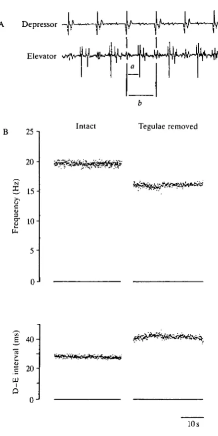
Electromyograms (EMGs) were recorded either from forewing muscles 83 (tergosternal muscle) and 97 (first basalar muscle) or from hindwing muscles 113 and 127 (see Fig. 2A; recording from the hindwing muscles 113 and 127). The placement of the electrodes and the recording procedure were the same as those described in Pearson and Wolf (1987). The animals were tethered by the pronotum to a rigid rod and held in an upright position facing the opening of a wind tunnel. The tethering rod was free to rotate, enabling the animals to make yaw adjustments in the windstream. Flight was initiated and maintained by constant wind from the wind tunnel. The wind velocity was about 2.5 ms"1. The EMG recordings were first passed to a window discriminator with thresholds adjusted so as to generate pulses for all the muscle potentials. These pulses were then passed into monostable circuits that gave single pulses corresponding to the first action potential in each burst of elevator and depressor activity. The times of onset of elevator and depressor activity were stored and analyzed using a general purpose computer (DEC LSI 11/23). The time resolution for the analysis of the events was 0.1ms. The following parameters of the flight motor pattern were evaluated: (i) the wingbeat frequency, calculated from the cycle period of the depressor activity; (ii) the time interval between the activity of the depressor and the elevator motoneurones (D-E interval), calculated from the first spike in the depressor EMG to the first in the elevator EMG; (iii) the phase of the elevator activity in the depressor cycle. WBF and D - E intervals for single trials were calculated as the average values for a continuous 30 s of stable flight starting 30 s after flight had been initiated (see Fig. 2B). The average phase value for each trial was calculated as the ratio of the average D - E interval to the average cycle period.
To compare the flight motor pattern of groups of animals in different experimental situations the average WBF and D - E interval were calculated as the arithmetic means of these parameters for all animals within a group. With respect to the phase of elevator activity, the average phases of elevator activity for each group of experimental animals were calculated using circular statistics after Horsmann et al. (1983). The phase value of each animal on a given day was taken as a unity vector in the circular period between successive depressor spikes. Then the average phase value (P) was evaluated using the formula given in Horsmann
etal. (1983). The length (R) of the mean vector was also calculated to give a
measure of the scatter. As R increases in length (0=SR^l) the scatter decreases.
Intracellular recordings
ganglion. The neurones were identified by the location of their somata, by their activity pattern according to a reference EMG taken from the first basalar muscle, and by their morphology determined by injecting them with Lucifer Yellow dye. The soma recordings were very stable and allowed the transection of meso- and metathoracic nerves 1 and 6 while the recording was maintained.
Surgical removal of the wing tegulae and recording procedure during recovery
The animals were tethered by the pronotum to a rigid rod and placed above a cork platform. The forewings were unfolded and pinned to the platform. The exposed hindwing tegulae were ablated by slightly lifting each with forceps and then removing them with microscissors. To remove the forewing tegulae the animals were turned almost sideways to the dissection microscope and the junction between the meso- and metathoracic segments was enlarged to expose the forewing tegulae. The tegulae were then held with forceps and transected at their base. Recordings from nerve 1C showed that the wing nerve (lClb) was not damaged by this operation (see also below). The procedures used to expose the tegulae had no significant effect on the flight ability and the flight pattern (see Fig. 6D-F, controls), as shown by sham-operated animals that were treated identically except for removal of the wing tegulae.
For the purpose of identification each animal was uniquely marked with white ink on the pronotum after the operation. The animals were kept together in a cage throughout the period of the investigation and the EMG electrodes were implanted for each recording session. The animals were tested in four different groups, (i) For the investigation of the hindwing flight motor pattern, one group of animals (N=28) was tested before and immediately after the removal of the wing tegulae. During the 2 weeks after the operation (see Figs 6,7) every animal in this group was tested 2-6 times, four times on average. These trials were randomly distributed over the group to avoid always testing the same animals on the same day. No other experiments were carried out with these animals. A control group (N=8) was only sham-operated and then treated in the same way as groups i and ii. (ii) A group of 14 animals was tested in the same way for the investigation of the forewing flight motor pattern, (iii) Another group of animals ( N = l l ) was tested only once, 2 weeks after the removal of the wing tegulae. (iv) A fourth group of animals (N=4) was tested before and after removal of the tegulae and then every 2 h for the first day after the operation. A control group of animals (7V=4) was only sham-operated and then treated in the same way.
Deafferentation of the flight system
In some experiments (see Figs 9, 10) the flight system was deafferented by severing the meso- and metathoracic nerves 1 distal to their bifurcations with nerves 6. This was done by cutting small holes in the ventral cuticle of the thorax and, after removing as little overlying tissue as possible, cutting the nerves. The holes in the cuticle were then closed with silicone grease.
Staining of tegulae and wing afferents
Afferents in nerve 1C were stained in intact control animals and in animals 14 days after removal of the wing tegulae by backfilling them with CoCl2.
Prep-arations were kept for about 18 h at 4°C. After cobalt sulphide precipitation, the ganglia were fixed and intensified according to Bacon and Altman (1977). Staining of the metathoracic nerve 1C afferents was performed to confirm that removal of the tegulae caused degeneration of the afferents from these receptors. A comparison of seven stained preparations from intact animals with seven stained preparations from recovered animals revealed that the prominent dorsal lateral branches of nerve 1C afferents in the lateral neuropile, representing the tegula afferents (Tyrer and Altman, 1974; Braunig et al. 1983), were absent (N=4, Fig. 1) or strongly reduced (N=3, a few arborizations in this region) in animals 14 days after removal of the tegulae (see arrowheads in Fig. 1). Furthermore, fewer axons were found to run in the fibre tract located dorso-medially in the ganglion (see arrowheads in Fig. 1). These data indicate that tegula afferents had degenerated by 14 days after removal of the tegulae. In addition, control experiments showed that no afferents in nlC could be activated by touching the former location of the tegulae. Thus, anatomical and electrophysiological data indicated that the flight motor system received no input from tegula afferents during the 14 days of the experiments.
Statistics
Depending on the sample sizes, significance was assessed with the Student's Mest (Mest) or with a modified Student's Mest (mt-test) after Dixon and Massey (1969). The latter test allows the comparison of sample sizes of less than 30 and with unequal variances. Samples were considered to be significantly different throughout this study at P<0.02.
Results
Effects of removal of the tegulae on the flight pattern
Intact 14 days after removal of the tegulae
[image:6.451.40.424.65.407.2]200//m Fig. 1. Projections of the afferents in nerve 1C in the metathoracic ganglion of an intact and a recovered animal (one preparation out of seven in each case), stained with CoCl2 backfills. Note that the extensive arborizations in the lateral neuropile of the
metathoracic ganglion are no longer visible in the recovered animal. In addition, the median dorsal branches are less dense in the recovered animal. The arrowheads mark these regions.
1985). Since a quantitative description of these effects is necessary for the assessment of adaptive capabilities in the flight system, we first established the changes in the flight motor pattern produced by removal of tegulae from the forewing, then from the hindwing and then from both wings.
Depressor
Elevator
25
20-
15-Intact
5
-0J
Tegulae removed
B
40-m
i
O 0J
10 S
[image:7.451.106.327.91.530.2]320 A. BUSCHGES AND K. G. PEARSON
25 1
20-15
% 10
N=55
Intact Removed Intact Removed Intact Removed
B
50-N=l
Intact Removed Intact Removed Intact Removed
50
~ 40
.5 30
u.
N=14
N=l
Intact Removed
la
Intact Removed
[image:8.451.74.387.94.540.2]Intact Removed
These data are similar to those of Pearson and Wolf (1988), in which only the hindwing tegulae were removed, indicating that the forewing tegulae have a much weaker influence on the flight motor pattern than the hindwing tegulae. This was confirmed by recording the EMG patterns of the forewing and the hindwing elevator and depressor muscles following the removal of the forewing tegulae (Fig. 3, middle column). Removal of the forewing tegulae had almost no effect on the measured flight motor pattern of the fore- and hindwings. Neither the WBF nor the D - E interval of the hindwing muscles was significantly changed after removal of the forewing tegulae. Only the D - E interval of the forewing muscles was significantly influenced by the ablation of the forewing tegulae, increasing by 2.9±1.8ms. In contrast, the removal of the hindwing tegulae led to a significant decrease in the WBF and a significant increase in the D - E intervals in the forewing and in the hindwing muscles (Fig. 3, right column). Interestingly, the effects of the forewing and hindwing tegulae on the flight motor pattern appeared not to be simply additive, because removal of all the wing tegulae had a greater effect than the sum of the single effects caused by the removal of either the forewing or the hindwing tegulae. This was especially obvious for the D - E intervals (Fig. 3).
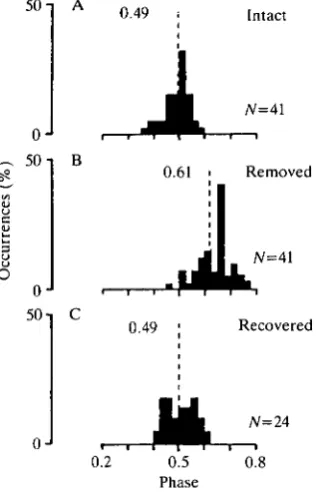
The increase in the D - E intervals after the removal of all wing tegulae was not simply a consequence of the decrease in WBF because the phase of the onset of the elevator activity in the depressor cycle increased after removal of the wing tegulae (for the effects of removal of the hindwing tegulae, see also Pearson and Wolf, 1988). Circular statistics showed that after removal of all tegulae, the value of the average phase of the elevator activity for the hindwing muscles changed from P=0.49 (R=0.94, JV=41) to P=0.61 (R=0.91) (Fig. 4). For the forewing muscles the phase increased from P=0.39 (R=0.97, N=14) in intact animals to P=0.49 (R=0.95) in animals with all tegulae removed.
Recovery of the flight pattern following removal of the tegulae
322 A. BUSCHGES AND K. G. PEARSON
50 -i A
0
50 i
0J
0J
0.49 i
0.49
0.2 0.5
Phase
Intact
/V=41
Removed
N=4l
Recovered
/V=24
[image:10.451.147.306.83.330.2]0.8
Fig. 4. Histograms of the phase values of the elevator activity in the depressor cycle recorded from the hindwing muscles. (A) Intact flying animals (7V=41), R=0.94; (B) the same animals as in A after removal of the wing tegulae, R=0.91; (C) 24 of the 41 animals were tested 2 weeks after removal of the tegulae (tested on day 14,15 or 16), R=0.94. The length R (given in the histograms) of the mean vector is shown by the dashed line, which is the length of the ordinate when R equals 1.
animal the average WBF initially decreased to 75 % (15.2Hz) of its intact value and after 16 days it had recovered to 102% (20.6Hz); the D - E interval for the hindwing muscles was 190% (48.7ms) of its intact value after the operation; 16 days after the operation it had recovered to 100% (25.7 ms).
The WBF and the D - E interval in 30 % out of a group of 24 animals (see also Fig. 5) recovered to values that were ±10% of their intact values. Since the flight motor pattern of a locust can vary to some extent from day to day (Kutsch, 1973), we regard this recovery as complete. The other animals showed an incomplete return towards the intact values of the WBF and/or the D - E interval: 50 % of the animals returned to values within ±20 % of the intact values, while the remaining 20 % recovered only one of the two parameters to within ±20 % of the intact values.
oJ
oJ
0J
k
RemovedIntact10 20
Frequency (Hz)
Recovered
[image:11.451.64.389.86.307.2]20 30 40 50 60 D-E interval (ms)
Fig. 5. Recovery of the wingbeat frequency (WBF) (A) and the D-E interval (B) of the hindwing muscles after removal of the wing tegulae. A group of 24 animals was tested before (upper histograms), immediately after (middle histograms) and 2 weeks (tested on day 14, 15 or 16) after removal of all the wing tegulae (lower histograms). The mean values of all tested animals are indicated by arrows (see also text).
tegulae the average WBF decreased from 20.4±1.6Hz (7V=24) to 15.2±1.7Hz. Two weeks after the operation the average WBF of this group of animals had risen significantly to 18.5±1.6Hz, but it remained significantly less than its values before removal of the tegulae (Fig. 5A). The average D - E interval also recovered to values close to normal (see Fig. 5B). For the hindwing muscles the average D - E interval was 24.6±2.7ms (iV=24) in intact animals. It was 41.9±7.6ms (7V=24) immediately after removal of the tegulae and 27.2±3.1ms (N=24) 2 weeks later. Similarly the D - E interval in the forewing muscles returned towards normal values: in intact animals it was 19.8±2.1ms (N=14), immediately after the operation it was 32.0±2.9ms (N=14) and 14 days after removal of the tegulae it was 19.4±1.0ms (N=5, the other animals were tested on days other than day 14). Finally, the phase of the onset of elevator activity in the depressor cycle returned completely to its intact value: for the hindwing muscles the phase was 0.49 (R=0.94) in intact animals, 0.61 (R=0.91) immediately after the operation and 0.49 (R=0.94) 2 weeks later (see Fig. 4A,B,C). This recovery of phase is shown in Fig. 6C (left side) for a single animal and in Fig. 4 for the group of tested animals.
324 A. BUSCHGES AND K. G. PEARSON
u_
25
20
15
10
251
20
15
10 i
D
00 1 10 15 0 0 1 5 10 15
50
in
^ 40
in 30
i Q
20
L
T H T ,
-00 1
50
40
30-20
A A Control • O Experimental
10 15 0 0 1 10 15
0.7
0.3 i
0.7
0.5
0.3
0 0 1 10 15 0 0 1
Time (days)
10 15
[image:12.451.92.385.74.513.2]significantly different from the final values of the group of 24 animals (see Fig. 5) that were tested repeatedly during the recovery period.
Time course of the recovery of the flight pattern
Fig. 6A-C shows the values of WBF, D - E interval and phase in a single animal over the period of 2 weeks after the removal of all wing tegulae (EMG recording from hindwing muscles). The first point to note is that the WBF was lower and the D - E interval was larger on the first day after the operation than it was immediately after the operation (Fig. 6A,B). Starting on the second day after removal of the tegulae there was a progressive increase in the WBF and a progressive decrease in the D - E interval for the single animal as well as for a group of 28 animals (Fig. 6, compare left-hand and right-hand column).
Most of the recovery of the WBF and the D - E interval for single animals, as well as for the group of 28 animals, occurred within the first week after the operation (Fig. 6D,E). Thereafter, only small changes were detectable. These almost paralleled the slight changes in the control animals occurring during the same period. This is even more obvious when data from the tested animals are normalized. All values obtained for the WBF and the D - E interval throughout the 14-day period were therefore calculated as a percentage of the intact value and arithmetically averaged. This procedure showed that 6 days after the operation the experimental animals reached, on average, 90 % of their intact WBF and 110 % of their intact D - E interval. In addition, we found that the elevator phase in the depressor cycle showed a progressive return to its intact value. Starting on the first day after removal of the tegulae the phase value returned towards normal (Fig. 6C,F). On the seventh day after the operation the phase value had returned to its intact value.
The small changes in the average values of the WBF and the D - E interval detectable from day 6 to day 14 after the operation were partly due to individual differences in the recovery time course. Some animals recovered quickly, e.g. the WBF in one animal returned to 96% of its intact value within 6 days, whereas others showed a much slower increase over the same time, e.g. another animal reached only 79 % of its intact value of WBF by the tenth day. After 2 weeks all animals had reached their final values and we did not observe any further significant time-dependent changes in the WBF, the D - E interval or the phase. Experiments on 14 animals showed that the time courses of recovery of the D - E interval and phase were similar for the forewing muscles.
326
A. BUSCHGES AND K. G. PEARSON25 -i
20-
15-
10-/ 0 3 6 9 12 24
20 -\
Time (h)
[image:14.451.34.418.63.291.2]/ 0 3 9 12
Fig. 7. Changes in the average wingbeat frequency (WBF) and the average D-E interval in the hindwing muscles during the first 24 h after removal of the wing tegulae. Bars indicate S.D. ( • ) Experimental animals (four animals); (O) controls (four animals).
removal of the tegulae are not due to the repeated testing procedure during this interval. This is consistent with the observation that most animals not flown repeatedly in the first 24 h showed a lower WBF and a larger D - E interval than did animals immediately after removal of the tegulae (Fig. 6). Between 12 and 24 h after the operation we observed no significant changes in the flight pattern.
Pattern of elevator activity in the recovered animals
The depolarization pattern of the elevator motoneurones and its dependence on proprioceptive input to the flight system have been investigated in detail by Wolf and Pearson (1988). They showed that the elevator motoneurones are rapidly depolarized following depressor activity, that this rapid depolarization is visible over a wide range of WBFs and that it is initiated by tegula activity. This initial rapid depolarization underlies the short D - E interval in the intact animal. Because recovered animals have a D - E interval close to normal, we wished to determine whether the pattern of synaptic input to elevator motoneurones in recovered animals resembled that in intact animals.
and without forewing tegulae (see also Fig. 3, showing that removal of the forewing tegulae has no effect on the hindwing D - E interval). After removal of the hindwing tegulae the initial rapid depolarization was not generated and the onset of the first elevator spike was delayed, resulting in an increase in the D - E interval (Fig. 8A,B; Fig. 4 in Wolf and Pearson, 1988). At all WBFs the depolarization pattern showed only a burst of activity late in the flight cycle. In recovered animals the pattern of synaptic input to hindwing elevator
moto-A Forewing tegulae removed
EMG
B All tegulae removed
C Recovered (13 days)
5mV
50 ms
328 A . B U S C H G E S AND K. G. PEARSON
neurones (Fig. 8C) resembled that in animals with intact hindwing tegulae. The initial depolarization in elevator motoneurones was more rapid in recovered animals than in animals immediately after removal of the tegulae. In addition, at lower WBFs a second late component occurred in the elevator depolarization similar to that reported for intact animals (see Fig. 2 in Wolf and Pearson, 1988).
No obvious changes were found in the depolarization profile of depressor motoneurones immediately after removal of the tegulae, and this profile was similar to that in recovered animals.
Effects of complete deafferentation on the flight activity in recovered animals
[image:16.451.71.381.454.589.2]A possible mechanism for the recovery of the flight motor pattern following removal of the tegulae could be alterations in the central elements of the motor pattern-generating network. In this case a total deafferentation of the flight system in a recovered animal should lead to a different flight pattern from that of an intact animal immediately after deafferentation. Contrary to this prediction, we found no significant differences in the average WBF and the average D - E interval in deafferented recovered animals (Fig. 9, left and middle histograms) compared with deafferented normal animals. Furthermore, the average elevator phase in the depressor cycle was similar for both situations (deafferented normal, 0.58; deafferented recovered, 0.59). We also found no significant differences in the flight duration (Fig. 9C) for the deafferented animals in both situations. Deaffer-entation of the flight system in the recovered animals led to a drastic change in the elevator activity pattern (Fig. 10), comparable to that occurring in normal animals after deafferentation. In both cases the rapid depolarization in the elevator motoneurones was abolished and bursts of elevator activity were generated following a relatively slow initial depolarization.
A Recovered
EMG
-JU^
B Recovered deafferented
5mV
50 ms
Fig. 10. Intracellular recordings from hindwing tergosternal motoneurones (113) of a recovered animal show that deafferentation of the flight system caused a drastic change in the depolarization profile. (A) Depolarization profile in a tergosternal elevator motoneurone during flight in a recovered animal. (B) Depolarization profile in the same motoneurone after deafferentation. The bottom traces in A and B show the EMG recording of a hindwing first basalar motoneurone. Results were confirmed in 10 preparations.
Discussion
Adaptive changes in the flight motor pattern following removal of the tegulae
responsible for the deterioration of the motor pattern during the 12 h after removal of the tegulae.
The main result of the present investigation was that the effects of the removal of the tegulae on the WBF, the D - E interval and the elevator phase were not permanent. Starting on the first day after the operation, all the parameters recovered towards their intact values. Some of the animals (30%) recovered completely. The major part of this recovery occurred during the first 6 days after removal of the tegulae (Fig. 6) and final values were reached between 10 and 14 days. After 7 days the phase of the elevator activity in the flight cycle returned to its intact value (Fig. 6). The observed recovery in the motor pattern was not due to maturational processes because at the time of removal of the tegulae all animals had developed beyond the age (3 weeks) when maturation influences the flight motor pattern (Kutsch, 1973, 1974).
Because we measured only three parameters of the flight motor pattern, the question arises of whether other parameters are influenced by the removal of the wing tegulae and whether these also return towards normal during the recovery process. That is, is there a recovery of the whole flight motor pattern? In the absence of additional data we cannot answer this question. However, several behavioural observations indicate that the whole flight behaviour is influenced and does recover. First, when animals were flown on a roundabout the flight speed and the flight duration decreased immediately after the removal of the wing tegulae, but both returned to their intact values during the time course of the recovery (A. Buschges and K. G. Pearson, unpublished results). Second, most of the animals were able to fly freely across a room and generate lift 2 weeks after the operation. No animal was able to do this during the first days after removal of the tegulae (A. Buschges and K. G. Pearson, unpublished results).
An interesting aspect of the recovery process is that it occurred in the absence of active flight behaviour. This was shown most clearly for a group of 11 animals that were not flown during the 14-day recovery period. The average values of the parameters we measured were close to normal (average WBF, 18.7±1.7Hz; average D - E interval of the hindwing muscles, 29.3±5.6ms) and not significantly different from those in the group of animals that were flown repeatedly during the recovery period. Although this result demonstrated that flight activity is not required for the recovery, we have not yet ruled out the possibility that the rate of recovery could be influenced by active flight behaviour. The results of Mohl (1988, 1989) on short-term adaptations in the steering muscles of the flying locust indicate that this could occur.
have observed is not unique and is similar to that found in systems where the functional relevance for recovery is clear. Since the generation of the flight motor pattern depends strongly on proprioceptive feedback (Wolf and Pearson, 1988), it is conceivable that the adaptation following removal of the tegulae is a reflection of the fact that the flight system is continuously using proprioceptive information to modify the motor pattern to make it appropriate for the current state of the flight apparatus (e.g. muscle strength, cuticular hardness, receptor transduction) and for body characteristics (e.g. weight, shape). All these factors change in the course of adult development and there is no a priori reason for expecting that sensory feedback would be utilized in the same manner throughout the adult life of an animal. It is quite conceivable that during adult development the strength and/or timing of tegula input is altered (owing to changes in wing mechanics and/or the properties of the tegulae themselves) and this must be compensated for by alterations in other elements of the pattern-generating network. It would be of interest, therefore, to examine the timing and strength of tegula input throughout adulthood. If these changes occur then it is to be expected that the immediate effects of removal of the tegulae may vary depending on the age of the animal.
Mechanisms underlying the adaptive properties in the flight system
Although the objectives of this study did not include the determination of the mechanisms underlying recovery, some data are relevant to this issue. The major change to occur during recovery was a change in the timing of the synaptic input to the elevator motoneurones. Immediately after removal of the tegulae the initial depolarization in the elevator motoneurones was slowed (Fig. 8B). In recovered animals the rate of depolarization in the elevator motoneurones was fast and comparable to that in intact animals (Fig. 8C). What causes this increase in the depolarization rate during the recovery?
tegulae does involve alteration of some properties of the central network generating elevator activity, but that these alterations are only expressed in the presence of afferent input from the wing proprioceptors.
We would like to thank Drs J. M. Ramirez and J. Schmitz for reading the manuscript critically. This work was supported by a grant from the Medical Research Council to K.G.P. and by a grant from the Alberta Heritage Foundation for Medical Research to A.B.
References
BACON, J. P. AND ALTMAN, J. S. (1977). A silver intensification method for cobalt-filled neurons in wholemount preparations. Brain Res. 138, 359-363.
BRAUNIG, P., PFLUGER, H.-J. AND HUSTERT, R. (1983). The specificity of central nervous projections of locust mechanoreceptors. J. comp. Neurol. 218, 197-207.
COMER, C. AND CAMHI, J. M. (1984). Behavioral compensation for altered cereal position in the cockroach. J. comp. Physio). A 155, 31-38.
COTMAN, C. W (ed.) (1978). Neuronal Plasticity. New York: Raven Press.
DIXON, W. J. AND MASSEY, F. J., JR (1969). Introduction to Statistical Analysis. 3rd edition. New York: McGraw Hill.
HORSMANN, U., HEINZEL, H.-G. AND WENDLER, G. (1983). The phasic influence of self-generated air current modulations on the locust flight motor. J. comp. Physiol. A 150, 427-438.
HUBER, F. (1987). Plasticity in the auditory system of crickets: phonotaxis with one ear and neuronal reorganization within the auditory pathway. /. comp. Physiol. A 161, 583-604.
HUBER, F., KLEINDIENST, H.-U., WEBER, T. AND THORSON, J. (1984). Auditory behavior of the cricket. III. Tracking of male calling song by surgically and developmentally one-eared females, and the curious role of the anterior tympanum. J. comp. Physiol. A 155, 725-738.
JENKINS, W. M. AND MERZENICH, M. M. (1987). Reorganization of neocortical representations after injury: a neurophysiological model on the basis of recovery from stroke. Prog. Brain. Res. 71, 249-266.
KAAS, J. H., KRUBITZER, L. A., CHINO, Y. M., LANGSTON, A. L., POLLEY, E. H. AND BLAIR, N.
(1990). Reorganization of retinotopic cortical maps in adult mammals after lesions of the retina. Science 248, 229-231.
KNUDSEN, E. I. AND KNUDSEN, P. F. (1990). Sensitive and critical periods for visual calibration of sound localization by barn owls. J. Neurosci. 10, 222-231.
KUTSCH, W. (1973). The influence of age and culture-temperature on the wing-beat frequency of the migratory locust, Locusta migratoria. J. Insect Physiol. 19, 763-772.
KUTSCH, W. (1974). The influence of the wing sense organs on the flight motor pattern in maturing adult locusts. J. comp. Physiol. A 88, 413-422.
LISBERGER, S. G. (1988). The neural basis of learning of simple motor skills. Science 242, 728-735.
MOHL, B. (1988). Short-term learning during flight control in Locusta migratoria. J. comp. Physiol. A 163, 803-812.
MOHL, B. (1989). 'Biological noise' and plasticity of sensorimotor pathways in the locust flight system. J. comp. Physiol. A 166, 75-82.
NEUMANN, L. (1985). Experiments on tegula function for flight coordination in the locust. In Insect Locomotion (ed. M. Gewecke and G. Wendler), pp. 149-156. Berlin, Hamburg: Paul Parey.
OPTICAN, L. M., ZEE, D. S. AND CHU, F. C. (1985). Adaptive response to ocular muscle weakness in human pursuit and saccadic eye movements. J. Neurophysiol. 54, 110-122.
PEARSON, K. G. AND WOLF, H. (1987). Comparison of motor patterns in the intact and deafferented flight system of the locust. J. comp. Physiol. A 160, 259-268.
SCHILDBERGER, K. AND HUBER, F. (1988). Post-lesion plasticity in the auditory system of the cricket. In Post-Lesion Neural Plasticity (ed. H. Flohr), pp. 565-575. Berlin, Heidelberg: Springer-Verlag.
SCHILDBERGER, K. AND KLEINDIENST, H.-U. (1989). Sound localization in intact and one-eared crickets. J. comp. Physiol. A 165, 615-626.
SCHMITZ, B. (1989). Neuroplasticity and phonotaxis in monoaural adult female crickets {Gryllus bimaculatus de Geer). J. comp. Physiol. A 164, 343-358.
TYRER, N. M. AND ALTMAN, J. S. (1974). Motor and sensory flight neurones in a locust demonstrated using cobalt chloride. /. comp. Neurol. 157,117-138.
VARDI, N. AND CAMHI, J. M. (1982a). Functional recovery from lesions in the escape system of the cockroach. I. Behavioral recovery. J. comp. Physiol. A 146, 291-298.
VARDI, N. AND CAMHI, J. M. (1982ft). Functional recovery from lesions in the escape system of the cockroach. II. Physiological recovery of the giant interneurones. J. comp. Physiol. A 146, 299-309.
WIESEL, T. (1982). Postnatal development of the visual cortex and the influence of environment. Ataure 299, 583-591.
WOLF, H. AND PEARSON, K. G. (1987). Intracellular recordings from interneurons and motoneurons in intact flying locusts. /. Neurosci. Meth. 21, 345-354.