J. Exp. Biol. (1970), 52, 109-124 I 0Q
With II text-figures Wrinted in Great Britain
OPTIC AND STATIC CONTRIBUTIONS TO OCULAR
COUNTER-ROTATION IN CARP
BY A. B. TRAILL AND R. F. MARK
Department of Physiology, Monash University, Clayton, Victoria, Australia
(Received 9 May 1969)
INTRODUCTION
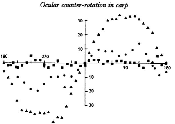
A detailed knowledge of eye movements is useful for the analysis of sensori-motor mechanisms and for the study of visual preception in creatures with mobile eyes. Both these topics can be pursued by experimenting with animals from the lower end of the vertebrate scale because the arrangement of eye muscles (Walls, 1942) and the basic processes of visuo-motor behaviour appear fairly similar all through the phylogenetic series (Sperry, 1950). For these reasons as much as for their immediate significance we have made a study of some factors controlling eye position in the goldfish and carp
(Carassius auratus and Carassius carassius).
In common with most vertebrates these fish hold their eyes more or less horizontal when their body rotates round the longitudinal or transverse axis. The compensatory eye movements, known as counter-rotation, have previously been shown to stem largely from the activity of labyrinthine receptors (Maxwell, 1923; Dusser de Barenne, 1934). This paper describes an accurate method for measuring eye rotation when a fish is turned passively 3600 around the transverse axis. Measurements made on labyrin-thectomized or blinded animals confirm the importance of the vestibular apparatus but show that there is a large optic component as well. The regulation of eye position by the interplay of optic and vestibular factors is compared with the way the same two sensory mechanisms operate on whole body orientation.
A preliminary account of this work has been published (Traill, 1968).
METHODS
(a) Apparatus
Counter-rotation was measured in the apparatus shown schematically in Fig. 1. It consisted of a jig to hold the fish in any desired position in an aquarium so that the eye could be viewed from the side through a binocular microscope. The eye-piece of the microscope was fitted with a graticule consisting of a series of parallel lines which were superimposed over the eye of the fish. By rotation of the eye-piece these lines could be aligned with any marker on the fish's eye. Goldfish usually have a pair of fine equatorial lines on the iris which are very suitable and have the added advantage of only minor variation in orientation from fish to fish. The position of the fish with
n o A. B. TRAILL AND R. F. MARK
respect to horizontal was measured in degrees from the graduations on the jig, an position of the eyes with respect to horizontal was measured in degrees from the pro-tractor scale mounted on the eye-piece, the counter-rotation of the eye being the difference between the two readings. The clamping system used was a pair of inflatable rubber fingers which lightly held the fish belly downwards inside a glass tube.
Marks on goldfish eye Marks aligned with graticule
Fixed scale
Rotating eye-piece with pointer
Ocular counter-rotation in carp 111
Bscape backwards was prevented by a stopper, and a Perspex collar prevented the fish Trom escaping forwards. The size of the collar is important. If too small the eyes are obscured, if too large the fish has difficulty in breathing owing to interference with gill movement, or it may be able to escape. Some difficulty was found in developing a suitable clamping mechanism as even a slight pressure on the body resulted in many fish dying a day or so afterwards. This system permitted the fish to struggle moment-arily, after which the resilience of the inflatable fingers returned it to its original position. No fatalities occurred with this technique.
(b) Methods of measurements
The fish was clamped in the jig in the horizontal position and left alone for a settling-in period ranging from several minutes to an hour or more in the case of an excitable fish. Four readings of the eye position with the fish horizontal were obtained and averaged—this average being used to re-set the eye-piece scale to make it direct reading. The fish was then rotated head over tail in ten degree steps and a set of no less than four readings of eye position was taken at each step. The measurements took about i to i | min. at each position. Readings were taken to the nearest degree, the graticule being moved and re-aligned for each reading. Usually the readings for any one orientation differed by only two or three degrees.
The eye is continuously moving backwards and forwards in a series of jerks (Johnstone & Mark, 1969) and thus for the purposes of comparison the readings were always taken with the eye in the most backwards position, i.e. when the fish was looking to one side. It is possible to distinguish two types of saccadic movement. The first is a series of forward and backward movements with a frequency of approxi-mately once per second, and the second type is very abrupt and occurs when the fish is looking at something. It may be held for a few seconds. Readings were not taken if it was thought that the fish was fixating in this manner. As a check to see whether the fish had slipped in the jig during the experiment readings were obtained for the posi-tion of the eye with the fish again horizontal. If the average of the two sets of readings differed by more than three or four degrees slippage was suspected and the experi-ment was repeated. Such an occurrence was rare. The averages of both sets are recorded on the graphs.
i i 2 A. B. TRAILL AND R. F. MARK
(c) Surgical procedures (i) Labyrinthectomy
As one of the sensory inputs contributing to counter-rotation in man and other animals comes from the labyrinth, the labyrinths were removed from a number of fish.
The technique for the removal of the labyrinths was as follows.
The fish was anaesthetized with MS 222 (Sandoz) at an approximate concentration of 0-4 g/1. The skin and bone of the skull were cut through using a sharp and pointed scalpel. One incision was along the longitudinal axis slightly to one side of the mid-line and nearer to the labyrinth to be removed. This incision was straight through at right angles to the skin and bone. Another incision, which in contrast to the first was slightly undercut, passed from the posterior end of the first incision forwards and laterally. The resulting V of bone was gently prised upwards—hinging on the anterior uncut section. The labyrinth was then removed with a pair of straight jewellers forceps.
Although the final approach to the vestibular apparatus was blind it was successful. Using the bone as a guide the tips of the forceps were slid down between the inside of the skull and the outer rear edge of the tectum. The utricule and saccule complex to-gether with a length of nerve and pieces of the semi-circular canals were often pulled out on the first attempt. The operation was considered a success after the utricule and saccule were removed together with a length of nerve. The incision was closed by pressing the flap of bone back into the undercut when it clicked into place. The survival rate was high especially when accompanied by some bleeding as the clot acted as a seal. A second operation was performed at a later date when a bilateral labyrinthectomy was required because fish usually died if both labyrinths were removed at once. Removal of either or both labyrinths had remarkably little effect on the behaviour of the animal. The fish would lie on its operated side for only a few minutes while recovering from the anaesthetic, after which it would swim in the usual manner. The bilaterally labyrinthectomized fish also appeared normal, swimming at unusual angles only under some special circumstances. The time between operation and testing varied from a few days to 2 years.
(ii) Blinding
Goldfish can be reversibly blinded for about 3 weeks by crushing the optic nerve. The fish was anaesthetized and the optic nerve was exposed by cutting through the conjunctiva and underlying connective tissue and rolling the eye to one side. The nerve was crushed under direct vision with a pair of fine artery forceps and the eye was returned to its former position. Fish were also blinded by cutting the optic nerve in the cranium as it joined the tectum so as not to disturb the oculomotor nerves and muscles. The same incision was used as for labyrinthectomy.
RESULTS
Ocular counter-rotation in carp
[image:5.451.57.396.142.588.2](i) Normal fish
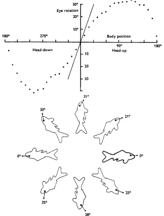
Figure 2 shows the counter-rotation curve of a normal fish. The fish is horizontal at o°, head-up at 900, upside down at 1800, head down at 2700 and horizontal again at 3600 or o°. The amount of counter-rotation is shown on the vertical scale. If there was
180°
i |
•
•
270°
Head-down
30°
Eye rotation 20°
10°
i i
J
.' j
* " /
"A'
/
- 10- 20
- 3 0
• • •
Body position •
i i i i i
90° 18 Head-up
31°
30°
21°
25°
35'
Fig. 2. Ocular counter-rotation curve of the right eye of a normal carp. Horizontal axis, position of fish; vertical axis position of the eye. The diagonal line through the origin shows where the curve would pass if the eye remained perfectly horizontal as the fish was turned head-up (to the right of 0°) or head-down (to the left of o°). The lower part of the figure shows the same thing diagrammatically. The arrows show the direction of the eye at each body position.
A. B. TRAILL AND R. F. MARK
no counter-rotation the graph would be a horizontal straight line passing through thej origin, whereas if the eye remained horizontal as the fish rotated (perfect counter-rotation) the graph would be represented by the diagonal line. The lower half of Fig. 2 shows the same thing diagrammatically. The arrows indicate the orientation taken by the eyes, and the figures represent the amount of counter-rotation. This graph is typical of those obtained, as can be seen from Fig. 3, which is a collection of curves from normal fish. The amount of counter-rotation is variable but the form of the curve is very similar from fish to fish. In each case the graph is straight round about
180
1. 1
' .
270 1 . 1 1
*
30 20
10
1
-* -* -* -* • 1 1 1 1 1 1
-10 90 V
- 2 0 -30 -40
J—I I I I • I I - 1 — I — L _ l
Ocular counter-rotation in carp 115
|the origin and the average slope in this region is 0-59, which gives a measure of the overall gain of the counter-rotation mechanism.
(ii) Labyrinthectomized fish
Experiments were performed to determine what sensory inputs contribute to counter-rotation, and the first procedure was to remove the labyrinths. Figure 4 shows the effects in four fish of removing the right labyrinths on the counter-rotation of the
180
i i
270
1 1 1 *
302 0 1 0
-1 • |
-10 -20 - 3 0
• -. * i 90 * * • i i *i
180
I I I- I I
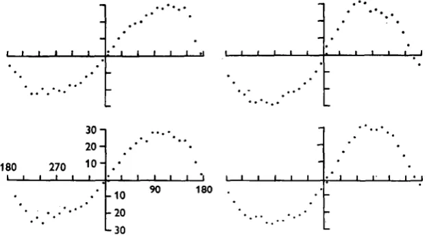
Fig. 4. Ocular counter-rotation curves of the right eye of four fish after removal of the right labyrinth. The average slope of the central region of the curve is 0^44 compared with 0-59 for normal fish.
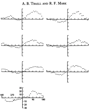
right eye. Removal of one labyrinth leads to a slight reduction of the counter-rotation for the eye on the same side. The average slope of the curve round the central region of the graph is 0-44 compared with 0*59 in normal fish. In one experiment only, the effect on the left eye was examined as well. Over the range o + 6o° (horizontal to head-up or head-down) the two curves were the same. Beyond these limits there was slightly less counter-rotation of the right eye than of the left. Whether this difference is significant or not we do not know because we do not have enough data on the normal symmetry of ocular counter-rotation. Removal of both labyrinths produces a considerable reduction of counter-rotation. In Fig. 5 are plotted curves from seven animals after bilateral labyrinthectomy. Compensatory eye movements were much less marked particularly in the head-down position although considerable rotation still occurred when the animal was head-up. The average slope round the origin gives a gain of 0-20.
(iii) Effects of direction of illumination
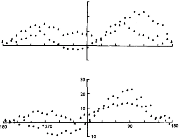
Although removal of the labyrinth reduces the amount of counter-rotation of the eye it does not abolish it entirely. By reversing the direction of illumination and lighting a fish from below while measuring counter-rotation it can be shown that most of the residual movement depends on the direction of incident light. Figure 6 shows two curves obtained from different labyrinthectomized fish with light coming from above or
u 6 A. B. TRAILL AND R. F. MARK
180 270 30 20 10
V * . « ' * '
-10 20
L
30
[image:8.451.68.378.42.420.2]90 180
Fig. 5. Ocular counter-rotation curves of seven fish after removal of both labyrinths. There is now very little compensatory eye rotation in the head-down position. The average slope round the origin is O-2O compared with 0-44 in fish with one labyrinth removed (Fig. 4) and 0-59 in normal fish (Fig. 3).
below. The curves can be reversed in some body positions by illumination from below but they are not completely reversed in others. Possible reasons for this are discussed later.
An effect of light direction on counter-rotation can also be seen in normal fish as is shown in Fig. 7. Light coming from below reduces the amplitude and the slope round the origin of the counter-rotation curve.
(iv) Effects of blinding
Ocular counter-rotation in carp 117
linded by crushing the optic nerve while the right eye was blinded by crushing the uptic nerve in the cranial cavity. An example is shown in Fig. 8. The slope and amplitude of counter-rotation of the right eye was greatly reduced in otherwise normal fish. Blinding of labyrinthectomized fish resulted in no counter-rotation at all.
30 r
20
,4 4
10
180 •270
* »
1
90 *180
L
[image:9.451.80.380.130.363.2]1 0
Fig. 6. The effect of light direction upon ocular counter-rotation curves of the right eye of two fish from which both labyrinths had been removed. Circles, light from above; triangles, light from below. Note that counter-rotation is reduced in the head-up position and reversed in the head-down position.
(v) Other influences on counter-rotation
The counter-rotation curves obtained from one usually excitable but otherwise normal fish showed a gain of unity, that is perfect counter-rotation, during the first few minutes of an experiment—however, at the end of the experiment the slope was nearer that usually seen (Fig. ga). This finding prompted further experiments in which a small amount of powdered food was mixed with the water after a counter-rotation curve had been obtained. Further readings were then taken and Fig. gb shows a pair of curves obtained in this manner. The addition of the ground-up food was found to increase the slope of the counter-rotation curve, particularly in the head-down position.
(vi) Time course of counter-rotation
laby-u 8
A. B. TRAILL AND R. F. MARKrinths show a drift towards lesser counter-rotation when facing the light. Counter rotation of labyrinthectomized fish is less than half of that seen in normal fish whereas the drift is as large or larger than in normal animals. The proportional loss of counter-rotation with time is therefore much larger in labyrinthectomized fish.
I i
A A
A
. 4
30 T
2 0 • •
1 0
-180 270
• A . A
, A . A * » A A A .
90
A* +10
* * A * * A * * i * + 2 0
• +30
Fig. 7. Effect of light direction on ocular counter-rotation of the right eye of two normal fish. Circles, light from above; triangles, light from below. Note that illumination from below reduces the amount of counter-rotation.
DISCUSSION
Ocular counter-rotation in carp
119Fig. 8. Effect on ocular counter-rotation of blinding normal and labyrinthectomized fish. Triangles, normal fish; filled circles, same fish after blinding both eyes; squares, another animal blinded and labyrinthectomized. Counter-rotation is reduced by blinding and is absent after blinding and labyrinthectomy.
in normal fish under the usual conditions of these experiments was 0-59 (range 0-55-0-67). When the fish is rotated past these limits in the standard head-over-tail direction used in these experiments, the eye position tends to remain fixed with respect to body until about 1500, when the eye begins to rotate in the same direction and faster than the body, so the eye now leads. At about 1800 both the eyes and the body are upside down. At about 2200 the position again reverses and the body leads, the eye catching up when horizontal is reached once more. Strictly speaking the eye is only counter-rotating when the rotation of the body leads that of the eye, but this distinction is not usually drawn. These results are the same as those obtained by Benjamins (1918).
At least two sensory channels contribute to the direction of counter-rotation, the vestibular apparatus and the eyes. The stimulus to the vestibular apparatus is the static orientation of the fish with respect to gravity, that for the eyes appears to be the direc-tion of incident light. The final posidirec-tion that the eyes take up depends on informadirec-tion supplied by both these pathways modified by a third factor dependent on the mood or level of excitability of the fish ('Umstimmung* of von Hoist (1948)). An increase in excitability shows up spontaneously by the fish struggling in the jig or can be induced by dissolving food in the water, and presumably there are other means. The result is a short-term increase in the amount of counter-rotation in a given body position and can be most easily thought of as a temporary increase in gain of the counter-rotation system. We have never seen the gain exceed unity, that is the eyes turn further towards, but never past horizontal. The results of all these experiments are summarized in Fig. 11, which shows the eye orientations obtained under varying conditions for a fish restrained in a typical foraging position.
1 2 0 A. B. TRAILL AND R. F. MARK
(a)
I I
• • •
Fig. 9. Changes in counter-rotation curves in the course of measurement, (a) Spontaneous change in slope. The diagonal line shows the slope of the counter-rotation curve when measure-ment was begun. By the time the fish had been rotated head-over-tail and the origin of the graph was approached from the head-down position the slope of the curve was less marked. (6) Changes induced in the slope round the origin by adding ground food to the water. Filled circles, initial measurements; triangles, after food power was mixed with the water. Note that the slope approaches 1 -o after food was added.
Ocular counter-rotation in carp
1 2 14 blinded fish
7 normal fish
6 labyrinthectomized fish
- • 10
180 270 Degrees Head-down
Drift to more counter rotation Degrees Head-up
90 180
-UJJ
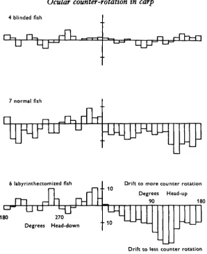
[image:13.451.77.376.54.421.2]Drift to less counter rotation Fig. io. Histograms of the difference between the first and last readings of right-eye position in groups of blinded, labyrinthectomized and normal fish plotted against body position. In blinded fish the drifts are small and show no tendency to be always in one direction. Normal and labyrinthectomized fish show larger drifts and in the head-up position facing the light the tendency is always for the eye to drift towards lesser counter-rotation.
sensory inputs. With such a deficit in sensation fish are very inactive and the gain of the counter-rotation mechanism may have fallen so low that minor inputs may fail to produce any change in eye position. Other sensory pathways from the lateral line and swim bladder could carry information about body position but their contribution to eye position must be very small.
1 2 2 A. B. TRAILL AND R. F. MARK
Lightt No labyrinth
Axis of body
Horizontal axis
Light! labyrinths+food
Light! labyrinths
Lightt labyrinths
Lightt no labyrinths
Fig. I I . Eye positions taken up under various conditions described in this paper when a fish is held restrained in the normal head-down foraging position.
drift in blinded fish is random in all body positions. In all three categories the illumina-tion is from above. Thus the drift, when seen, occurs when the fish is facing the light. Fish without labyrinths show the most drift, whereas no significant drift is seen in blinded fish. From these results three assumptions can be made—firstly that the direction of the incident light is measured by the eyes. Secondly as more drift is shown by the fish without labyrinths (which show less counter-rotation than normal fish) the drift is not due to muscular fatigue but is more likely to be due to some process of adaptation to the incident light—the drift being greatest when the light intensity is maximum. The third point is that when the labyrinths are working normally they provide information which not only contributes to the counter-rotation but also counteracts the tendency of the eyes to drift towards the light.
Ocular counter-rotation in carp 123
The optic component which was first studied by von Hoist (1935) in the wrasse
(Crenilabrus rostratus) shows an interesting change with time. When illuminated from
one side the fish tended to incline its dorsal surface towards the light (dorsal light reaction), io° after 10 min., nearly 300 after 40 min. and the inclination reached 580 after 4 days. When the light was turned off there was a latency of about 5 min., after which the fish gradually returned to its normal orientation over the next 35 min. Our results from normal goldfish show there is a drift (Fig. 10) towards lesser counter-rotation when the animal is directed towards the light. Perhaps the increased response of the dorsal light reaction of the unrestrained body with time is related to lessening of ocular counter-rotation.
The interactions of the optic and static components have been investigated (von Hoist et al. 1950) and the fish were found to align their dorso-ventral axis along some resultant of light direction and gravity. Normally both act in the same direction and the fish swims vertically, but with the light from one side the fish leans towards that side. Goldfish with only one labyrinth orient more by light direction than do normal gold-fish, but like normal goldfish do not swim upside down when illuminated from below. If either gravity or light direction is kept constant it can be shown that the turning tendencies produced by both components follow a sine function: in one case the sine of the angular deviation of the animal from vertical and the other the sine of the angular deviation of illumination from the animal's dorso-ventral axis.
Optic and static factors appear to combine in a similar way in regulating eye posi-tion. Denervation experiments show that both retina and labyrinth provide information on which counter-rotation is based, and experiments with lighting from below show that the two components are additive. We do not yet know the mathematical relation-ship connecting each of these factors to eye position. This can only be determined by experiments similar to those of von Hoist in which one sensory stimulus is varied and the other held constant. In our experiments on intact fish both the direction of gravitational pull and light direction changed together. The resulting ocular counter-rotation curves look rather like sine curves but no firm conclusions can be drawn from this. It is most interesting that in spite of the complex, mixed sensory input an ap-parently linear relationship between eye and body position emerges which holds good over the normal range of body tilt.
SUMMARY
1. This paper describes a method for measuring static eye reflexes in restrained but unanaesthetized fish.
2. Ocular counter-rotation in response to movement round the transverse axis of the body was studied in detail by means of denervation experiments and changes in the direction of incident light.
3. Both body position, measured by the labyrinths and light direction, measured by the eyes, contribute to counter-rotation.
4. Round about the normal attitude of the fish ocular counter-rotation is propor-tional to the angular deviation of the body from horizontal.
124 A. B. TRAILL AND R. F. MARK
REFERENCES
BENJAMINS, C. E. (1918). Contribution a la connaissance des reflexes toniques des muscles de l'oeil.
Archs nderl. Physiol. 2, 536-44.
DUSSER DE BARENNE, J. G. (1934). The labyrinthine and postural mechanisms. In Handbook of General
Experimental Psychology, Volume 1, pp. 219-20. Ed. C. Murchison. London: Humphrey Milford;
Oxford University Press.
VON HOLST, E. (1935). Uber den Lichtruckenreflex bei Fischen. Publ. Staz. zool. Napoli 15, 143-58. VON HOLST, E. (1948). Quantitative Untersuchungen uber Umstimmungsvorgange in
Zentralnerven-system. Z. vergl. Physiol. 31, 134-48.
VON HOLST, E., KAISER, H., SCHOEN, L., ROEBIG, A. & GOLDNER, G. (1950). Die Arbeitsweise des
Statolithenapparates bei Fischen. Z. verg. Physiol. 32, 60-120.
JOHNSTONE, J. R. & MARK, R. F. (1969). Evidence for efference copy for eye movements in fish optic lobe. Comp. Biochem. Physiol. 30, 931-939.
MAXWELL, S. S. (1923). Labyrinth and Equilibrium. Philadelphia and London: Lippincott. PFEIFFER, W. (1964). Equilibrium orientation in fish. Int. Rev. Gen. Exptl Zool. 1, 77-111.
SPERRY, R. W. (1950). Neural basis of the spontaneous optokinetic response produced by visual inver-sion. J. Comp. physiol. Psychol. 43, 482-9.
TRAILL, A. (1968). Ocular counter-rotation in goldfish. Aust. J. exp. Biol. med. Sci. 46, 13.