Aneurysm Hemodynamics: An Experimental Study
Charles M. Strother,1'2 Virgil B. Graves,1 and Alan Rappe1
Purpose: To study the flow of blood in aneurysms. Methods: A canine model was used to study
the hemodynamics of lateral, bifurcation, and terminal aneurysms with angiography and color
Doppler techniques. Findings: Flow within experimental aneurysms, although not laminar, is
seldom if ever turbulent, but rather is highly predictable, varying primarily according to the relationship of the aneurysm to its parent artery. Conclusons: These studies support earlier in vitro work and provide further evidence that not all aneurysms share similar stresses. A more complete understanding of these hemodynamic features will be useful in the establishment of criteria that allow recognition of those aneurysms that are more or less likely to rupture, to grow, or to thrombose.
Index terms: Aneurysm, hemodynamics; lnterventional neuroradiology, experimental; Cerebral
angiography
AJNR 13:1089-1095, Jul/ Aug 1992
The occurrence, growth, thrombosis, and rup-ture of intracranial saccular aneurysms can all be directly related to the effect of hemodynamic forces. Strong evidence favors the notion that aneurysms of this nature occur because of a hemodynamically induced degenerative vascular injury (1). Although less direct, the data associ-ating the enlargement, thrombosis, and rupture of saccular aneurysms with the effects of hemo-dynamic forces is also convincing. In spite of extensive in vitro studies over the last decade, however, the exact mechanisms of the action of these stresses remain to be fully understood and, guidelines for determining the likelihood that a particular aneurysm will rupture, grow, or throm-bose do not exist (2-4).
Ferguson, in a series of studies using glass models, direct phonocatheter recordings taken from the surface of both human intracranial ar-teries and aneurysms, and pressure measure-ments taken from within aneurysms during
sur-Received August 27, 1991; revision requested December 9; revision accepted January 20, 1992.
1 Department of Radiology, University of Wisconsin, Clinical Science
Center, Madison, WI. 2
Address reprint requests to Charles M. Strother, MD, Department of Radiology E 1/311, University of Wisconsin Center for Health Sciences, 600 Highland Ave, Madison WI 53705-0001.
AJNR 13:1089-1095, Jui/August 1992 0195-6108/92/1304-1089
© American Society of Neuroradiology
gery, demonstrated the presence of increased
hemodynamic stresses at arterial bifurcations (5, 6). These early studies also established the oc-currence of non laminar flow within the lumen of
saccular aneurysms. More recently, Perk told et
al. used numerical calculations and computer simulations to predict flow fields and particle
paths is an axisymmetrical aneurysm model (7, 8). These results indicated the presence of com-plex but consistent intra aneurysmal flow fields
with the occurrence of varying shear stresses in
different portions of an aneurysm.
Flow conditions, velocities, and hydrodynamic stresses have recently been evaluated using a
laser-Doppler technique in both glass and silastic aneurysm models made to simulate the geometry
of lateral, bifurcation, and terminal aneurysms (3,
4, 9). In these investigations, the geometrical relationship between an aneurysm and its parent artery was found to be the principal factor that determined the intraaneurysmal flow pattern. Flow instabilities representing intermediate stages between laminar flow and turbulence, ie, full
chaotic motion, were observed in all three types
of aneurysm geometries.
Clinical studies of aneurysm growth, rupture
and thrombosis have given little attention to the
importance of the geometrical relationship
be-tween an aneurysm and its parent vessel as a
factor that determines the degree of stress
in-duced by hemodynamic forces. Using a canine
1090
model, we have studied the flow characteristics of lateral, bifurcation, and terminal type aneu-rysms by standard angiography, selective angiog-raphy done by injection of contrast medium within the aneurysms, and color Doppler tech-niques.
Materials and Methods
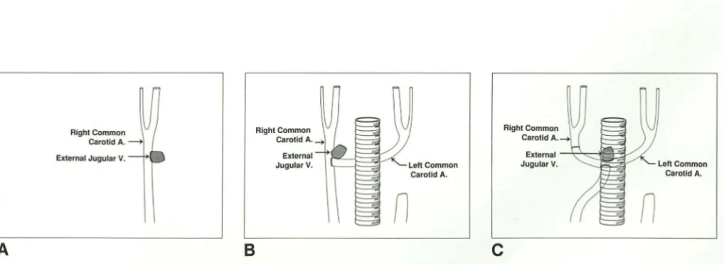
Aneurysms were created in adult male mongrel dogs using modifications of a technique first described by Ger-man and Black and further developed and modified in our laboratory over the last 3 years (1 0, 11 ). All procedures were performed under sterile conditions and with general anesthesia. In all instances, aneurysms were constructed from a venous pouch made from a 2-cm segment of excised external jugular vein, one end of which had been closed with 7-0 proline suture. The aneurysm pouch was connected to the parent artery directly following creation of a 5-mm diameter arteriotomy with a circular Hancock vascular punch. Three types of aneurysms were con-structed: 1) lateral, 2) bifurcation, and 3) terminal (Fig. 1 ). Lateral aneurysms were constructed in the midcervical segment of a common carotid artery by attaching a venous pouch to the artery in an end to side anastomosis. Three lateral aneurysms were made in each of 7 dogs, two on one common carotid and one on the other.
Bifurcation aneurysms were constructed by first ligating the midsection of the left common carotid artery and then transferring the distal segment of the vessel beneath the trachea and anastomosing it end to side into the midportion of the right common carotid artery. A venous pouch was then incorporated into the junction of this anastomosis. A total of four bifurcation aneurysms were made.
Terminal aneurysms were made by first ligating and then dividing both common carotid arteries in their mid-cervical segment. The distal segment of the left common carotid artery was then transferred beneath the trachea and was anastomosed end to end with the distal segment of
A
Right Common
Carotid A. --->
External Jugular V.
8
Right Common Carotid A.4
External Jugular V.
AJNR: 13, July/August 1992
the right common carotid artery. To complete the construc-tion, the proximal segment of the right common carotid artery was anastomosed end to side into the undersurface of the U formed by this linkage and a vein pouch was attached at the site of an arteriotomy made immediately above this anastomosis. Four aneurysms of this type were made.
Following completion of the surgical procedure, a base-line transfemoral angiogram was done to determine the patency of the aneurysm pouch. The animals were allowed to recover and were maintained in the Animal Care Facility of the University of Wisconsin for at least 2 weeks prior to further study. The long-term patency rate in these aneu-rysms was high, with only three of the 21 lateral aneuaneu-rysms showing any evidence of spontaneous thrombosis.
Angiographic studies were obtained following both the power injection of 8 mL of contrast medium through a 4-F single-end hole catheter placed 5-8 em below the level of the aneurysm and by the hand injection of 2 mL of contrast medium through a variable stiffness 2.2-F single-end hole catheter (Tracker-18; Target Therapeutics, San Jose, CA) placed directly into the aneurysm lumen. Both tape recordings of video fluoroscopy and film screen an-giograms were used for angiographic analysis.
Duplex color Doppler examinations were carried out using an Acuson 128 computed sonography system (Acu-son Corporation, Mountain View, CA) with a 7-MHz linear array transducer. The technical factors were set in each case to maximize the direction of flow (red toward the transducer and blue away from the transducer) and also to display velocity. In every case, color flow examinations were recorded with static color images. Pulsed Doppler waveform information was analyzed from both videotape and hard-copy images.
Results
Lateral Aneurysms
When contrast medium was injected into a parent artery proximal to a lateral aneurysm, the
left Common Carotid A.
c
Right Common
CarotidA. 4
External ----l.-.1...----"1-=Piil. / Jugular V.
n
Left Common
Carotid A.
[image:2.614.56.565.545.730.2]AJNR: 13, July/ August 1992
inflow into the aneurysm occurred at the distal
extent of the aneurysm ostium. Opacification of
the lumen then proceeded in a cranial-to-caudal
fashion and the outflow developed at the proximal
extent of the ostium. In every instance, a central vortex opacified and cleared slowly. Stagnation
of contrast medium within the lumen of these
aneurysms was pronounced, with contrast me-dium remaining with the aneurysm sac for as
long as several minutes after the injection. These
angiographic characteristics were noted in both
single and tandem lateral aneurysms. Although
they were most easily observed by reviewing
injections recorded on videotape, they could also,
however, clearly be seen on examinations done
with standard film screen techniques (Fig. 2).
The color Doppler examinations of all lateral
aneurysms demonstrated a similar pattern of
in-flow, central vortex, and outflow. In addition, the
color Doppler images revealed the inflow
circu-lation zone to be comprised of at least two
com-ponents with a central higher velocity region
being encompassed in an area of lower velocity
(Fig. 3). Disturbed laminar flow was also noted within a segment of the parent artery adjacent to
and just beneath the aneurysm ostium. There
was no difference noted between the color
Dop-pler findings of tandem lateral aneurysms as
compared to those of single lateral aneurysms.
Bifurcation and Terminal Aneurysms
The angiographic analysis of the flow
charac-teristics of the bifurcation and terminal type
aneu-rysms yielded similar findings. When contrast medium was injected into the parent artery at a
point well proximal to one of these types of
aneurysms, it was impossible to identify a distinct
inflow or outflow circulation. Unlike the lateral
aneurysms, the circulation inside the bifurcation
and terminal aneurysms was rapid and there was never any angiographic evidence of vortex for-mation or stasis.
When contrast medium was injected directly
into one of these types of aneurysms, the outflow
from the aneurysm was always seen to pass·
entirely into one of the two "branches." This
feature was noted regardless of where the con-trast medium was injected within the aneurysm lumen and it also appeared to be independent of
the speed of injection (Fig. 4). As measured from
the angiographic studies, there was 1 mm or less difference in the diameter of the arterial "branches" related to all of these aneurysms. Our
1091
technique did not allow accurate determination of the angle between either an aneurysm and its
parent artery, ie, the stem, or of the angles
between an aneurysm and its "branches."
There was not in any instance a consistent difference in the color Doppler findings of the bifurcation aneurysms as compared to those of
the terminal aneurysms. Without exception,
how-ever, both of these two types of aneurysms had color Doppler features that were distinctly differ-ent from those of the lateral aneurysms.
In all of the bifurcation aneurysms, the color
Doppler examinations revealed that the inflow
occurred at the edge of the ostium closest to the
long axis of the parent artery, ie, stem, while outflow occurred through the opposite corner of
the ostium. The Doppler studies, like the
angie-graphic studies made following selective
injec-tions of contrast medium into the aneurysms,
also showed that the outflow occurred exclusively into one of the two "branches" associated with the aneurysm. This was always into the branch opposite to the side of the ostium through which
the inflow occurred. Rapid flow was always
pres-ent in these aneurysms (Fig. 5). Within the lumen,
flow was rotatory in the direction of the outflow branch.
In the terminal aneurysms, the inflow occurred
at the side of the ostium that was closest to a
straight line drawn through the center of the
parent artery, ie, stem. Outflow was at the other
extreme of the ostium and was seen to pass exclusively into the "branch" nearest to the out-flow portion of the ostium. Like the bifurcation aneurysms, flow within these aneurysms was also rapid and rotatory (Fig. 6).
Discussion
Over the last two decades, there has been
considerable in vitro investigation and computer simulation/mathematical modeling of the hemo-dynamic features of saccular aneurysms. There
has been, however, little in vivo inquiry into these
phenomena. Although our canine aneurysm
model and laboratory techniques do not allow
quantitative analysis of flow characteristics,
ve-locities, or vascular stresses, the phenomena
ob-served in our studies do contribute information
that correlates with the more quantitative studies.
The flow characteristics of the various geo
1092 AJNR: 13, July/ August 1992
-
-A
8
c
DFig. 2. Angiographic study of lateral aneurysm done following contrast injection into parent artery.
A, Early film just before opacification of the aneurysm begins. Note slight deflection of contrast column along ostium of the aneurysm (arrows).
B, Subsequent film showing inflow of contrast medium into the aneurysm with opacification of the lumen in a cranial to caudal fashion.
C, Slightly later, the entire lumen is opacified.
D, Late film showing the central vortex and stagnation characteristic of this type of aneurysm.
Fig. 3. Color Doppler examination of lateral aneurysm showing the inflow, outflow, central vortex, and zone of high-velocity flow in the inflow circulation.
A
8
Fig. 4. A, Angiogram of bifurcation aneurysm done following injection of the contrast medium into the parent artery, ie, stem. The flow within this aneurysm is rapid and no differences in opacification of the "branches" related to it could be recognized.
B, Angiogram of same aneurysm made after hand injection of contrast into the aneurysm. The outflow of contrast medium from the aneurysm appears to be exclusively into one of the two "branches."
artery and branches, are of importance to those applying endovascular techniques for the treat-ment of patients with these lesions. Decisions such as the type and configuration of catheter that may most easily be used for aneurysm
cath-eterization, the location within an aneurysm
[image:4.612.60.564.77.246.2] [image:4.612.314.562.311.535.2] [image:4.612.56.298.348.702.2]6
Fig. 5. Color Doppler examination of bifurcation aneurysm.
A, Inflow is seen in the portion of the ostium nearest the long axis of the parent artery, ie, stem.
B, Image from a slightly different plane shows the outflow passing through the opposite portion of the ostium and exclusively into one of the two branches. These images also illustrate the rotatory flow present in bifurcation aneurysms.
[image:5.614.54.558.39.753.2]1094
which can best be used for treatment can all be logically based on an understanding of these hemodynamic features (12).
As discussed below, the observations from our study also provide some possible additional in-sight into the mechanisms involved in the growth,
rupture, and thrombosis of saccular aneurysms.
Importance of Hemodynamic Factors in Determining Growth of Saccular Aneurysms
A prominent angiographic characteristic of all of the lateral aneurysms in our study was stasis of contrast medium within the aneurysm sac. This is not a new observation, the phenomenon first having been described by German and Black
in 1954 (13). Stagnant flow has also been
re-ported in in vitro flow experiments as well as in computer simulation/mathematical models of flow in lateral aneurysms, as well as in one type
of terminal aneurysm, ie, the aneurysm fundus is
in the plane of the stem of the parent artery and there is balanced outflow into the efferent vessels (3, 4, 7, 8). Stasis is also a commonly observed feature in angiographic studies of many large and giant intracranial aneurysms in humans.
The common analogy of an aneurysm to a weak spot in an inner tube that will necessarily rupture when the strength of its thinning wall can no longer contain the tension upon it is not accurate, since the wall of an aneurysm is alive
and can, under certain conditions, reinforce itself.
Enlargement of an aneurysm does not thus nec-essarily imply thinning of its wall (14).
Large and giant aneurysms most commonly
involve the internal carotid artery (59%) or the
tip of the basilar artery (16%) ( 15). Aneurysms
of the cavernous and supraclinoid segments of the internal carotid artery are the geometric equivalent of the lateral aneurysm used in our canine experiments. Large and giant basilar tip
aneurysms in our experience most often occur
when there is a close symmetry in size between the two proximal segments of the posterior cer-ebral arteries and a near perpendicular orientation
of the aneurysm sac to its parent artery, ie, stem.
As already discussed, it is in these two
geomet-rical types of aneurysms that in vitro studies have
shown that stasis is prominent and flow is
sluggish (3, 4).
Stagnation of blood flow is known to result in
the accumulation of both platelets and leukocytes
along intimal surfaces, as well as in an impairment
of the diffusion of oxygen and metabolites from
AJNR: 13, July/August 1992
blood to the vascular wall. Either together or separately, these two factors may operate to cause intimal damage and thus lead to both thrombus formation and wall thickening (16, 17). Both of these events, ie, thrombus formation and wall thickening, have been proposed as factors
important in the growth of an aneurysm (18).
We have found it impossible to duplicate the terminal aneurysm geometry associated with stasis in our canine model, since in all of our terminal aneurysms there was significant angu-lation between the aneurysm sac and its parent
artery, ie, stem. Terminal aneurysms of this
ge-ometry are characterized in vitro by the absence of stasis (3).
One possible explanation, which to our knowl-edge has not been previously proposed, for the predilection of large and giant aneurysms to in-volve the internal carotid artery and the tip of the basilar artery may be, therefore, simply that these sites favor a geometrical configuration between the parent artery and aneurysm that results in the presence of hemodynamic conditions favor-ing stagnation of flow within the aneurysm from its inception. This stagnation then results in an environment that promotes thickening and rein-forcement of the aneurysm wall and thereby favors overall growth of the aneurysm over its rupture. Again, such conditions appear most likely to occur either in aneurysms with lateral configurations or in terminal type aneurysms that have balanced branches and a near perpendicular orientation of the aneurysm sac to the parent artery.
Importance of Hemodynamic Factors in Determining Rupture of Saccular Aneurysms
The concept of turbulent flow within the lumen of saccular intracranial aneurysms dates from the
work of Ferguson ( 19) in 1972, prior to the
establishment of the current notion of turbulence as representing a state of fully random motion. Simkins and Stehbens, and later Steiger and Reulen, have provided strong in vivo and in vitro evidence for the presence of intraaneurysmal flow patterns that are not turbulent but that rather represent intermediate stages between laminar
flow and turbulence (9, 20). Our studies of flow
in aneurysms of various geometries further doc-uments that the circulation within aneurysms is regular and is predictable primarily according to the geometrical relationship between the
AJNR: 13, July/August 1992
Steiger has also shown in a variety of aneurysm
geometries that the maximum shear stresses and
velocity gradients are not found at the dome of
an aneurysm but rather near the aneurysm neck
(3). Fluctuations in flow are known to induce
added mechanical stress, vibrations, and perhaps
even resonance, all of which may contribute to
aneurysm rupture (3, 9, 19). Our Doppler studies,
while not quantitative, also clearly demonstrate
in lateral, bifurcation, and terminal aneurysms a
zone of unstable high-velocity flow at the region
of the aneurysm ostium where inflow occurs. It
thus appears that in some instances greater
hemodynamic, ie, mechanical, stress is
transmit-ted to the tissue at an aneurysm's ostium than to
that at its dome. The significance of this requires
additional investigation.
Currently, the main criteria used to judge the
risk of a particular aneurysm rupturing is its size. The increasing availability and usefulness of
transcranial Doppler techniques, magnetic reso
-nance angiography, and quantitative/ dynamic techniques of digital subtraction angiography make it evident that it will soon be possible to
determine, with little risk, the hemodynamic
char-acteristics of intracranial aneurysms in a manner
greatly superior to that which has been previously
possible. Such data combined with an under-standing of the stresses and strains associated
with altered hemodynamics should allow a much
more rational decision to be made about the
chance of growth, rupture, or thrombosis of any particular aneurysm.
In conclusion, although our studies are
quali-tative, they provide new in vivo observations that
support the pioneering work of German and
Black, Ferguson, Stehbens, Steiger, Liepsch, and
their colleagues. Flow within these experimental
aneurysms, although not laminar, is seldom if
ever turbulent, but rather is highly predictable,
varying primarily according to the relationship of
an aneurysm to its parent artery. A more
com-plete understanding of these hemodynamic
fac-tors may be useful in establishment of criteria
that allow recognition of those aneurysms that
are more or less likely to rupture, grow, or
throm-bose. Such an understanding will also help in
1095
optimizing future attempts at the endovascular
treatment of saccular intracranial aneurysms.
References
1. Stehbens WE. Etiology of intracranial berry aneurysms. J 1'/eurosurg
1989;70:823-831
2. Sekhar LH, Heros RC. Origin, growth, and rupture of saccular
aneu-rysms: a review. Neurosurgery 1981; 18:248-260
3. Steiger HJ, Liepsch DW, Poll A, Reulen HJ. Hemodynamic stress in
terminal saccular aneurysms: a laser-Doppler study. Heart Vessels
1988;4: 162-169
4. Liepsch DW, Steiger HJ, Poll A, Reulen HJ. Hemodynamic stress in
lateral saccular aneurysms. Bioreheology 1987;24:689-710
5. Ferguson GG. Physical factors in the initiation, growth, and rupture
of human intracranial saccular aneurysms. J 1'/eurosurg 1972;37:
666-677
6. Ferguson GG. Direct measurement of mean and pulsatile blood
pressure at operation in human intracranial saccular aneurysms. J
1'/eurosurg 1972;36:560-563
7. Perktold K, Gruber K, Kenner T, Florian H. Calculation of pulsatile flow and particle paths in an aneurysm model. Basic Res Cardia/ 1984; 79:253-261
8. Perktold K. On the paths of fluid particles in an axisymmetrical
aneurysm. J Biomech 1987;20:311-317
9. Steiger HJ, Reulen HJ. Low frequency flow fluctuations in saccular
aneurysms. Acta 1'/eurochir (Wein) 1987;83: 131-137
10. German WJ, Black PW. Experimental production of carotid
aneu-rysms. 1'1 Eng/ J /VIed 1954;250: 104-106
11. Graves VB, Partington CR, Rufenacht DA, Rappe AH, Strother CM.
Treatment of carotid artery aneurysms with platinum coils: an ex
-perimental study in dogs. AJI'IR 1990; 11 :249-254
12. Graves VB, Strother CM, Rappe A. Flow dynamics of lateral carotid
artery aneurysms and their effects on coils and balloons: an expe
ri-mental study in dogs. AJI'IR 1992; 13:186-196
13. German W, Black SPW.Intra-aneurysmal hemodynamics: turbulence.
Trans Am Neural Assoc 1954;79: 163-165
14. Crompton MR. Mechanism of growth and rupture in cerebral artery
aneurysms. Br /VIed J 1966; 1:1138-1142
15. Weir B. Aneurysms affecting the nervous system. Baltimore: Williams & Wilkins, 1987:188
16. Crompton MR. The pathogenesis of cerebral infarction following the
rupture of cerebral berry aneurysms. Brain 1964;87:491-510
17. Nakatani H, Hashimoto N, Kang Y, et al. Cerebral blood flow patterns at major vessel bifurcations and aneurysms in rats. J 1'/eurosurg 1991; 74:258-262
18. Artmann H, Vonofakos D, Muller H, Grau H. Neuroradiologic and
neuropathologic findings with growing giant intracranial aneurysm:
review of the literature. Surg 1'/euro/1984;21 :391-401
19. Ferguson GG, Roach MR. Flow conditions at bifurcations as deter-mined in glass models with reference to the focal distribution of
vascular lesions. In: Berge! VH, Derek H, eds. Cardiovascular fluid
dynamics. London: Academic Press, 1 972: 141-156
20. Simkins TE, Stehbens WE. Vibrations recorded from the advential
surface of aneurysms and arteriovenous fistulas. Vase Surg