Chiari III Malformation: Imaging Features
Mauricio Castillo, 1 Robert M. Quencer,2
and Rodrigo Dominguez3
Purpose: To analyze and discuss the MR and CT features of Chiari type Ill malformations. Patients and Method: MR and CT studies in nine neonates born at term with Chiari type Ill malformations were retrospectively reviewed. Results: High cervical/low occipital encephaloceles were present in all cases. Hypoplasia of the low and midline aspects of the parietal bones was seen in four patients. The encephaloceles contained varying amounts of brain (cerebellum and occipital lobes, six cases; cerebellum only, three cases), ventricles (fourth, six cases; lateral, three cases), cisterns, and in one case, the medulla and pons. Associated anomalies included: petrous and clivus scalloping (five cases/nine cases), cerebellar hemisphere overgrowth (two cases/nine cases), cerebellar tonsillar herniation (three cases/seven cases), deformed midbrain (nine cases), hydro-cephalus (two cases/nine cases), dysgenesis of the corpus callosum (six cases/nine cases), posterior cervical vertebral agenesis (three cases/eight cases), and spinal cord syrinxes (two cases/seven cases). In four patients who underwent surgical resection and closure, aberrant deep draining veins and ectopic venous sinuses within the encephaloceles were found. Pathology examination of the encephalocele (four cases/nine cases) showed multiple anomalies (necrosis, gliosis, heterotopias, meningeal fibrosis) that were not demonstrable by either MR or CT. The marked disorganization of the tissues contained within the cephalocele may account for the lack of MR sensitivity to these abnormalities. Conclusion: Preoperative determination of the position of the medulla and pons is essential and is easily accomplished by MR. To avoid surgical complications, the high incidence of venous anomalies should be kept in mind.
Index terms: Brain, hernia; Chiari malformations
AJNR 13:107-113, January/February 1992
Encephaloceles occur in approximately one out of every 4000 to 5000 newborns (1). Those located in the occipital region are the most com-mon type in the western world. In 1891, Chiari described the pathologic features of what today is known as the type I and type II Chiari malfor-mations (2). This paper also contains a single case in which cervical spina bifida compined with multiple cerebellar and brain stem anomalies was present. This spectrum of abnormalities has been called a Chiari type III malformation. Recently,
Received April 16, 1991; revision requested July 7; revision received July 25; final acceptance August 1.
1 Section of Neuroradiology, University of Texas Medical School at
Houston, Houston, TX. Address reprint requests toM. Castillo, Department of Radiology, LBJ General Hospital, 5656 Kelley Street, Houston, TX 77026.
2
Section of Neuroradiology, University of Miami School of Medicine,
Miami, FL 33101.
3 Section of Pediatric Radiology, University of Texas Medical School at Houston, TX.
AJNR 13:107-113, Jan/Feb 1992 0195-6108/92/1301-0107
© American Society of Neuroradiology
107
the definition of Chiari type III malformations has been expanded to include those patients with herniation of the hindbrain into a low occipital (below the inion) and/or high cervical encepha-locele in combination with pathologic and imag-ing features of Chiari II malformations (3). For the purpose of this paper, we analyzed the magnetic resonance imaging (MR) and computed tomog-raphy (CT) studied obtained in nine patients who harbored a combination of abnormalities con-forming to both the classic and the more recent definitions of Chiari type III malformations.
Materials and Methods
MR scans of nine patients with Chiari lll malformations were reviewed retrospectively (Table 1). All patients were neonates born at term (three males and six females). Five
patients had prenatal ultrasounds. The patients underwent
108
all cases. Axial spin-echo proton density and T2-weighted
(2000-2400/30-80/1) were also obtained. Two patients
underwent preoperative evaluation with only noncontrast
axial (5 mm) CT sections. One CT study included images
of upper five cervical vertebrae. The upper cervical spine
was also included in the saggital and axial MR T1-weighted images in all cases. In one case, the cervical spine was not
evaluated.
The MR and CT studies were then assessed for the
contents, location and signal abnormalities of the enceph-aloceles, presence of lacunar skull, scalloping of the
pos-terior aspect of the petrous pyramids and the clivus,
over-growth of the cerebellar hemisphere, towering of the
cere-bellum through a wide tentorial incisura, deformities of the
midbrain (specifically beaking of the tectum and/or fusion
of the colliculi), hydrocephalus, morphology of the corpus
callosum, cerebellar tonsillar herniation, and abnormalities
in the cervical spine and cervical spinal cord. The MR and CT findings were then compared to the surgical findings in four cases.
Results
In five instances, prenatal sonography revealed
encephaloceles, and all of these patients were born by cesarean section. Apgar scores were available in six cases, and all but two were ab-normally low (the median score was 3 at 1 minute
and 5 at 5 minutes). At birth, microcephaly (head
circumference below the fifth percentile for age)
was present in five newborns. Four patients were
considered normocephalic, two of these patients
TABLE 1: Imaging features of Chiari III malformation
Case Imaging Encephalocele Enceph- Lacunar Bone Cerebellar alocele
Scallo-No. Study Contents'
Location• Skull' pingd Overgrowth
1 MR B, V,CSF HC,O
2 MR B, V HC,O +
3 MR B, V, CSF, M HC, 0, P
4 CT B, V 0, p P, C Medial and anterior
5 CT B, V 0, p
6 MR B, V HC,O + P,C
7 MR B, V HC, 0, P P,C
8 MR B, V,CSF 0 + P,C Medial and anterior
9 MR B,CSF 0 P,C
AJNR: 13, January/February 1992
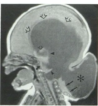
Fig. 1. Case 7: Sagittal spin echo 750/20 slightly off-center image shows a large low occipital/high cervical encephalocele. The CSF density (*) inside the cephalocele is believed to be a markedly dilated fourth ventricle, with the roof of the fourth ventricle displaced superiorly (small arrowhead). The third ventri-cle (large arrowhead) and massa intermedia (small open arrow) are mildly prominent. The corpus callosum (large open arrows) is thin and the splenium is absent. Residual cerebellar tissue (small arrows) are present inside the encephalocele. Note that there is absence of the posterior elements of C 1, C2, and C3. Scalloping (curved arrow) of the clivus is present. There is marked dilatation of the lateral ventricles.
had hydrocephalus. Surgical resection of the aberrant tissues contained within the encephalo-celes was performed in four cases. At surgery, these four patients had abberantly located venous sinuses and deep cerebellar veins and one patient
Towering
Hydro- Corpus Tonsillar Cervical Cervical Cere- Midbrain
cephal us Callosum• Herniation' Spine• Cordh bellum
Deformed PA + Syrinx
Deformed CA +
Deformed CA
Deformed CA NE NE
Deformed PA NE NE NE
Deformed CA + + Syrinx
Deformed + THIN +
Deformed + NL + +
Beaked NL
'B, brain; V, ventricles; CSF, cerebrospinal fluid containing spaces different than ventricles; M, medulla.
• HC, high cervical; 0, occipital; P, parietal.
'Evaluated with plain films. d P, petrous bones; C, clivus.
• PA, partial agenesis; CA, complete agenesis; THIN, corpus callosum is thinned but present; NL, normal.
' NE, not evaluated.
9 Refers to agenesis of the posterior elements of Cl through C3.
[image:2.613.357.514.76.246.2]AJNR: 13, January/February 1992
Fig. 2. Case 1: Midline sagittal spin echo 700/20 image dem-onstrates a low occipital encephalocele containing cerebellar tis-sues. The cystic portions (•) within the herniated cerebellum are of uncertain etiology. The posterior aspect of the corpus callosum
(arrows) is not clear and is probably dysgenetic. The third ventricle is not seen, but the massa intermedia (m) is very prominent. The tectum is deformed and not readily identified. The fourth ventricle
(arrowhead) is deformed and displaced posteriorly. A syrinx
(curved arrows) is present in the mid to lower cervical spinal cord.
Fig. 3. Case 3: Midline saggital spin echo 800/20 image in a patient with a large occipital and parietal encephalocele. The encephalocele contains a large amount of brain tissues and dilated ventricles (•). The intracranial contents are dysmorphic and the corpus callosum is not identified. Note the presence of a venous structure (arrowhead) inside the encephalocele. The medulla
(arrow) and pons (curved arrow) herniate into the encephalocele.
Recognition of these structures is critical for the surgical planning.
109
had aplasia of the posterior falx cerebri. These vascular structures were preserved in all cases.
All patients required duraplasties and skin grafts. Two patients died in the immediate postoperative period; one to diffuse edema probably due to thrombosis of the superior saggital sinus and one of aspiration. One patient who had surgery is alive, but severely retarded. The remaining five patients were lost to follow-up. In four cases, pathologic examination of the tissue removed from the encephalocele showed cerebral and cer-ebellar tissues with areas of pressure necrosis, ventricles with normal choroid plexuses, gray matter heterotopias, small focal areas of gliosis,
meningeal inflammation and fibrosis, and reactive astrocytosis.
Imaging studies showed that all encephaloceles involved the occipital bones; five extended into the high cervical (C1-C3) spine (Fig. 1). In four cases, the low and midline parietal bones were also involved (Table 1). All encephaloceles con-tained brain (cerebellum, nine cases; occipital,
five cases; and parietal lobes, one case). Portions
of the ventricular system (lateral ventricles, three cases; fourth ventricle, six cases) extended into six encephaloceles. Three encephaloceles con-tained no ventricles (Fig. 2). In one case, the brainstem and medulla herniated into the
enceph-alocele (Fig. 3). Three encephaloceles contained large cerebrospinal fluid (CSF) spaces unrelated to the ventricular system and, although these are
of uncertain etiology, we believe they could rep-resent distorted and enlarged basilar cisterns.
Lacunar skull was present on plain films in three patients. The abnormal venous structures identi-fied during surgery in four patients were only seen by MR in two cases (Figs. 3 and 4A). Angiograms were not performed on any patients.
The multiple pathologic findings (necrosis, grey matter, heterotopias, gliosis, reactive
astrocyte-sis, and meningeal inflammation and fibrosis) found within the encephaloceles could not be individually identified preoperatively by MR on either T1- or T2-weighted images, possibly due to the marked disorganization of these tissues.
Specifically, the encephaloceles did not show abnormal areas of increased signal intensity (cor-responding to gliosis) on the T2-weighted images. Five patients had scalloping of the posterior pet-reus pyramids and the clivus (Figs. 4A and 48).
[image:3.612.58.257.83.244.2] [image:3.612.49.302.359.656.2]110
Fig. 4. A, Case 8: Axial spin echo 700/
20 image at the level of the mid posterior
fossa shows scalloping (short arrows) of the
posterior aspect of the petrous bones. The cerebellar hemispheres (*) show medial and anterior overgrowth with respect to the brainstem (long arrow). Also seen are aber-rant paired venous structures (arrowheads) bordering the skull defect. Note the posterior
encephalocele.
8, Case 4: axial CT scan (bone windows) shows scalloping (arrows) of both petrous bones. Also note the encephalocele (*).
A
Fig. 5. Case 2: Sagittal spin echo 750/20 image in a
micro-cephalic patient with a high cervical/low occipital encephalocele.
There is herniation of the occipital lobes (arrow) into the defect. The cerebellum is not seen and the tectum is not clearly identified. The midbrain (*) is deformed and bulbous and is displacing the aqueduct (arrowhead) posteriorly. The splenium of the corpus callosum is absent. Agenesis of the posterior elements of C 1 through C3 is present.
Towering of cerebellum (supratentorial extension
of cerebellar tissues through a widened tentorial
incisura) was not documented in any case. The cerebellum was not identified in one patient (Fig.
5). The midbrain was considered abnormal if it
was beak-like in configuration (n = 1), or if the
colliculi could not be identified individually (n =
AJNR: 13, January /February 1992
B
8) (Fig. 5). A deformed midbrain was seen in eight cases (Fig. 5).
Hydrocephalus (dilation of the intracranial ven-tricular system) was seen in two cases (Fig. 1).
Colpocephaly (localized enlargement of the atria and occipital horns of the lateral ventricles) was seen in one case. Abnormalities of the corpus callosum included: partial agenesis (always in-volving the splenium), two cases; complete agen-esis, four cases; and extreme thinning probably secondary to marked hydrocephalus, one case (Table 1) (Figs. 1, 2, and 5). A normal corpus callosum was present in two cases. Extension of the cerebellar tonsils downward below the fora-men magnum was present in three cases (Fig. 2). In one case of cerebellar aplasia, the tonsils were not identified. The position of the tonsil could not be evaluated in two patients who underwent only preoperative CT. In three patients, the position of the cerebellar tonsils was considered normal. Ab-sence of the posterior elements of the first three cervical vertebrae was identified in three cases (Figs. 1 and 5). Spinal cord fluid filled cavities were seen in two cases; in one of these, the syrinx extended into the pons (Figs. 2 and 6). Two patients died due to surgical complications in the immediate postoperative period.
Discussion
[image:4.613.230.562.77.345.2] [image:4.613.119.557.88.503.2]AJNR: 13, January/February 1992
Fig. 6. Case 6: Axial spin echo 800/20 image through the low occipital regions clearly shows the encephalocele. Notice that the tissue within the encephalocele are markedly distorted and it is not possible to recognize any normal structures. A syrinx (arrow) is present in the medulla. In this patient, the syrinx extended into the pons. Note the cerebellar tonsils (*)displaced inferiorly.
Ill malformation is rare and only scattered cases (included in larger series of Chiari type I and II malformations) are found in the literature (4, 10-12). The Chiari III malformation includes displace-ment of the medulla, cerebellum, occipital lobes, and meninges into a high cervical and/ or low occipital cephalocele (13). The above features in combination with those seen in Chiari II malfor-mations are believed to be characteristic of the type Ill malformations (14). A skull defect asso-ciated with herniation of the intracranial contents is termed a "cephalocele" (14). If the herniation contains both brain and meninges, it is referred to as an "meningoencephalocele"; herniation of only the meninges and CSF is called a "menin-gocele"; herniation of only brain tissues is called an "encephalocele" (15). Encephaloceles are be-lieved to be slightly more common than menin-goceles ( 1 ). In Chiari Ill malformations, an en-cephalocele (which may or may not contain men-inges) must be present in combination with the brain anomalies present in the Chiari II malfor-mations. Occipital encephaloceles can be iso-lated, or be associated with the Chiari malfor-mations, Dandy-Walker complex, cerebellar
dys-111
plasias, diastematomyelia, and Kippei-Feil syn-drome (14).
The embryologic origin of a low occipital (base-of-skull) cephaloceles probably involves the fail-ure of induction of enchondral bone by incom-plete closure of the neural tube, or failure of the ossification centers to fuse completely (16). A posterior cephalocele may involve the supraoc-cipital and parietal bones (membranous origin).
This is likely to result from faulty induction or from pressure erosion by the adjacent mass of herniated tissues (16). It has been noted that supraoccipital encephaloceles occurring below the inion are the most common type seen in Chiari Ill malformations ( 1 ). The main features found in our nine patients with Chiari Ill malfor
-mations are discussed separately.
Cephaloceles, Skull, Dural Partitions, and
Venous Anomalies
In all cases, the skull defects were located in the high cervical/ occipital regions and extended to the parietal bones in four cases (Table 1 and Fig. 1). These encephaloceles contained cerebel-lum and occipital lobes in all cases. One enceph-alocele also contained the medulla and pons. In another case, only the cerebellum was herniated.
The masses of herniated tissues were markedly distorted and we were unable to recognize any individual structures within them (Fig. 6). In four cases, surgical resection and pathology exami-nation showed multiple areas of pressure necrosis in the herniated brain, gray matter heterotopias, gliosis, meningeal inflammation, and fibrosis.
[image:5.612.55.256.78.339.2]ac-112
companied the brain parenchyma in three
in-stances; in six of these, the fourth ventricle also
herniated. In four cases, the herniated ventricles
were dilated out of proportion to the intracranial ventricular system (Fig. 3). A possible explanation
is that these ventricles contained choroid plexus
and, therefore, produced CSF. Constriction of the
encephalocele at the level of the skull defect could
have accounted for lack of normal CSF drainage
from the ventricle leading to dilatation, and
sec-ondarily resulting in gliosis and atrophy of the
herniated tissues. Lacunar skull ("luckenschadel")
was present in three cases. "Luckenschadel" is
also commonly seen with isolated encephaloceles
and with Chiari II malformations (3). Scalloping
of the posterior petrous bones and clivus was
present in five of our patients (Fig. 48). In the
type II malformation, this latter finding is believed
to be secondary to pressure erosion from the
cerebellum as it attempts to grow within the
confines of a small posterior fossa (4). In the Chiari Ill malformation, the posterior skull defect
should decompress the posterior fossa and,
there-fore, pressure erosion is an unlikely cause for
bone scalloping. A more likely explanation is that this scalloping is a bone dysplasia similar to
"luckenschadel." Aplasia of the posterior aspect
of the falx cerebri was found at surgery in one
patient.
Hindbrain, Midbrain, and Cervical Spine
Downward herniation of the cerebellar tonsils
was present in three of our patients (30% ).
Trac-tion of the hindbrain into the encephalocele prob-ably accounted for this relatively low number of
cases with this finding , since only those patients
with relatively small encephaloceles showed
ton-sillar herniation. Medial and anterior overgrowth of the cerebellar hemispheres can be seen in up
to 75% <;>f patients with Chiari II malformation
(Fig. 4A) (4). In our series, only two cases showed
this feature (Table 1). The lack of cerebellar
confinement in a small posterior fossa could have
accounted for this relatively small number of
cases. Also, the same hypothesis can help explain
the absence of "towering" of the cerebellum
through the tentorial incisura in our population.
This finding is commonly noted in the type II
malformation (5). In one patient, the cerebellum
was not identified (Fig. 5). Extreme hypoplasia of
the cerebellum is known to occur in patients with
Chiari II (9). Beaking of the midbrain was clearly
identified in only one of our patients. However,
AJNR: 13, January/February 1992
eight patients had a deformed quadrigeminal plate (probably due to fusion or distortion of the colliculi) and, therefore, were considered to have
an abnormal midbrain (Fig. 5).
In three cases, the posterior elements of C 1,
C2, and C3 were absent (Figs. 1 and 5). In one
case, the upper cervical spine was not evaluated.
This anomaly is also commonly encountered in the type II malformation in which close to 70% of cases may show incomplete fusion of the posterior arch of C 1 and other cervical vertebrae
(16). In two of our patients, cord cavities were
documented (Figs. 2 and 6). In one case, the syrinx extended into the pons (syringobulbia). Even though we do not have a good explanation for these findings, a possible hypothesis involves the failure of normal formation of the foramina of Luschka and Magendie (as part of the hindbrain dysgenesis) with subsequent alterations of the normal CSF dynamics at the level of foramen
magnum leading to syrinx formation. These CSF
flow abnormalities are also believed to be present in the type II Chiari malformations (18). Since the entire spine was not studied in our patients, it is conceivable that the incidence of syrinxes may have been even higher.
Ventricular System and Corpus Callosum
Marked intracranial dilatation of the ventricular system was seen in two instances (Fig. 1). Both of these patients had a deformed midbrain. The aqueduct of Sylvius was not clearly identified in these two patients (Fig. 5). Aqueductal stenosis can then be considered as a reason for the
hydro-cephalus. The septum pellucidum was present in
all cases. Seven of our patients had dysgenesis
of the corpus callosum (complete agenesis, four
cases (50%); partial agenesis (always involving
the splenium), two cases (25% ); marked thinning
(probably secondary to severe hydrocephalus),
one case (12.5%)) (Table 3) (Figs. 1-3 and Fig.
5). Dysgenesis of the corpus callosum is com-monly seen in the Chiari II malformations and can be seen in patients with isolated encephaloceles
(19, 20). A normal corpus callosum was present
in only two cases.
AJNR: 13, January /February 1992
constant escape of CSF through the open neural
tube defect during intrauterine life (21). This leads
to failure of distension of the primitive ventricles with subsequent development of a small skull. As
the brain attempts to grow in the reduced space,
multiple cerebral abnormalities arise. We believe
that this hypothesis can be used to explain the
findings in the Chiari Ill patients. We also feel that
it is appropriate to mention here that, even if our
patients had no obvious source of CSF leakage
at birth, CSF leakage in utero cannot be excluded.
This may have lead to formation of the multiple abnormalities also present in the type II malformation.
In summary, Chiari Ill is a rare developmental
condition with a very poor prognosis. Due to its
distinct clinical and imaging features, it is easily
distinguished from the more common Chiari I and II malformations and isolated encephaloceles. Due to its multi planar capability, MR is more useful than CT to study preoperatively the content of
the cephalocele. MR can readily establish the
presence of solid tissues and of ventricular
exten-sion into the cephalocele. In our experience, MR
was unable to distinguish many subtle
abnormal-ities in the herniated brain; however, this issue is
not critical because the tissues within the enceph-alocele are believed to be nonfunctioning. Never-theless, the preoperative identification of the brain stem and medulla is mandatory to preserve respiratory function; this is easily accomplished
with MR. To prevent complications when surgery
is comtemplated, it should be kept in mind that anomalies of venous drainage are commonly
present, but often not identified on MR studies. If
surgery is performed in a patient with an
enceph-alocele, resection of the herniated brain with preservation of the herniated venous structures accompanied by duraplasty and skin grafts is
mandatory (GL Clifton, personal communication).
Resection of the posterior two-thirds of the
su-perior sagittal sinus is almost always fatal.
113
References
1. Diebler C, Dulac 0. Pediatric neurology and neuroradiology. Berlin: Springer-Verlag, 1987:51
2. Chiari H. Uber ueranderungen del kleinitirns infloge von Hydrophalie des grosshirns. Dsch Med Wochenschr 1891; 17:1172-1175 3. Barkovich AJ. Pediatric neuroimaging. New York: Raven, 1990:113 4. Naidich TP, Pudlowski RM, Naidich JB, eta!. Computed tomographic
signs of Chiari II malformation. I. Skull and dural partitions. Radiology 1980:134:65-71
5. Naidich TP, Pudlowski RM. Naidich JB. Computed tomographic signs of Chiari II malformation. II. Midbrain and cerebellum. Radiology 1980;134:391-398
6. Naidich TP, Pudlowski RM, Naidich JB. Computed tomographic signs of Chiari II malformation. Ill. Ventricles and cisterns. Radiology
1980; 134:657-663
7. Naidich TP, McCione DG, Fulling KH. The Chiari II malformation. IV. The hindbrain deformity. Neuroradiology 1983;25: 179-197 8. El Gamma! T, Mark EK, Brooks BS. MR imaging of Chiari II
malfor-mation. AJNR 1987;8:1037-1044
9. Wolpert SM. Anderson M, Scott RM, et al. Chiari II malformation: MR imaging evaluation. AJNR 1987;8:783-792
10. Mayr U, Aichner F, Menardi G, Hager J. Computer tomographical appearances of the Chiari malformations of the posterior fossa. Z
Kinderchir 1986;41 :33-35
11. Dyste GN, Menezes AH, VanGilder JC. Symptomatic Chiari malfor-mations: an analysis of presentation, management, and long-term outcome. J Neurosurg 1989; 71:159-168
12. Kuharik MA, Edwards MK, Grossman CB. Magnetic reson(!nce eval-uation of pediatric spinal dysraphism. Pediatr Neurosci 1985; 12:213-218
13. Ramsey RG. Neuroradiology. 2nd ed. Philadelphia: WB Saunders,
1987:441-442
14. Emery JL, Kalhan SC. The pathology of exencephalus. Dev Med Child Neurol1970;12:51-64
15. Diebler C. Dulac 0. Cephaloceles: clinical and neuroradiological ap-pearance. Neuroradiology 1983;25: 199-216
16. Blaauw G. Defect in posterior arch of atlas in myelomeningocele. Dev Med Child Neural 1971 ; 25: 113-115
17. Chuang SH, Edwards MK, Barkovich AJ. Congenital brain malfor-mations of the brain. In: Cohen MD, Edwards MK, eds. Magnetic resonance imaging in children. Philadelphia: Decker, 1990:151 18. Banna M. Syringomyelia in association with posterior fossa cysts.
AJNR 1988;9:867-873
19. Barkovich AJ, Norma D. Anomalies of the corpus callosum: correla-tion with further anomalies of the brain. AJNR 1988;9:493-501 20. Kendall BE. Dysgenesis of the corpus callosum. Neuroradiology
1983;25:239-256