Sexual reproduction is critical for genetic exchange and is almost universal among animals and plants. Many organisms spawn their gametes into the environment and fertilization then occurs externally. During external fertilization, factors that influence sperm–egg encounter rates can include fluid flow (Mead and Denny, 1995), gamete age (e.g. Lillie, 1915; Brown and Knouse, 1973; Pennington, 1985; Levitan et al. 1991), parentage (Levitan, 1993) and gamete sensory capabilities (e.g. Miller, 1979, 1985a,b; Ward et al. 1985). Because most of these factors affect sperm motility, an analysis of locomotory patterns is fundamental to an understanding of sperm–egg interactions. Nearly all previous studies of motility placed sperm in small volumes of fluid on microscope slides. In several cases, only sperm trapped along the fluid–air or fluid–surface interfaces were considered (e.g. Gray, 1955; Miller 1978, 1979; Ward et al. 1985). Regrettably, studies conducted in this manner ignored important physical phenomena, including viscous wall effects.
Theoretical work during the last 20 years permits an accurate evaluation of wall effects on uniflagellar motion. According to physical principles, a sperm flagellum can be treated as a finite
number of straight units (slender bodies) (Katz et al. 1975). The translational velocity of each unit is then separated into normal and transverse components. Each component has an associated resistance, or drag, coefficient (Gray and Hancock, 1955; Katz et al. 1975; Lighthill, 1975; Brennan and Winet, 1977), and integration extends this analysis to the combined sperm head and flagellum as a whole. The overall ratio of transverse to normal resistance determines both the swimming speed and direction (Katz and Blake, 1975; Lighthill, 1975). Theoretically, a sperm moves faster within approximately 10 body lengths of a solid boundary or wall (Katz et al. 1975).
Recognizing the disparity between the theoretical work regarding flagellates near solid surfaces and the protocols used in most studies of sperm swimming, we examined empirically the effects of walls on gamete motility. Previous work on external fertilization and early development in the sea urchin Arbacia punctulata motivated our selection of this species. As with any study involving sex cells, the source of gametes must also be considered a relevant variable (Levitan, 1993). Research on a number of other species has demonstrated that sperm age affects fertilization success (e.g. Lillie, 1915; Brown Printed in Great Britain © The Company of Biologists Limited 1997
JEB0836
The measurement of sperm motility is critical when studying fertilization kinetics and chemotaxis. Analysis of motility has traditionally been carried out on cells in small fluid volumes on microscope slides. Several theoretical treatments suggest that drag forces significantly affect flagellar motion within 10 sperm body lengths of the slide surface. Understanding how sperm move in the absence of surface drag is crucial when considering natural locomotory patterns. To examine the effects of solid surfaces, motile sperm from sea urchins (Arbacia punctulata) were placed in a Plexiglas chamber (69 mm×45 mm×15.5 mm; length × width × height). A system was constructed to minimize convective flow by limiting temperature differences within the chamber to less than 0.1 °C. The movement of sperm was video-recorded at two levels: <100µm (3 body lengths) and 5 mm (150 body
lengths) below the chamber lid. When swimming speeds were measured using a computerized video motion-analysis system, a highly significant difference (P<0.0001) between cells at the two depths was found. Cells nearest the lid swam at 174.6±5.9µm s−1 (mean ± S.E.M.), whereas those
farther away slowed to only 111.1±9.9µm s−1 (mean ± S.E.M.). Swimming speed was also found to be significantly
(P<0.01) affected by paternity, but not by sperm age. We conclude that viscous wall effects must be carefully considered in studies of sperm motility and chemotaxis. The analysis of sperm on a microscope slide may substantially exaggerate swimming speed.
Key words: sperm, sea urchin, Arbacia punctulata, swimming, motility, motion analysis, wall effects, drag.
Summary
Introduction
THE EFFECTS OF WALLS, PATERNITY AND AGEING ON SPERM MOTILITY
CHRISTOPHER C. GEE1,* ANDRICHARD K. ZIMMER-FAUST2
1Marine Science Program, and Belle W. Baruch Institute for Marine Biology and Coastal Research, University of South Carolina, Columbia, SC 29208, USA and 2Department of Biology, University of California,
Box 951606, Los Angeles, CA 90095-1606, USA
Accepted 24 September 1997
and Knouse, 1973; Pennington, 1985; Levitan et al. 1991). In this report, we assess whether proximity to walls, sperm ageing and paternity affect gamete motility.
Materials and methods Gamete collection
Adult Arbacia punctulata (Lamarck) were collected at Huntington Beach State Park in Murrells Inlet, South Carolina, between June and September 1994. The animals were held in the laboratory in recirculated sea water (21–23 °C, 32–35 ‰ salinity, 14 h:10 h L:D) for less than 3 weeks. During this time, they were fed agar-based ‘urchin cakes’, consisting of boiled eggs and Tetramin fish food (Lutz and Inoué, 1986). We sexed each individual urchin by withdrawing a sample of gonadal material. The gametes were obtained by inserting an 18 gauge syringe needle through the test (eggs are dark maroon to purple in color, while semen is white). Eggs were collected by inducing a randomly selected female to spawn by injecting 1 ml of 0.5 mol l−1 potassium
chloride (KCl) through the test and inverting the spawning female over a 250 ml glass beaker filled with sea water filtered through a 0.45µm filter (Tyler and Tyler, 1966). Eggs were added to filtered sea water until a concentration of 200 eggs ml−1 was achieved. To minimize sperm ageing,
semen harvesting occurred after the egg mixture had been made. We collected semen in a dry beaker after a randomly selected male had been injected with KCl as described for the female. Semen was added to filtered sea water to produce a sperm concentration, assessed using a hemocytometer, of 2×10⁵cells ml−1. To prevent complications associated with
employing gametes of differing maturity, sperm were only used if the injected male provided abundant (>2 ml), pure white semen. Additionally, fertilization assays were conducted to establish the competency of the harvested sperm (see below for explanation).
Experimental chamber and video imaging
Sperm swimming in a 69 mm long ×45 mm wide ×15.5 mm deep Plexiglas chamber were video-taped at a magnification of 41.25× with a Sony SLV-686HF video cassette recorder at 30 frames s−1using a NEC TI 23A CCD video camera mounted
on an Olympus BH-2 compound light microscope. The chamber was completely filled with sperm solution, and all head space was removed to eliminate evaporation. Convective flow within the chamber was substantially reduced by minimizing temperature fluctuations. A peristaltic pump delivered water (40 ml min−1) held at constant temperature
from a reservoir to an insulated Plexiglas water bath (202 mm long ×118 mm wide ×33 mm deep) placed on the microscope stage. Water flowed around the outside of the experimental chamber, which was placed in the center of the bath. Two water-filled containers supported by pieces of Plexiglas were placed over the top of the bath so that only the middle section of the lid, with an approximate area of 12.5 cm2, was left
uninsulated; this space accommodated the microscope
objective. A Campbell Scientific CR10 data logger was used to record temperature simultaneously from sensors (24 gauge type T copper/constantan thermocouples) in the head tank, in the air above the microscope stage, in the water bath and at two separate points within the experimental chamber. The temperature at each sensor was recorded every minute for the duration of each experiment.
Video images of sperm were digitized and processed over 30 s time intervals at 30 frames s−1 using a computer-video
motion analysis system (model VP 110 and Expert Vision software, Motion Analysis Corporation) interfaced with a Sun SPARC IPC computer work station. To eliminate the effects of convective flow on our measurements of speed of swimming, we added heat-killed (approximately 40 min at 60 °C) sperm to each suspension. These dead sperm served as passive particles allowing visualization of the background fluid movement experienced by live gametes. To correct for fluid flow, mean velocities (speed and direction) for the dead sperm paths were subtracted from live sperm paths on a frame-by-frame basis using a customized Expert Vision batch program (available upon request). We estimated our depth of field to be approximately 100µm, or fewer than 3 sperm body lengths. To avoid mistaking vertically for horizontally moving cells, short paths that consisted of computer images with sperm changing more than 20 % in apparent size were discarded. All paths that could be followed for at least six frames were included in the analysis.
Examination of age and adult effects on sperm swimming Separate trials were performed for the sperm from five adults. Sperm ‘age’ was defined here as the amount of time following final gamete dilution in sea water. Cells from each adult were video-recorded in 3–5 sessions at 5 mm depth below the chamber lid, with each session spaced approximately 30–60 min apart. The sperm were maintained in the experimental chamber between recording sessions.
Examination of the effects of proximity to walls on the speed of swimming of sperm from different individuals To examine the effects of walls, we video-recorded swimming by sperm at two depths within the chamber (<100µm and 5 mm below the lid). All trials were performed with sperm released from the gonads between 1 and 1.5 h previously. Replicate trials were conducted with sperm from each of eight adults.
Fertilization assay
Fertilization (at 23 °C) was assayed by putting 3.5 ml samples of the egg mixture into replicate 13 mm×100 mm borosilicate glass culture tubes and adding 3.5 ml of sperm mixture to each tube (1000 sperm per egg) (Pennington, 1985; Dinnel et al. 1987). At 10, 30, 60 or 120 min, fertilization was stopped by adding two drops of 10 % formalin and gently shaking. Assays were run when filming began, 3 h after filming started, and once filming was complete, with 3–5 replicates per time period. To control for egg age, freshly shed sperm were added to aged samples of the egg mixture in a few cases. The fertilization rates were nearly identical between freshly diluted and aged eggs. We estimated the percentage fertilization in each replicate by counting the number of raised fertilization membranes among the first 100 eggs observed under a compound light microscope at 125×(Pennington, 1985).
The sperm and egg mixtures used in the fertilization assay were kept in aerated beakers at room temperature. Once filming was finished, we compared the fertilizing capabilities of the sperm held in the aerated beakers with those in the chamber. Chamber and aerated sperm mixtures were separately and simultaneously incubated with eggs for three time intervals (30, 60 or 120 min) with four replicates per incubation period. Finally, diluted samples of the heat-killed sperm were assayed for fertilization to confirm that cells had indeed died before use in flow visualizations. In each trial, five replicate samples of heat-killed sperm were incubated with eggs for more than 120 min.
Results
Temperature and water flow within the experimental chamber
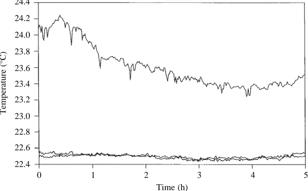
Data recorded from the thermocouples illustrate the degree to which temperature variations within the chamber were controlled (Fig. 1). Despite changes in air temperature (upper line), measurements recorded by sensors mounted at opposite ends of the chamber (two lower lines) never differed by more
than 0.1 °C across all experiments. Different trials were run between 21.7 and 22.9 °C.
Temperature constancy resulted in a slow, steady flow within the experimental chamber. The streamlines within the field of view, as marked by the heat-killed sperm paths, were nearly always parallel, indicating laminar flow. The uniformity of the flow field in each 30 s video recording is evident from the small standard error of the mean speed (typically less than 1.42µm s−1) and the small angular deviation (less than 4.79 ° overall) seen among dead sperm paths. Not surprisingly, no measurable flow was recorded within 100µm of the lid. While flow speeds at 5 mm depth ranged from 5.2 to 140.8µm s−1 depending upon the trial, they were typically less than 20µm s−1. Considering that flow speeds seen in the experimental chamber prior to insulation were approximately 1 mm s−1, the temperature-stabilizing system used in this study dramatically reduced convection to a tractable level.
Water flow effects on sperm swimming
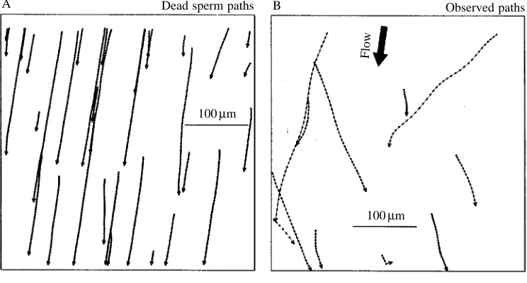
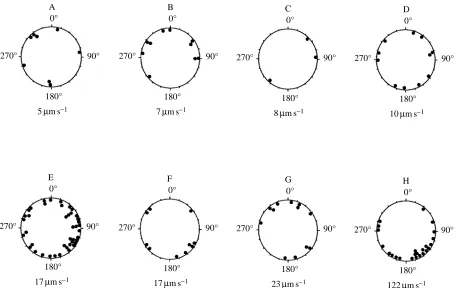
The paths swum by live sperm were clearly distinguishable from those of their dead counterparts (Fig. 2). Swimming speed and direction were determined by subtracting the mean flow vector frame-by-frame from each live sperm path. Water flow speed had no effect on the mean corrected swimming speed (Fig. 3, and linear regression: F=0.05, d.f.=1,6, P=0.8313). Similarly, flow direction had no effect on swimming direction in these same sperm (Fig. 4, and Rayleigh test: P>0.10 in all cases for both unimodal and bimodal distributions). The combined results provide no evidence for a link between flow and either the direction or speed of sperm swimming in this investigation.
Age and adult effects on swimming of sperm following release from the gonads
To investigate the effects of both sperm ageing and paternity on sperm swimming, 197 cells from five adults were examined. These gametes were assayed between 1.2 and 3.8 h after dilution in sea water. Two-factor analysis of variance
24.4
24.2
24.0
23.8
23.6
23.4
23.2
23.0
22.8
22.6
22.4
Temperature (°C)
0 1 2 3 4 5
[image:3.609.252.566.546.741.2]Time (h) Fig. 1. Temperature recorded at three
revealed a highly significant effect of adult on swimming speed (Fig. 5; F=3.56, d.f.=4,166, P=0.0082). Neither age (F=1.33, d.f.=5,166, P=0.3422) nor the adult × sperm age interaction (F=0.54, d.f.=9,166, P=0.8462) was found to be significant.
The effects of proximity to walls and of variations between individuals on sperm swimming
The influences of walls and of paternity on sperm motility were considered using 224 cells from eight adults. In total, 120 gametes were video-taped while swimming 5 mm below the chamber lid, and another 104 gametes were video-taped within 100µm of the lid. All sperm at the 5 mm depth swam in horizontally directed helical paths. Among the 104 sperm swimming near the chamber lid, 58 moved in horizontally directed helical paths and 46 moved in vertically directed circles, with 82.5 % (38 cells) spinning clockwise (G-test, G=21.217, d.f.=1, P<0.0001). Circular paths were generated by cells swimming head-on in contact with the lid. A
one-A Dead sperm paths
100µm
B Observed paths
100µm
Flow
C Corrected paths
100µm
Flow
D Observed and corrected paths
100µm
Flow
[image:4.609.186.558.75.275.2]Observed Corrected Fig. 2. Sperm paths video-taped
during a single 30 s time clip at 5 mm depth. (A) The movement of dead sperm showing the direction of fluid flow. (B) The observed pattern of movement of live sperm. (C) The pattern of sperm movement shown in B corrected for fluid flow (see Materials and methods). (D) The corrected pattern of movement superimposed on the observed pattern. The mean flow speed in these figures is 119.5µm s−1. Dots correspond to
[image:4.609.187.559.76.690.2]video images captured at 0.033 s intervals. Arrowheads correspond to the directions of travel for individual cells.
Fig. 3. Mean (± S.E.M.) corrected swimming speed as a function of flow speed. Each mean value (N=3–42) is for sperm drawn from one of eight adults. All speeds were measured at a depth of 5 mm below the chamber lid. The regression line and equation are shown.
200
180
160
140
120
100
80
60
Mean corrected swimming speed (
µ
ms
−
1)
0 10 20 30 40 110 120
factor analysis of variance indicated that the swimming speeds for sperm moving in circular and in helical paths at the chamber lid did not differ significantly (F=2.36, d.f.=1,102, P=0.1197 ). Grand mean speeds across all eight adults were 161.5±5.7µm s−1(mean ± S.E.M.) for circular-swimming and
174.6±5.9µm s−1(mean ±S.E.M.) for helical-swimming sperm
at the shallower depth, as opposed to 111.1±9.9µm s−1 (S.E.M.)
for helical-swimming sperm at 5 mm. A two-factor analysis of variance with adult and depth as class variables was conducted using speeds for the 178 sperm swimming in helical paths and video-taped at the two depths. Very highly significant differences in speeds were found among the sperm from different adults (F=32.91, d.f.=7,178, P<0.0001) and between depths (F=31.78, d.f.=1,178, P<0.0001). The adult × depth A
0°
180°
90°
5µm s−1
B 0°
180°
90° 270°
7µm s−1
C 0°
180°
90° 270°
8µm s−1
D 0°
180°
90° 270°
10µm s−1
E 0°
180°
90° 270°
17µm s−1
F 0°
180°
90° 270°
17µm s−1
G 0°
180°
90° 270°
23µm s−1
H 0°
180°
90° 270°
122µm s−1
[image:5.609.84.541.70.358.2]270°
Fig. 4. Polar plots of sperm swimming directions. Each plot is for sperm drawn from one of eight adults, video-taped within 1.5 h of sperm dilution. Angles are measured relative to the direction of water flow (flow direction 0 °), with flow speed listed below each plot. Bimodal Rayleigh statistics are: (A) r=0.2694, z=0.5806, P>0.50; (B) r=0.2493, z=0.6217, P>0.50; (C) r=0.7542, z=1.7066, P>0.50; (D) r=0.057,
z=0.036, P>0.50; (E) r=0.1632, z=1.1188, 0.20<P<0.50; (F) r=0.3271, z=1.070, 0.20<P<0.50; (G) r=0.3912, z=1.8364, 0.10<P<0.20; (H) r=0.2507, z=1.5087, 0.20<P<0.50. Unimodal Rayleigh statistics are less significant.
180
160
140
120
100
80
60
40
20
0
Mean corrected swimming speed (
µ
ms
−
1)
1.0 1.5 2.0 2.5 3.0 3.5 4.0
Sperm age (h) Fig. 5. Mean (±S.E.M.) corrected swimming speed
as a function of sperm age. Each symbol represents the mean values (N=2–36) for sperm drawn from one of five individual adults. There is no evidence for either an age effect (F=1.33, d.f.=5,166,
[image:5.609.258.563.546.739.2]interaction term was nonsignificant (F=1.24, d.f.=7,178, P=0.286).
Fertilization success
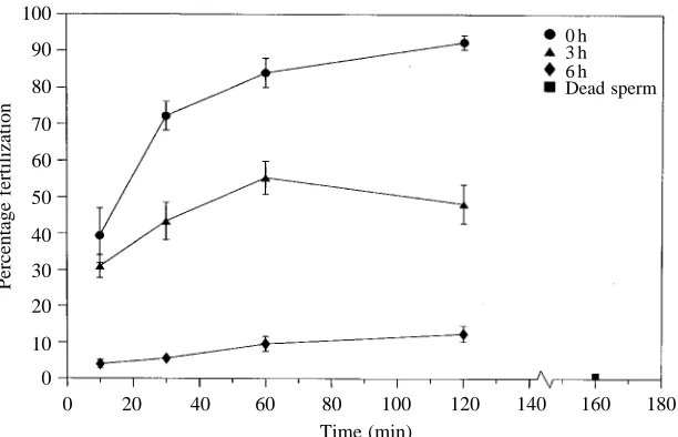
The percentage of fertilized eggs decreased as a function of sperm ageing following release from the gonads. Fig. 6 provides data that are representative of the trends seen in fertilization success across all experiments. After 2 h of incubation, 92 % of the eggs were fertilized by freshly diluted sperm, 46 % were fertilized by 3 h sperm, and 23 % were fertilized by sperm drawn from the experimental chamber 6 h after dilution. The numbers of eggs fertilized by sperm taken from the experimental chamber and sperm maintained on the laboratory bench never differed by more than 3 %. The percentage of eggs fertilized in the heat-killed sperm treatment did not exceed 1.5 % (e.g. Fig. 6).
Discussion
This study investigated the consequences of walls, paternity and age on sperm motility. We were particularly interested in viscous wall effects, since virtually all previous investigations employed sperm in small water volumes on microscope slides (e.g. Miller, 1975, 1977, 1985b; Ward et al. 1985; Ishijima et al. 1992; but see Crenshaw, 1996, for an exception). Adult sea urchins spawn their eggs and sperm into the ocean environment, and fertilization occurs naturally where walls such as those imposed by microscope slides are absent. Because measurements of sperm motility are critical to studies on fertilization kinetics and chemotaxis, it is important to know how walls influence swimming speeds. Several theoretical treatments suggest that drag forces significantly affect flagellar motion within approximately 10 sperm body lengths of a microscope slide surface (Gray and Hancock, 1955; Reynolds, 1965; Katz, 1974; Katz and Blake, 1975; Lighthill, 1975). According to physical principles, movement by a flagellum rotating in an oscillatory manner is described by both
transverse and normal resistance coefficients. A cell moves forward as long as the ratio of normal to transverse resistance is greater than unity. Significantly, near a wall, the coefficient of normal resistance is predicted to increase in magnitude faster than the coefficient of transverse resistance. The propulsive force should thus become larger and drive the cell forward at a faster rate. To test this theory, we video-recorded sea urchin sperm in a large chamber at two depths: <100µm (3 body lengths) and 5 mm (150 body lengths) below the lid. Our experimental results were consistent with predictions, since cells near the lid swam 1.5 times faster than those farther away. From these results, wall effects must be carefully considered in studies on sperm motility, and the analysis of sperm on a microscope slide may substantially exaggerate swimming speed.
Although our results support the theoretical considerations well, it cannot be assumed that sperm motility near an egg is strictly governed by physics alone. On the contrary, a cell might show a behavioral response as it approaches an egg due to contact with chemical attractants released by the jelly layer (Kopf et al. 1979; Hansbrough and Garbers, 1981; Suzuki et al. 1981; Garbers et al. 1982; Ward et al. 1985; but see Holland and Cross, 1983, for alternative observations). The relative importance of physical surfaces and chemical attractants in regulating sperm motility and encounter rates with eggs remains to be resolved. On the basis of the methods described here, a series of experiments can be designed to address this issue while eliminating the effects of microscope slide surfaces. Specifically, sperm motility near eggs with, or without, egg jelly coats can be video-recorded in large, temperature-controlled chambers and the results then compared. Further experiments can be performed using Couette flow devices to determine the impacts of hydrodynamic shear forces on sperm–egg interactions (Mead and Denny, 1995).
Previous investigators have often observed that sperm swim head-on in circular paths at microscope slide surfaces. Several 100
90
80
70
60
50
40
30
20
10
0
Percentage fertilization
0 20 40 60 80 100 120 140 160 180 Time (min)
[image:6.609.255.561.71.268.2]0 h 3 h 6 h Dead sperm
Fig. 6. Percentages of eggs with elevated fertilization membranes as a function of contact time. Assays were started at the time of dilution (d), 3 h after dilution (m) and 6 h after dilution (r). Each point is the mean percentage (±S.E.M.) of fertilized eggs for five
authors attributed this behavior to thigmotaxis, but all reports noted movements by sperm of a given species favoring a particular direction (clockwise or counterclockwise) (Miller, 1977, 1985b; Ward et al. 1985; Punnett et al. 1992; Hamel and Mercier, 1995). Gray (1955) proposed an alternative hypothesis to explain the circular paths on microscope slides. He argued that, when a helical path is compressed lengthwise from three dimensions to two dimensions, a circular path is obtained. A sperm traveling in this type of condensed path appears to move clockwise or counterclockwise simply depending upon the handedness of the helix and the perspective of the observer. Prior investigators failed to present evidence disproving Gray’s hypothesis, and his explanation appears to fit our observations. In the absence of any apparent directional cue, Arbacia punctulata sperm usually swam in a left-handed circle at the chamber lid surface (82.5 % of all cells observed; P<0.0001).
Levitan (1993) found a significant association between sperm swimming speed and paternity for each of three sea urchin species (Strongylocentrotus spp.). Our results demonstrate a similar trend for A. punctulata. Although the current study does not specifically address the cause of this relationship, future investigations can be used to distinguish between genetic and environmental factors in controlling gamete motility.
Physical theory predicts that sperm should swim faster near walls. We constructed a large chamber for water on a microscope stage in which the effects of walls could be determined. This apparatus was built to regulate water temperature and to reduce convective mixing, allowing us to measure both the speed and direction of swimming sperm. Whereas walls and paternity were each found to affect motility significantly, ageing of sperm after collection did not have a measurable impact on locomotion. Sperm as they aged were observed to become less effective in fertilizing eggs. Degradation of the acrosomal reaction, chemotaxis or some factor other than sperm swimming characteristics must be responsible for causing this limitation to fertilization success.
This work was supported by grants from the National Science Foundation (OCE 94-16749), the Slocum-Lunz Foundation and the Marine Science Program at the University of South Carolina. We thank Christen M. Herren for technical assistance, Holmes Finch for guidance in statistical analysis and N. Dean Pentcheff for helping with computer programs. Comments by Sarah A. Woodin, Richard Showman and two anonymous reviewers greatly improved the manuscript.
References
BRENNAN, C. ANDWINET, H. (1977). Fluid mechanics of propulsion
by cilia and flagella. A. Rev. Fluid Mech. 9, 339–498.
BROWN, G. G. AND KNOUSE, J. R. (1973). Effects of sperm
concentration, sperm aging and other variables on fertilization in the horseshoe crab, Limulus polyphemus L. Biol. Bull. mar. biol.
Lab., Woods Hole 144, 462–470.
CRENSHAW, H. C. (1996). A new look at locomotion in microorganisms: Rotating and translating. Am. Zool. 36, 608–618. DINNEL, P. A., LINK, J. M. AND STOBER, Q. J. (1987). Improved methodology for a sea urchin sperm cell bioassay for marine waters. Arch. env. Contam. Toxicol. 16, 23–32.
GARBERS, D. L., WATKINS, H. D., HANSBROUGH, J. R., SMITH, A. AND
MISONO, K. S. (1982). The amino acid sequence and chemical synthesis of speract and of speract analogues. J. biol. Chem. 257, 2734–2737.
GRAY, J. (1955). The movement of sea-urchin spermatozoa. J. exp. Biol. 32, 775–801.
GRAY, J. ANDHANCOCK, G. J. (1955). The propulsion of sea-urchin
spermatozoa. J. exp. Biol. 32, 802–814.
HAMEL, J.-F. AND MERCIER, A. (1995). Prespawning behavior,
spawning and development of the brooding starfish Leptasterias
polaris. Biol. Bull. mar. biol. Lab., Woods Hole 188, 32–45.
HANSBROUGH, J. R. ANDGARBERS, D. L. (1981). Speract. Purification and characterization of a peptide associated with eggs that activates spermatozoa. J. biol. Chem. 256, 1447–1452.
HOLLAND, L. Z. ANDCROSS, N. L. (1983). The pH within the jelly coat
of sea urchin eggs. Dev. Biol. 99, 258–260.
ISHIJIMA, S., HAMAGUCHI, M. S., NARUSE, M., ISHIJIMA, S. A. AND
HAMAGUCHI, Y. (1992). Rotational movement of a spermatozoon around its long axis. J. exp. Biol. 163, 15–31.
KATZ, D. F. (1974). On the propulsion of micro-organisms near solid boundaries. J. Fluid Mech. 64, 33–49.
KATZ, D. F. AND BLAKE, J. R. (1975). Flagellar motions near walls. In Swimming and Flying in Nature, vol. 1 (ed. T. Y.-T. Wu, C. J. Brokaw and C. Brennan), pp. 173–184. New York: Plenum Press.
KATZ, D. F., BLAKE, J. R. ANDPAVERI-FONTANA, S. L. (1975). On the movement of slender bodies near plane boundaries at low Reynolds number. J. Fluid Mech. 72, 529–540.
KOPF, G. S., TUBB, D. J. ANDGARBERS, D. L. (1979). Activation of
sperm respiration by a low molecular weight egg factor and by 8-bromoguanosine 3′5′-monophosphate. J. biol. Chem. 254, 8554–8560.
LEVITAN, D. R. (1993). The importance of sperm limitation to the
evolution of egg size in marine invertebrates. Am. Nat. 141, 517–36.
LEVITAN, D. R., SEWELL, M. A. ANDCHIA, F.-S. (1991). Kinetics of fertilization in the sea urchin Strongylocentrotus franciscanus: Interaction of gamete dilution, age and contact time. Biol. Bull.
mar. biol. Lab., Woods Hole 181, 371–378.
LIGHTHILL, J. (1975). Mathematical Biofluiddynamics. Philadelphia: Society for Industrial and Applied Mathematics. 281pp.
LILLIE, F. R. (1915). The fertilizing power of sperm dilutions of
Arbacia. Proc. natn. Acad. Sci. U.S.A. 1, 156–160.
LUTZ, D. G. ANDINOUÉ, S. (1986). Techniques for observing living gametes and embryos. In Echinoderm Gametes and Embryos,
Methods in Cell Biology, vol. 27 (ed. T. E. Schroeder), pp. 89–110.
New York: Academic Press.
MEAD, K. S. ANDDENNY, M. W. (1995). The effects of hydrodynamic shear stress on fertilization and early development of the purple sea urchin Strongylocentrotus purpuratus. Biol. Bull. mar. biol. Lab.,
Woods Hole 188, 46–56.
MILLER, R. L. (1975). Chemotaxis of the spermatozoa of Ciona
intestinalis. Nature 254, 244–245.
MILLER, R. L. (1977). Chemotactic behavior of the sperm of chitons (Mollusca: Polyplacophora). J. exp. Biol. 202, 203–212.
release of a sperm chemo-attractant by the egg of the leptomedusan,
Orthopyxis caliculata. J. exp. Zool. 205, 385–392.
MILLER, R. L. (1979). Sperm chemotaxis in the Hydromedusae. I. Species-specificity and sperm behavior. Mar. Biol. 53, 99–114. MILLER, R. L. (1985a). Sperm chemo-orientation in the metazoa. In
Biology of Fertilization, vol. 2 (ed. C. B. Metz and A. Monroy),
pp. 275–337. New York: Academic Press.
MILLER, R. L. (1985b). Demonstration of sperm chemotaxis in
Echinodermata: Asteroidea, Holothuroidea, Ophiuroidea. J. exp.
Zool. 234, 383–414.
PENNINGTON, J. T. (1985). The ecology of fertilization of echinoid eggs: The consequences of sperm dilution, adult aggregation and synchronous spawning. Biol. Bull. mar. biol. Lab., Woods Hole 169, 417–430.
PUNNETT, T., MILLER, R. L. AND YOO, B.-H. (1992). Partial
purification and some chemical properties of the sperm chemoattractant from the forcipulate starfish Pycnopodia
helianthodes (Drandt, 1835). J. exp. Zool. 262, 87–96.
REYNOLDS, A. J. (1965). The swimming of minute organisms. J. Fluid Mech. 23, 241–260.
SUZUKI, N., NOMURA, K., OHTAKE, H. AND ISAKA, S. (1981).
Purification and primary structure of sperm-activating peptides from the jelly coat of sea urchin eggs. Biochem. biophys. Res.
Commun. 99, 1238–1244.
TYLER, A. ANDTYLER, B. S. (1966). The gametes: Some procedures
and properties. In Physiology of Echinodermata (ed. A. Boolootian), pp. 639–682. New York: Interscience Publishers. WARD, G. E., BROKAW, C. J., GARBERS, D. L. ANDVACQUIER, V. D.