β-Adrenergic regulation of the fish heart is a central mechanism in maintaining normal cardiac function and in adapting cardiac muscle to changing physiological and environmental conditions. Adrenaline and noradrenaline are involved in supporting cardiac contractility in resting fish (Graham and Farrell, 1989), in enhancing cardiac performance during exercise and in protecting cardiac function during anoxia and acidosis (Gesser et al. 1982; Farrell, 1985). Adrenergic mechanisms are also intimately involved in adapting cardiac function to changing ambient temperature (Graham and Farrell, 1989; Keen et al. 1993). There are, however, prominent species-specific differences in the extent to which adrenergic stimulation is able to regulate the contractility of the fish heart. For example, in salmonid hearts, the inotropic effects of β-adrenergic stimulation are relatively
strong, while in the crucian carp heart adrenergic agonists produce only a modest increase in the force of contraction (Vornanen, 1989; Keen et al. 1993; Ask et al. 1997). It is not clear how these species-specific differences in β-adrenergic regulation of the heart are associated with the activity pattern of the fish and with the environmental factors to which the fish will be subjected. Furthermore, it is largely unknown to what extent the interspecies differences in β-adrenergic regulation are related to the differences in number and/or affinity of adrenergic receptors or to the differences in the signal transduction cascade from receptor occupation to intracellular second messengers. Species-specific differences in the cellular effectors, e.g. the excitation–contraction coupling mechanism, could also be involved.
The L-type Ca2+current (ICa) is an essential component of
JEB1256
A patch-clamp analysis of L-type Ca2+ current in
ventricular myocytes of cold- and warm-acclimated rainbow trout (Oncorhynchus mykiss) and crucian carp (Carassius carassius) hearts was performed. Trout were acclimated at 4 and 17 °C and carp at 4 and 24 °C for a minimum of 4 weeks. Ventricular myocytes were isolated by enzymatic dissociation using collagenase and trypsin. Marked species-specific differences were noted in Ca2+
current density and its β-adrenergic regulation. The density of basal Ca2+ current in crucian carp
(6.9–7.4 pA pF−1) was almost double that of trout
(4.2–4.5 pA pF−1) ventricular myocytes. Maximal
β-adrenergic stimulation increased Ca2+ current by
approximately 2.3-fold in trout but by only 1.4-fold in crucian carp, so that Ca2+ current densities in the
presence of 10µmol l−1isoprenaline were almost equal in
trout (8.6–10.5 pA pF−1) and carp (9.6–10.4 pA pF−1)
cardiac cells. Direct activation of adenylate cyclase by forskolin (10µmol l−1) was also associated with similar
interspecies differences in the stimulation of Ca2+
current.
Thermal acclimation did not change either the density or the kinetics of L-type Ca2+ current in crucian carp
ventricular myocytes. In trout cardiac cells, thermal acclimation had no effects on the density of Ca2+current,
but the rate of current inactivation was accelerated after acclimation to cold temperature. As a consequence of faster current decay, the contribution of sarcolemmal Ca2+
current to total cellular [Ca2+] was smaller in
cold-acclimated than in warm-cold-acclimated trout. The responses of Ca2+ current to maximal β-adrenergic stimulation by
isoprenaline or to direct activation of adenylate cyclase by forskolin were not changed by thermal acclimation in either species. It is concluded (1) that the density of sarcolemmal Ca2+ current is not increased after
acclimation to cold, (2) that sarcolemmal Ca2+ influx
through L-type Ca2+ channels can make a significant
contribution to contractile [Ca2+] in both teleost species
studied and (3) that β-adrenergic stimulation of Ca2+
current is more important in modulating cardiac contractility in trout than in carp.
Key words: fish heart, ventricular myocyte, L-type Ca2+current,
β-adrenergic stimulation, isoprenaline, forskolin, sarcolemmal Ca2+
influx, temperature acclimation, rainbow trout, crucian carp,
Carassius carassius, Oncorhynchus mykiss.
Summary
Introduction
L-TYPE Ca
2+CURRENT IN FISH CARDIAC MYOCYTES: EFFECTS OF THERMAL
ACCLIMATION AND
β
-ADRENERGIC STIMULATION
MATTI VORNANEN*
Department of Biology, University of Joensuu, PO Box 111, FIN-80101 Joensuu, Finland
*e-mail: [email protected]
excitation–contraction coupling in vertebrate cardiac myocytes (Reuter, 1979). In myocytes of mammalian and avian hearts, ICa serves mainly as a trigger for Ca2+-induced Ca2+ release from the sarcoplasmic reticulum (SR) (Fabiato, 1983; Callewaert, 1992), while in ectothermic vertebrates it is thought to provide a significant trans-sarcolemmal entry of Ca2+ during the relatively long plateau phase of the cardiac action potential (Näbauer et al. 1989; Vornanen, 1997). One of the hallmarks of vertebrate L-type Ca2+ channels is their susceptibility to modulation by various physiological factors. Therefore, Ca2+ entry into the myocyte and the force of myocardial contraction are controlled through the function of ICa by nervous stimulation, by circulating hormones, by cellular pH and by ATP supply (Sperelakis, 1995). Perhaps the most significant physiological regulation of ICaoccurs through
β-adrenergic receptors by local transmitter release from autonomic nervous endings or by circulating catecholamines.
Thus, it is evident that the β-adrenergic pathway is a central mechanism in the regulation of cardiac function, while the L-type Ca2+ current of cardiac myocytes is the target of modulation by multiple extrinsic and intrinsic factors, most notably by catecholamines. Because of their vital role in the regulation of cardiac contractility and in the adaptation of cardiac function to changing environmental conditions, ICaand its β-adrenergic control warrant a detailed study in different teleost species. As a first step to this end, we have compared ICain two teleost species that use different strategies to adapt to low environmental temperature and which possibly also differ in regard to their excitation–contraction coupling mechanism. L-type Ca2+ current and its β-adrenergic regulation were measured in thermally acclimated trout (a cold-active species) and crucian carp (a species entering cold torpor) to investigate the importance of ICa in normal excitation–contraction coupling and in the adaptation of cardiac contractility to low ambient temperature.
Materials and methods Animals
Crucian carp (Carassius carassius L.) were caught with weirs from ponds near the University of Joensuu between July and September. Rainbow trout [Oncorhynchus mykiss (Walbaum)] were obtained from a local fish farm throughout the year. In the laboratory, the fish were maintained in 500 l aquaria in aerated tap water. The photoperiod was a 12 h:12 h light:dark cycle. The fish were randomly divided into two groups for thermal acclimation at two ambient temperatures. The lower acclimation temperature was 4 °C for both species. The higher acclimation temperature was 17 °C for trout and 24 °C for carp. A minimum of 4 weeks acclimation was allowed before starting the experiments. During this period, the fish were fed daily on commercially available trout pellets (Ewos, Sweden). The body masses of warm-acclimated and cold-acclimated trout were 88.7±20.8 g and 106.6±16.5 g, respectively. Crucian carp were somewhat smaller, with mean
body masses of 33.3±3.8 and 24.8±1.6 g (means ±S.E.M.) for warm-acclimated and cold-acclimated fish, respectively.
Cell preparation
Fish were stunned by a blow on the head, the spine was cut and the heart was carefully removed. Single ventricular cells were obtained by enzymatic dissociation. A cannula was inserted through the bulbus arteriosus into the ventricle, and the heart was perfused from a height of 50 cm, first with nominally Ca2+-free low-Na+ solution to disrupt Ca2+-dependent bonds between cells, and then with proteolytic enzymes to dissolve intercellular connective tissue. The composition of the perfusion solution was as follows (in mmol l−1): NaCl, 100; KCl, 10; KH2PO4, 1.2; MgSO4, 4; taurine, 50; glucose, 20; and Hepes, 10 (adjusted to pH 6.9 using KOH). Ca2+-free perfusion with this solution lasted for 8 min in both species. For enzymatic dissociation, collagenase (Type IA) and trypsin (Type III) together with fatty-acid-free bovine serum albumin (BSA) (all from Sigma) were added to the low-Na+ solution. For trout hearts, the concentrations of collagenase, trypsin and BSA were 0.75, 0.5 and 0.75 mg ml−1, respectively, and the hearts were perfused with this solution for 20 min. When perfusing carp hearts, the concentrations of collagenase and trypsin were double those used for trout (1.5 and 1.0 mg ml−1, respectively) and the enzyme perfusion was extended to 40 min for warm-acclimated carp and to 60 min for cold-warm-acclimated carp. After enzymatic treatment, the ventricle was cut into small pieces and triturated using a Pasteur-pipette in filtered (0.2µm) K+-based physiological solution (CsCl replaced with KCl, see below) containing 1.8 mmol l−1CaCl
2. The isolated cells were stored in this solution at room temperature.
Solutions and chemicals
The composition of the physiological solution used for recording Ca2+ current was as follows (in mmol l−1): NaCl, 130; CsCl, 5.4; MgSO4, 1.5; NaH2PO4, 0.4; CaCl2, 1.8; glucose, 10; and Hepes, 10 (adjusted to pH 7.6 with CsOH). For recording ICa, the pipette solution contained (in mmol l−1): CsCl, 130; MgATP, 5; tetraethylammonium chloride, 15; MgCl2, 1; oxaloacetate, 5; succinate, 5; EGTA, 5; Na2GTP, 0.03; and Hepes, 10 (adjusted to pH 7.2 with CsOH). The drugs used in these experiments were isoprenaline (ISO, a β-receptor agonist), forskolin (FOR, an activator of adenylate cyclase), tetrodotoxin (TTX, a selective blocker of fast Na+ channels), nifedipine (NIF, a selective blocker of L-type Ca2+channels) and caffeine. ISO was dissolved in 1 mmol l−1 ascorbic acid solution to make a 10 mmol l−1 stock solution, which was stored in small samples at −20 °C. FOR was dissolved in dimethyl sulphoxide as a 10 mmol l−1stock solution. TTX was dissolved in distilled water at 1 mmol l−1 and was stored at 5 °C. NIF was dissolved in ethanol and was stored in the dark at 5 °C. Caffeine was added as a solid directly into physiological solutions.
Experimental procedure
to the recording chamber (volume 0.5 ml) and allowed to settle to the bottom. Solutions were superfused through the bath at a rate of 2 ml min−1. Voltage-clamp experiments were performed at 22±1 °C in the whole-cell configuration using an Axopatch 1D amplifier with a CV-4 1/100 headstage (Axon Instruments, Foster City, CA, USA). Pipettes were pulled from borosilicate glass (Vitrex microhaematocrit tubing, Modulohm A/S, Denmark) using a vertical patch-pipette puller (Narishige PP-83) and had a resistance of 1.5–2.5 MΩwhen filled with the pipette solution. Junction potentials were zeroed prior to the formation of the seal. No correction was made for the small potential difference (−2 mV) between the bath solution and the pipette solution. The immersion depth of the pipette was kept as shallow as possible, and the pipette capacitance (4–5 pF) was compensated after the formation of a gigaohm seal. The membrane patch under the pipette tip was ruptured by delivering a short voltage pulse, and capacitative transients were eliminated by iteratively adjusting the series resistance and cell capacitance compensation circuits. The mean series resistance before compensation was 4.2±0.4 MΩ. Approximately 70 % of the series resistance was electronically compensated, so the final effective series resistance was less than 2 MΩ. Since the amplitude of ICawas generally less than 1 nA, the voltage drop due to series resistance was less than 2 mV. Currents were leakage-corrected using the P/N procedure of the software. The Ca2+currents elicited were low-pass-filtered at a frequency of 2 kHz and analysed off-line on a computer using pCLAMP 6.0 software (Axon Instruments). Membrane capacitance was measured using the calibrated capacity compensation circuit of the Axopatch amplifier.
Membrane potential was routinely depolarized for 500 ms every 10 s from a holding potential of −50 mV to 0 mV for the study of L-type Ca2+current. Inactivation and activation curves for ICawere obtained from double-pulse protocols: every 10 s, a 1000 ms prepulse to different potentials (−70 to +50 mV) was followed by a 500 ms test pulse to 0 mV. Prepulse and test pulses were separated by a 5 ms return to the holding potential. The voltage-dependence of steady-state inactivation f∞(V) was calculated by dividing the amplitude of the test current by the maximal current elicited. Activation voltage-dependence, d∞(V) was determined as normalized Ca2+ conductance
d∞(V)=gCa/gmax, where gmax is the maximum value of ICa conductance. The voltage-dependence of the peak conductance of Ca2+channels was calculated from the equation:
gCa = ICa/(V−Vrev) , (1)
where gCais the membrane Ca2+conductance, ICais the peak Ca2+current at a given potential (V) and V
rev is the apparent reversal potential obtained by extrapolating the ascending portion of the current–voltage (I–V) relationship to zero current. Steady-state kinetic parameters were obtained by fitting the data to Boltzmann equations:
d∞(V) = 1/{1 + exp[(V0.5−V)/k]} (2)
f∞(V) = 1 −1/{1 + exp[(V0.5−V)/k]} , (3)
where V0.5is the half-activation or half-inactivation potential and k is the slope factor. Current–voltage relationships were
obtained from prepulse depolarizations or from separate runs where ICa was triggered from −50 mV to test potentials between −40 and +60 mV for 500 ms.
The contribution of ICato total cellular [Ca2+] was calculated from the transferred charges and cell volume, as described previously (Vornanen, 1996, 1997). Charge transfer through L-type Ca2+ channels was determined by integrating the inactivating portion of the Ca2+ current for 500 ms voltage pulses from −50 mV to 0 mV. Cell volume was calculated from the measured cell capacitance and the experimentally determined surface-to-volume ratio of the cells (1.15). The myocytes were considered to be flat elliptical cylinders with an axis ratio of 1:2 for the elliptical cross section (Vornanen, 1997). The change in total cellular [Ca2+] due to Ca2+influx through L-type Ca2+channels was expressed as a function of non-mitochondrial myoplasmic volume or of myofibrillar volume. The volume densities of myofibrillar and mitochondrial compartments were determined from electron micrographs of single cells using a point-counting method.
Electron microscopy
A small sample of the myocyte suspension was pipetted into Eppendorf tubes. The cells were fixed by adding 4 volumes of 2 % glutaraldehyde in 0.1 mol l−1sodium cacodylate buffer (pH 7.5) to 1 volume of myocyte suspension. Myocytes were allowed to sediment on the bottom of the vial, the solution was drawn out using a Pasteur pipette and was replaced with an equal volume of fresh glutaraldehyde fixative. Postfixation was carried out in 2 % OsO4and 0.8 % K4Fe(CN)6buffered with 0.1 mol l−1sodium cacodylate (pH 7.5). Myocyte pellets were embedded in Epon, and thin sections were cut using an LKB ultramicrotome and stained with saturated uranyl acetate and 0.4 % lead citrate. Specimens were examined and photographed using a Zeiss 90 electron microscope at an operating voltage of 80 kV. Volume fractions of cellular organelles were obtained from paper prints of longitudinally sectioned myocytes using the point-counting method.
Statistics
All data are presented as means ±1 S.E.M., and statistical comparisons were made using a two-tailed Student’s t-test. Graphs and curve-fitting were performed using SigmaPlot software (Jandel Scientific).
Results Ventricular cells
to proteolytic enzymes than the hearts of warm-acclimated carp. The increased resistance of cold-acclimated carp hearts to enzymatic digestion is at least partly explained by their high content of collagenous proteins (Pelouch and Vornanen, 1996). The ventricular myocytes of the carp heart are typically 6µm in width and approximately 100µm in length, while those of the trout heart are somewhat bigger in both dimensions (8µm in width and 170µm in length). The membrane capacitance of
ventricular myocytes was approximately 20 pF in carp and 46–61 pF in trout (Table 1). The bigger size of trout myocytes is only partially explained by larger body size of the fish, since in similar-sized fish the whole-cell capacitance of trout myocytes was almost twice the average capacitance of carp myocytes. Electron microscopy revealed many similarities but also some prominent structural differences between trout and carp ventricular myocytes (Fig. 1, shown only for trout). In both N
M
[image:4.609.51.545.70.328.2]F
Fig. 1. Electron micrographs of enzymatically isolated ventricular myocytes of rainbow trout heart. (A) A longitudinal section through a small part of a ventricular myocyte. (B) A cross section of a ventricular myocyte. M, mitochondria; MF, myofibrils; N, nucleus; F, fat. Scale bars, 2µm.
1 nA
100 ms Control
Nifedipine, 2.5 µmol l−1
100 ms control
TTX, 1 µmol l−1
TTX-sensitive current Nifedipine-sensitive current
1 nA A
B
C
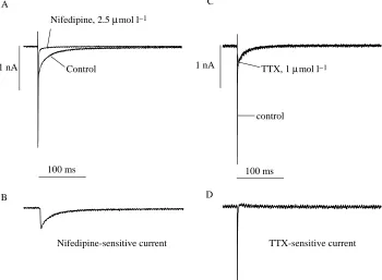
D Fig. 2. Two types of inward current are
present in trout ventricular myocytes. (A) Inward currents in the absence and presence of 2.5µmol l−1 nifedipine; the
nifedipine-sensitive difference current is indicated in B. (C) Inward currents in the absence and presence of 1µmol l−1
[image:4.609.207.557.457.714.2]species, the nucleus and mitochondria are in the middle of the cell, whereas myofibrils are located cortically just beneath the sarcolemma. The metabolic machinery as a whole (mitochondria, fat droplets and glycogen) constitutes approximately half of the cell volume in both fish, but has a quite different composition in the two species. Crucian carp myocytes are characterized by a huge glycogen content, a moderate number of mitochondria (22 % of cell volume) and by the total absence of fat, while trout ventricular myocytes have little glycogen, some fat droplets and a very high density of mitochondria (45 % of cell volume). Accordingly, the non-mitochondrial volume of the cell is larger in carp (78 %) than in trout (55 %). The myofibrillar volume density was approximately 40 % in both species. The average cell capacitance of cold-acclimated trout hearts was some 20 % larger than that of warm-acclimated trout hearts, which could be due to hypertrophic growth of the heart at low ambient temperature (Graham and Farrell, 1989).
Two inward currents in teleost ventricular myocytes In myocardial cells of both species, depolarizing pulses from a holding potential of −80 mV elicited two inward currents (shown only for trout); a fast, large-amplitude current that was completely blocked by 1µmol l−1 TTX, and a more slowly activating and inactivating current that was completely suppressed by 2.5µmol l−1 NIF (Fig. 2). These findings indicate the presence of TTX-sensitive Na+currents and NIF-sensitive L-type Ca2+ currents in teleost heart. The current–voltage relationship for depolarization from −80 mV to various voltage levels in the presence of 1µmol l−1TTX was similar to that elicited from −50 mV (N=8, results not shown), suggesting that T-type Ca2+ channels are not present in the
ventricular myocytes of trout heart. Previously we have shown that T-type Ca2+ currents occur very infrequently in crucian carp ventricular myocytes and, when present, are small in amplitude (Vornanen, 1997).
Comparison of ICain trout and carp
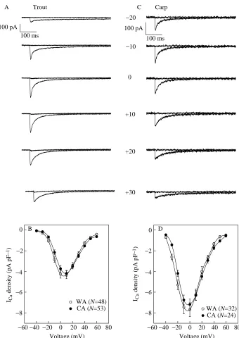
The voltage-clamp experiments were conducted under identical experimental conditions for both fish, allowing direct comparison between species. The most striking difference between trout and crucian carp ventricular myocytes was in the density of the basal ICa. In crucian carp myocytes, the L-type Ca2+current was 6.9–7.4 pA pF−1, and in trout cells the current was 4.2–4.5 pA pF−1(Fig. 3; Table 1). It is notable that thermal acclimation did not change Ca2+current density in either trout or carp ventricular myocytes.
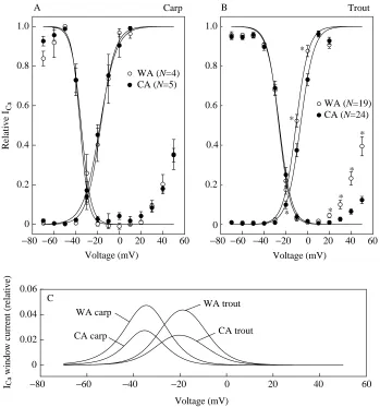
There was also a clear species-specific difference in voltage-dependence of ICa: in crucian carp ventricular myocytes, maximum current occurs at 0 mV, but in trout cells it occurs at approximately +10 mV (Fig. 3). This difference in voltage-dependence of ICais also evident in the steady-state activation and inactivation curves (Fig. 4). Voltages for half-maximal activation and inactivation of ICa are far more negative in carp than in trout myocytes (P<0.05) (Table 2). Because of the overlap of the inactivation and activation curves, a small window current is present in both species. The maximum window current occurs at approximately −30 mV in crucian carp ventricular myocytes and at approximately −20 mV in trout ventricular myocytes (Fig. 4). Thermal acclimation changed neither steady-state inactivation nor activation parameters in carp ventricular myocytes. In contrast, the activation curve of warm-Table 1. Characterization of trans-sarcolemmal Ca2+influx in ventricular myocytes of rainbow trout and crucian carp hearts
under control conditions and in the presence of β-adrenergic stimulation
Cell Charge Cell surface ∆[Ca2+]tot/ ∆[Ca2+]tot/
capacitance ICa density density area Cell volume MyoF NonM
(pF) (pA pF−1) (pC pF−1) (µm2) (µm3) (µmol l−1) (µmol l−1) N
Cold-acclimated trout
Control 61.4±2.6* 4.2±0.3 0.19±0.01* 3855±161* 3352±140* 44.2±2.32* 32.1±1.6* 53
ISO, 1µmol l−1 66.2±5.6 11.1±2.1 0.46±0.11 4159±353 3617±307 108.2±25.7 78.7±18.7 14
ISO, 10µmol l−1 58.9±4.7 10.5±0.8 0.44±0.03 3699±254 3217±255 104.0±6.7 75.6±4.8 16
Warm-acclimated trout
Control 46.0±2.1 4.5±0.3 0.27±0.02 2890±129 2513±112 63.0±3.9 45.8±2.6 50
ISO, 1µmol l−1 54.1±4.2 9.4±1.4 0.53±0.07 3396±263 2953±228 125.0±17.1 90.9±12.4 13
ISO, 10µmol l−1 47.7±4.0 8.6±0.6 0.48±0.04 2995±217 2603±220 114.0±6.7 83.3±6.3 12
Cold acclimated carp
Control 19.4±1.2 6.9±0.4 0.32±0.02 1215±79 1056±68 76.1±4.0 38.6±2.1 24
ISO, 10µmol l−1 20.9±3.2 9.6±1.0 0.39±0.03 1312±203 1141±172 93.0±8.2 47.2±4.2 7
Warm acclimated carp
Control 20.6±0.8 7.4±0.6 0.36±0.03 1296±53 1127±46 84.6±6.1 42.9±3.1 33
ISO, 10µmol l−1 22.7±2.1 10.4±1.4 0.44±0.06 1427±133 1241±116 105.0±13.1 53.3±6.7 6
∆[Ca2+]
tot/MyoF, change in total cellular [Ca2+] expressed as a function of myofilament space; ∆[Ca2+]tot/NonM, change in total cellular
[Ca2+] expressed as a function of non-mitochondrial cell volume.
[image:5.609.50.565.101.298.2]acclimated trout myocytes was shifted slightly, but significantly, to more negative voltages compared with that of cold-acclimated trout (Table 2). This accords with the leftward shift of the I–V curve in the cells of warm-acclimated trout. Furthermore, there was clear difference in steady-state inactivation curves between warm-acclimated and cold-acclimated trout cells at positive membrane potentials. In warm-acclimated trout cells, the inactivation of ICa was significantly attenuated between 0 and +50 mV, while in cold-acclimated trout the attenuation of ICa inactivation in this voltage range was significantly smaller.
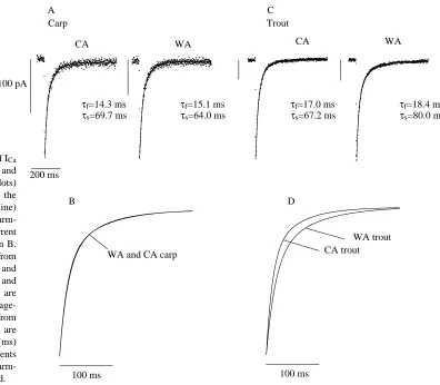
In agreement with a previous study on crucian carp (Vornanen, 1997), the inactivation rate of ICawas best fitted by the sum of two exponential curves (Fig. 5). The time
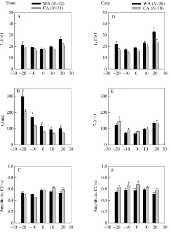
constant of the slow component of ICadecay exhibited a U-shaped voltage-dependence (Fig. 6B,E): the time constant was smallest at 0 mV in carp and at +10 mV in trout and increased at both more positive and more negative membrane potentials. Owing to the species-specific difference in current–voltage relationship of ICa, the right rising ‘arm’ of the Uis less clear in trout than in carp cells. The minimum for the fast time constant occurred at 0 mV in trout and at −10 mV and carp myocytes and clearly increased at more positive voltages in both species (Fig. 6A,D). The relative amplitude of the fast component was 50–60 % in trout and 55–65 % in carp (Fig. 6C,F). Thermal acclimation had no effect on the rate of ICa decay in carp myocytes, but the relative amplitude of the fast current component was slightly Voltage (mV)
−60 −40 −20 0 20 40 60 80 ICa
density (pA pF
−
1)
−8
−6
−4
−2 0
Voltage (mV)
−60 −40 −20 0 20 40 60 80
−8
−6
−4
−2 0
A C
B D
Trout Carp
−20
−10
0
+10
+20
+30
WA (N=48)
CA (N=53) WA (N=32)
CA (N=24) 100 ms
100 pA 100 ms
100 pA
ICa
density (pA pF
−
[image:6.609.216.556.71.551.2]1) Fig. 3. Current–voltage relationships of the
nifedipine-sensitive Ca2+current in trout (A,B)
and crucian carp (C,D) ventricular myocytes.
(A,B) Original current tracings of ICa at six
different membrane potentials before and after
treatment with nifedipine (5µmol l−1).
(B,D) Mean current–voltage curves for trout and carp cells, respectively. The values are
means ±S.E.M. of 24–53 myocytes as indicated.
Ca2+currents (ICa) were elicited from a holding
larger in warm-acclimated than in cold-acclimated crucian carp (Fig. 6D–F). In trout cardiac cells, the fast time constant of ICa was unaltered by thermal acclimation, but the time constant of the slow component was significantly longer in warm-acclimated than in cold-acclimated trout (Fig. 6B). When BaCl2 was used as a charge carrier, the inactivation rate of ICa was significantly reduced and the differences in inactivation rate between warm-acclimated and
cold-acclimated trout disappeared (Fig. 7). Caffeine (5 mmol l−1) did not change the inactivation rate of ICa(Fig. 8), suggesting that Ca2+ release from the SR has no major effect on the decay of ICain fish heart cells and that the difference in Ca2+ -dependent inactivation of ICabetween warm-acclimated and cold-acclimated trout is not explained by temperature-induced changes in the function of the SR (Aho and Vornanen, 1998).
Voltage (mV)
−80 −60 −40 −20 0 20 40 60
Relative I
Ca
0 0.2 0.4 0.6 0.8 1.0
−80 −60 −40 −20 0 20 40 60 0
0.2 0.4 0.6 0.8 1.0
Voltage (mV)
−80 −60 −40 −20 0 20 40 60 ICa
window current (relative)
0 0.02 0.04 0.06
WA trout
CA trout WA carp
CA carp
A B
C
Carp Trout
* *
* *
* *
*
WA (N=4) CA (N=5)
WA (N=19) CA (N=24)
[image:7.609.216.565.73.446.2]Voltage (mV) Fig. 4. Steady-state activation and
inactivation of ICain carp (A) and trout (B)
ventricular myocytes. (C) The ICawindow
current, is calculated as the product of steady-state activation and inactivation factors derived from Boltzmann equations for a voltage range from −70 to +50 mV. The results are means ±S.E.M. for 4–24 cells as indicated. An asterisk indicates a statistically significant difference (P<0.05) between acclimation groups. WA, warm-acclimated; CA, cold-acclimated.
Table 2. Steady-state activation and inactivation parameters of L-type Ca2+current in ventricular myocytes of rainbow trout and crucian carp heart
Trout Carp
Warm-acclimated Cold-acclimated Warm-acclimated Cold-acclimated
d∞Vh(mV) −10.55±0.87 −6.44±0.92* −17.14±2.15 −17.65±1.88 d k (mV) 5.74±0.39 5.81±0.15 5.82±0.05 7.25±0.54
f∞Vh(mV) −26.37±0.63 −26.01±0.99 −35.08±1.98 −36.20±1.06 f∞k (mV) 4.98±0.27 5.04±0.31 3.77±0.30 3.61±0.34
N 19 24 4 5
The results are means ±S.E.M.
An asterisk denotes a statistically significant difference (P<0.05) between acclimation groups.
[image:7.609.47.569.606.703.2]Effects of β-adrenergic stimulation and adenylyl cyclase activation
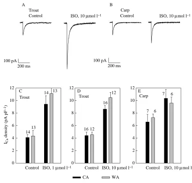
Since there was a marked difference in the density of the basal ICa, we wanted to determine whether the same difference persists in the presence of maximal β-adrenergic stimulation. We used either 1 or 10µmol l−1 ISO, which should cause maximal β -adrenergic stimulation in both species (Vornanen, 1989; Ask et al. 1997). There was large difference between trout and carp ventricular myocytes in the magnitude of the response to ISO. ISO-induced stimulation of basal ICawas approximately 2.3-fold in trout myocytes but only 1.4-fold in carp cardiac cells (Fig. 9; Table 1). Consequently, in the presence of maximal β -adrenergic stimulation, the density of ICawas approximately the same (8.6–10.5 pA pF−1) in the two species.
We also investigated whether the much stronger stimulatory effect of ISO in trout myocytes was due to species-specific difference in the coupling of β-receptors to adenylyl cyclase. To this end, we measured the stimulation of ICa by forskolin (10µmol l−1), an activator of adenylyl cyclase. Addition of FOR resulted in a stimulation of ICa to a similar extent to that obtained with ISO (Fig. 10). In crucian carp myocytes, FOR increased the peak ICaby approximately 1.4-fold, which closely matches the value for stimulation by ISO. In trout ventricular myocytes, the maximum ICa was increased by approximately 2.1-fold, which is only slightly less than the maximal
stimulation by ISO. However, the stimulation by FOR in trout cells was clearly voltage-dependent, being approximately sixfold at −20 mV and approximately twofold at +10 mV (Fig. 10D). This was associated with a shift of the activation curve, but not that of the availability curve, by 10 mV to the left (not shown). Thermal acclimation did not change the responsiveness of ICato either ISO or FOR in either species.
Contribution of ICato total cellular [Ca2+]
The increase in total cellular [Ca2+] was calculated from the integral of ICa and cell volume, the latter being obtained from the cell capacitance and the surface-to-volume ratio. The total cellular [Ca2+] was calculated for both the myofibrillar space, which is approximately 40 % of the total cell volume in both species, and for the non-mitochondrial cell volume, which is some 40 % larger in carp (78 %) than in trout (55 %) cells. If myofibrillar space is considered to be the Ca2+-accessible cell volume, then Ca2+ influx through Ca2+ channels increases intracellular [Ca2+] to a significantly greater extent in carp than in trout myocytes under basal conditions. However, in the presence of maximal β-adrenergic stimulation, this species-specific difference largely disappears. If non-mitochondrial cell volume is considered to be the space in which Ca2+is diluted, then ICa contributes almost equally to total cellular [Ca2+] in both species under basal conditions. However, under optimal β -Fig. 5. The rate of inactivation of ICa
in ventricular myocytes of trout and carp hearts. (A) Ca2+currents (dots)
and double-exponential fits of the current decay (continuous line) for cold-acclimated and warm-acclimated carp; the fits of current inactivation are superimposed in B. The voltage-clamp step was from
−50 to 0 mV. (C) Ca2+currents and
their fits for cold-acclimated and warm-acclimated trout; the fits are superimposed in D. The voltage-clamp step for trout cells was from
−50 to +10 mV. τf and τs are
inactivation time constants (ms) for the fast and slow components of ICa, respectively. WA,
warm-acclimated; CA, cold-acclimated.
A C Carp Trout
WA
CA CA WA
τf=18.4 ms
τs=80.0 ms
τf=14.3 ms
τs=69.7 ms
τf=15.1 ms
τs=64.0 ms
τf=17.0 ms
τs=67.2 ms
WA trout CA trout WA and CA carp
B D
100 ms 100 ms
[image:8.609.158.554.69.415.2]adrenergic stimulation, the increase in total cellular [Ca2+] is some 20 % greater in trout than in carp myocytes (Table 1).
In crucian carp heart, thermal acclimation had no effect on trans-sarcolemmal Ca2+influx through L-type Ca2+channels. In contrast to carp, acclimation to cold caused a significant reduction in sarcolemmal Ca2+influx in trout ventricular cells. This can be explained by the faster decay of ICa in cold-acclimated trout, which reduces the current integral and accordingly sarcolemmal Ca2+influx (Table 1).
Discussion
Species-specific differences in sarcolemmal Ca2+current
The present results show some remarkable species-specific
differences in the density of the basal L-type Ca2+current and its β-adrenergic regulation between crucian carp and rainbow trout ventricular myocytes. The density of ICawas almost twice as high in carp as in trout myocytes under control conditions but, because of stronger β-adrenergic stimulation in trout cells, this difference disappeared in the presence of a maximally effective concentration of ISO. The measured difference in the
β-receptor-mediated increase in sarcolemmal Ca2+current of single myocytes correlates well with the reported species-specific differences in inotropic responsiveness to adrenergic agonists in ventricular preparations from these two teleost fish. In trout ventricular strips, optimal adrenaline stimulation is able to increase the force of contraction 2.6- to threefold above the control level (Keen et al. 1993). In crucian carp ventricle,
−30 −20 −10 0 10 20 30 τ (mf
s)
0 10 20 30 40 50
−30 −20 −10 0 10 20 30
(m
s)
0 10 20 30 40 50
−30 −20 −10 0 10 20 30 0
100 200 300
−30 −20 −10 0 10 20 30 τ (ms
s)
0 100 200 300
−30 −20 −10 0 10 20 30
Amplitude,
f
/(
f+
s)
0 0.2 0.4 0.6 0.8 1.0
Voltage (mV)
−30 −20 −10 0 10 20 30
Amplitude,
f
/(
f+
s)
0 0.2 0.4 0.6 0.8 1.0 A
B
C
D
E
F *
*
*
* *
* *
Trout WA (N=32) Carp
CA (N=31)
WA (N=20) CA (N=18)
τf
Voltage (mV)
ss
τ (ms
[image:9.609.227.570.262.731.2]s)
Fig. 6. Mean values of the time constants (τ) of the fast (f) and slow (s) components of ICa
decay in trout (A,B) and carp (D,E) ventricular myocytes. (C,F) The relative amplitudes of the fast-inactivating component of ICa. The values
are means + S.E.M. for 18–32 cells, as
the maximal stimulation by ISO is only 1.4-fold above the the control value (Vornanen, 1989). Therefore, it is evident that the measured differences in the β-adrenergic regulation of ICa between trout and crucian carp myocytes are real and are not caused by the experimental conditions or by the enzymatic method used to isolate the cells. Forskolin, an activator of adenylyl cyclase, also had a markedly stronger stimulatory effect on ICa in trout than in carp ventricular myocytes. Therefore, the weak stimulation of ICain carp myocytes is not due to weaker coupling of β-adrenergic receptors to adenylate cyclase in carp compared with trout. The similarity of Ca2+ current density in the presence of ISO and FOR suggests that the number of Ca2+channels per unit membrane area does not differ in the two species. The simplest explanation for the species-specific differences is that the density of L-type Ca2+
channels is similar in the two species, but that the phosphorylation level of Ca2+channels under basal conditions is higher in carp cells than in trout myocytes. Such a difference could be due to low level of phosphatases or to weak Gi -protein-mediated tonic inhibition of ICa (Osaka and Joyner, 1992) in carp myocytes (see below). However, we cannot exclude the possibility that the Ca2+channels of trout hearts are slightly different from those in carp hearts.
Ligand binding and pharmacological experiments have indicated that the cardiac β-receptors of trout and common carp are almost exclusively of the β2 type (Temma et al. 1986; Gamperl et al. 1994). The β-adrenergic receptors act through the stimulatory G-protein (Gs) to activate adenylyl cyclase, thereby promoting phosphorylation of the channel by a cyclic-AMP-dependent protein kinase. Cyclic-cyclic-AMP-dependent
CA trout WA trout
A B
C
BaCl2
BaCl2
Voltage (mV)
−30 −20 −10 0 10 20 30 40
Tim
e constant
(m
s)
0 200 400 600 800 1000 1200
[image:10.609.245.551.74.417.2]WA (N=6) CA (N=7) 200 ms
Fig. 7. The effect of substitution of Ba2+for Ca2+as
the charge carrier on the rate of ICa inactivation in
trout ventricular myocytes. (A,B) Original current tracings for warm-acclimated (WA) and cold-acclimated (CA) trout, respectively, showing the slow rate of inactivation in the presence of 1.8 mmol l−1 BaCl2. The voltage step was from
−50 mV to 0 mV, which elicited maximal inward current in the presence of BaCl2. (C) Mean values (+ S.E.M.) of the time constant for the decay of the Ba2+
current at different membrane potentials. The decay of the Ba2+ current was fitted with an exponential
function.
A B WA trout CA trout
Control and caffeine Control and caffeine
200 ms 200 ms
Fig. 8. The lack of effect of caffeine (5 mmol l−1) on the
inactivation rate of ICain ventricular myocytes of
warm-acclimated (WA) (A) and cold-warm-acclimated (CA) (B) trout hearts. Current tracings in the absence and in the presence of caffeine were normalized to the same peak amplitude for easy comparison. Ca2+ currents were elicited by a
phosphorylation of Ca2+channels results in an increase in the mean open probability of the individual channels and thus in enhancement of the whole-cell ICa. The tight coupling between cellular cyclic AMP level and inotropic effectiveness, which is typical for β1 stimulation, is weaker under β2-adrenergic stimulation, suggesting that β1- and β2-adrenoceptors may use, to some extent at least, different signal transduction pathways in enhancing Ca2+current. In rat and dog cardiac myocytes,
β2-receptors are simultaneously coupled to Gs and to a pertussis-toxin-sensitive Gi-protein. It has been suggested that, under β2-adrenergic activation, the Gs-protein- and cyclic-AMP-dependent augmentation of ICais antagonized by the Gi -protein-dependent cascade, thus explaining the uncoupling between the accumulation of cyclic AMP and the stimulation of ICa (Xiao and Lakatta, 1993; Xiao et al. 1995; Brotto and Creazzo, 1996). The observed differences in the density of the basal ICa and in the response of ICa to ISO and FOR suggest that the phosphorylation of sarcolemmal Ca2+channels may be differently regulated in trout and carp ventricular cells. It is possible that, in crucian carp cardiac cells, the adenylate cyclase is under tonic activation. This could be due to a low proportion of Gi-proteins relative to Gs-proteins, which would favour stimulatory Gs pathways at rest (Mery et al. 1997). Further experiments are needed to resolve these issues.
Effects of thermal acclimation on ICa
To our knowledge, the effects of thermal acclimation on the amplitude and kinetics of ICain cardiac muscle have not been reported previously in any ectothermic vertebrate. The present results suggest that the thermal history of the animal can change the kinetics of ICa and that these effects are species-specific. Temperature acclimation had no effects on the density or kinetics of ICain carp ventricular myocytes. In trout cells, the density of ICa remained unchanged, but the inactivation parameters of ICawere altered after acclimation to cold. In the ventricular myocytes of cold-acclimated trout, the decay of ICa was accelerated, and in steady-state inactivation curves there was much less attenuation of ICainhibition at positive voltages in cold-acclimated than in warm-acclimated myocytes.
The inactivation of ICais both voltage- and Ca2+-dependent, although the majority of the inactivation seems to be due to Ca2+-mediated mechanisms (Adachi-Akahane et al. 1996; Sham, 1997). The Ca2+-specific inactivation site is located in the inner mouth of the Ca2+ channel and can be occupied by Ca2+entering through the channels themselves (Argibay et al. 1988) or by Ca2+released from the SR (Grantham and Cannell, 1996; Adachi-Akahane et al. 1996). In mammalian cardiac myocytes, the inactivation sites are thought to be located in diffusion-restricted microdomains adjacent to the junctional ICa
density (pA pF
−
1)
0 2 4 6 8 10 12
0 2 4 6 8 10 12
0 2 4 6 8 10 12 A B
C D E
Control ISO, 10 µmol l−1 Control ISO, 10 µmol l−1
Control ISO, 1 µmol l−1 Control ISO, 10 µmol l−1 Control ISO, 10 µmol l−1
Trout Trout Carp
CA WA
14
13 16 12
7 6 16
12 7 6
13 14
Trout Carp
100 pA 100 pA
[image:11.609.174.557.68.433.2]200 ms 200 ms
Fig. 9. Effects of isoprenaline (ISO) (1 and 10µmol l−1) on the
density of ICa in the ventricular
myocytes of trout and carp hearts. (A,B) Original tracings of peak ICa in the absence and in the
presence of 10µmol l−1
isoprenaline for trout and carp myocytes, respectively. The currents were normalized so that peak current amplitudes of ICaare
the same for trout and carp before isoprenaline application. The voltage step for carp myocytes was from −50 to 0 mV and that for trout myocytes was from −50 to +10 mV. (C,D) Effects (mean + S.E.M.) of 1 and 10µmol l−1ISO
in trout ventricular myocytes. (E) Effects (mean + S.E.M.) of 10µmol l−1 ISO in carp
cleft between the sarcolemma and the SR. Accordingly, the inactivation of ICais largely insensitive to the bulk cytoplasmic [Ca2+] (Adachi-Akahane et al. 1996) and relatively resistant to chelators of intracellular Ca2+. In fish cardiac myocytes, the rate of ICainactivation was markedly depressed when Ba2+was the charge carrier, suggesting that a greater part of the inactivation is explained by Ca2+-dependent mechanisms. Furthermore, in the presence of Ba2+, the difference in inactivation rate between warm-acclimated and cold-acclimated trout cells was almost abolished. Since the density of ICawas independent of thermal acclimation, the difference in inactivation kinetics between cold-acclimated and warm-acclimated trout cannot be ascribed to Ca2+influx through the channels themselves. The faster inactivation of ICa in cold-acclimated trout could be due to more extensive Ca2+release from the SR as a consequence of the cold-induced increase in the Ca2+-handling capacity of the SR (Aho and Vornanen, 1998). That variations in the amount of Ca2+release from the
SR are probably not involved is suggested by the inability of caffeine (which should impair the Ca2+-handling ability of the SR) to change the inactivation kinetics. Therefore, we have to assume that thermal acclimation may induce isoform changes in the components of the Ca2+ channel complex or alter the structure of the diffusion-restricted microdomains that regulate Ca2+-mediated inactivation of I
Ca. Also, the possibility remains that acclimation to cold increases Ca2+ entry through the Na+/Ca2+exchanger, which could result in the acceleration of ICa inactivation. The latter alternative is supported by the finding that the steady-state inactivation of ICa was only slightly facilitated between 0 and +50 mV in cold-acclimated trout cells. Generally, the attenuation of steady-state inactivation at positive voltages is considered to be due to reactivation of ICa owing to reduced Ca2+ entry through the channels as consequence of a reduced driving force. In contrast to entry through Ca2+ channels, Ca2+ influx through the Na+/Ca2+ exchanger is augmented at positive voltages, and
A
B
Control FOR, 10 µmol l−1
Control FOR, 10 µmol l−1
−40 −20 0 20 40 60
−12
−8
−4 0
−12
−8
−4 0
−40 −20 0 20 40 60 ICa
density (pA pF
−
1)
−12
−8
−4 0
−12
−8
−4 0
0 2 4 6 8
Voltage (mV) 2
4 6 8
CA WA CA
WA C
D
E
F
G
H Trout Carp
WA trout
N=6
CA trout
N=15
WA carp
N=7
CA carp
N=4
ICa
density (pA pF
−
1)
−40 −20 0 20 40 60
−40 −20 0 20 40 60 ICa
density (pA pF
−
1)
ICa
density (pA pF
−
1)
−40 −20 0 20 40 60
−40 −20 0 20 40 60 ICa
, forskolin/control
ICa
, forskolin/control
100 pA 100 pA
200 ms 200 ms
[image:12.609.210.557.302.744.2]Voltage (mV) Fig. 10. Effects of forskolin (FOR)
(10µmol l−1) on the density of ICa in
trout and carp ventricular myocytes. (A,E) Original current tracings in the absence and in the presence of FOR in trout and carp, respectively. The currents were normalized so that the peak amplitudes of ICa are the same for trout
and carp before the application of FOR. The voltage step for carp myocytes was from −50 to 0 mV and that for trout myocytes was from −50 to +10 mV. Mean current–voltage curves of ICa in the
absence (d) and presence (s) of FOR in trout (B,C) and carp (F,G) myocytes.
Values are means ±S.E.M., and values of
N are given on the figure. (D,H) The
voltage-dependent effect of forskolin on
ICa, calculated by dividing the current
Ca2+entering through the exchanger could have an inactivating effect on ICa if the channels and exchanger were sufficiently close to each other. Weak facilitation of ICa suggests that significant Ca2+entry might occur in this voltage range.
Contribution of ICato total cellular [Ca2+]
In mammalian cardiac cells, myofibrils are rather uniformly distributed throughout the cell, and the majority of the activator Ca2+is derived from the junctional or corbular SR. Because of the relatively homogeneous distribution of myofilaments, it is logical to express the trans-sarcolemmal Ca2+ influx as a function of non-mitochondrial cell volume in mammalian cells. When calculated this way, Ca2+influx through L-type channels in mammalian ventricular myocytes contributes 8–14µmol l−1 of [Ca2+], which is only 10–20 % of the total Ca2+needed to activate a normal twitch (Berlin et al. 1994; Negretti et al. 1995; Vornanen, 1996; Delbridge et al. 1996). Using non-mitochondrial space as the volume into which incoming Ca2+ is diluted, ICaincreases total cellular [Ca2+] by 32–46µmol l−1 in trout and by 39–43µmol l−1in carp ventricular cells. These estimations suggest that, in the absence of adrenergic tonus, ICa contributes 2–6 times more Ca2+ in fish than in mammalian hearts. Furthermore, it is notable that, using non-mitochondrial space as the reference volume, the increase in total cellular [Ca2+] is similar in both teleost species in spite of the much lower Ca2+current density in trout. This is because of the much higher mitochondrial content, and therefore smaller Ca2+ -accessible cell volume, in trout cardiac myocytes.
As in many other teleost species (Santer, 1985), the ventricular myocytes myofibrils in crucian carp and trout cells are located cortically, just beneath the sarcolemma. Accordingly, the incoming Ca2+ must pass through the myofibrillar sieve before being diluted in the central myoplasm. Therefore, it is reasonable to express the amount of sarcolemmal Ca2+influx as a function of myofibrillar volume in fish myocytes. In both fish species, myofibrillar volume density is approximately 40 %. This means that the contribution of ICa to total cellular [Ca2+] under basal conditions is 76–84µmol l−1 in carp and 44–63µmol l−1 in trout. These values again suggest that, in fish ventricular cells, trans-sarcolemmal Ca2+ influx through L-type Ca2+channels can provide an appreciable amount of activator Ca2+. Furthermore, these findings indicate that, under basal conditions, sarcolemmal Ca2+ influx is smaller in trout ventricular myocytes than in carp cardiac cells. However, even a small adrenergic tonus in resting trout myocytes could abolish this difference in sarcolemmal Ca2+ influx, owing to the much stronger β-adrenergic stimulation of ICa in this species.
Physiological significance
The present study indicates that there is no temperature-dependent compensation in the density of ICaor in the function of L-type Ca2+channels in crucian carp cardiac myocytes and that, in the trout heart cells, the inactivation kinetics of ICais accelerated after acclimation to cold. Consequently, the
trans-sarcolemmal Ca2+ influx is smaller in the heart of cold-acclimated than of warm-cold-acclimated trout. Since low temperature decreases the Ca2+-sensitivity of the myofilaments and reduces the peak amplitude of the Ca2+current (Cavalie et
al. 1985; Harrison and Bers, 1990), it could be anticipated that the force production of cardiac myocytes would be compromised in the cold. However, our analysis of Ca2+ current were made at 22 °C and using a constant voltage-pulse duration, which distorts the situation in nature. In intact fish, the duration of the action potential is greatly prolonged by low body temperature; the reduced peak amplitude of ICacould be compensated by slower inactivation of ICa, maintaining Ca2+ influx through non-inactivating channels (window current) and reopenings of individual Ca2+channels during the prolonged plateau phase of the cardiac action potential (Arreola et al. 1991; Rose et al. 1992; McDonald et al. 1994; Bouchard et al. 1995). The effects of acute temperature changes demonstrate that alterations in the density of Ca2+channels are not required to maintain the normal force production of the heart at low temperatures. A reduction in temperature from 17 to 2 °C does not significantly change the force of contraction in the atrial tissue of salmon or trout (Floysand and Helle, 1994; Ask et al. 1997), and lowering the bath temperature from 20 to 1 °C increases the force of contraction in crucian carp ventricle by 60 % (Vornanen, 1989). In all three species, the reduction in bath temperature was associated with a four- to fivefold increase in the time to peak force. It is evident that the long action potential duration at cold temperatures allows prolonged Ca2+influx through the same number of Ca2+channels and/or Na+/Ca2+ exchanger molecules, so that the integrated sarcolemmal Ca2+ influx remains relatively independent of ambient temperature. Furthermore, a longer Ca2+transient will allow more complete equilibration of Ca2+with myofilament proteins and thus more complete recruitment of contractile units.
Although the density of ICa was not altered by thermal acclimation, the possibility remains that extrinsic control of Ca2+channel function, e.g. by neurotransmitters or circulating hormones, is modified by the previous thermal history of the animal. In this regard, it is interesting to note that the sensitivity of the trout heart to β-adrenergic agonists is increased both following low-temperature acclimation and in response to acute temperature changes (Keen et al. 1993; Ask et al. 1997). These findings indicate that tonic β-adrenergic stimulation of contraction is essential for the normal function of the trout heart at low temperatures and that the β-adrenergic pathway is involved in adapting cardiac function to acute temperature changes (Graham and Farrell, 1989). The present experiments were conducted with a maximally effective concentration of ISO, and thus we are unable to determine whether the increased
crucian carp suggests that there is only a very limited up-regulation of cardiac contractility in this species. The high basal density of ICaand the weak β-adrenergic stimulation of ICa may indicate mainly negative modulation of cardiac contractility in crucian carp. These properties may be associated with the unusual ecophysiology of crucian carp, which is characterized by a long wintering period in seasonally anoxic ponds without biotic competition from other teleost species. Crucian carp are able to tolerate the long winter anoxia by metabolic down-regulation (Nilsson, 1992), which is associated with the inactivation of cardiac function (Matikainen and Vornanen, 1992). In the cold and anoxic environment typical of the habitat of crucian carp in winter, β -adrenergic activation of cardiac contractility might be unnecessary and could even be detrimental by causing increased energy consumption during β-adrenergic activation, which cannot be matched by increased energy production in the anoxic environment. Trout, as a cold-active species, may require efficient β-adrenergic stimulation to maintain adequate cardiac performance in the cold and to recruit the existing cardiac reserves during activity. Thus, the observed species differences in basal and adrenergically stimulated ICaand the effects of thermal activation on the kinetics of ICa may be associated with species-specific differences in the activity patterns of the two species at low ambient temperature.
This study was supported by the Academy of Finland (project no. 7641).
References
ADACHI-AKAHANE, S., CLEEMANN, L. ANDMORAD, M. (1996). Cross-signaling between L-type Ca2+channels and ryanodine receptors in
rat ventricular myocytes. J. gen. Physiol. 108, 435–454.
AHO, E. ANDVORNANEN, M. (1998). Ca2+-ATPase activity and Ca2+
uptake by sarcoplasmic reticulum in fish heart: effects of thermal acclimation. J. exp. Biol. 201, 525–532.
ARGIBAY, J. A., FISCHMEISTER, R. AND HARTZELL, H. C. (1988). Inactivation, reactivation and pacing dependence of calcium current in frog cardiocytes: correlation with current density. J. Physiol.,
Lond. 401, 201–226.
ARREOLA, J., DIRKSEN, R. T., SHIEH, R.-C., WILLIFORD, D. J. AND
SHEU, S.-S. (1991). Ca2+current and Ca2+transients under action
potential clamp in guinea pig ventricular myocytes. Am. J. Physiol. 261, C393–C397.
ASK, J. A., STENE-LARSEN, G. ANDHELLE, K. B. (1997). Temperature effects on the β2-adrenoceptors of the trout atrium. J. comp. Physiol. 143, 161–168.
BERLIN, J. R., BASSANI, J. W. M. ANDBERS, D. M. (1994). Intrinsic
cytosolic calcium buffering properties of single rat cardiac myocytes. Biophys. J. 67, 1775–1787.
BOUCHARD, R. A., CLARK, R. B. ANDGILES, W. R. (1995). Effects of action potential duration on excitation–contraction coupling in rat ventricular myocytes. Action potential voltage-clamp measurements. Circulation Res. 76, 790–801.
BROTTO, M. A. P. AND CREAZZO, T. L. (1996). Ca2+ transients in
embryonic chick heart: contributions from Ca2+channels and the
sarcoplasmic reticulum. Am. J. Physiol. 270, H518–H525.
CALLEWAERT, G. (1992). Excitation–contraction coupling in mammalian cardiac cells. Cardiovasc. Res. 26, 923–932.
CAVALIE, A., MCDONALD, T. F., PELZER, D. AND TRAUTWEIN, W. (1985). Temperature-induced transitory and steady-state changes in the calcium current of guinea pig ventricular myocytes. Pflügers
Arch. 405, 294–296.
DELBRIDGE, L. M., BASSANI, J. W. M. AND BERS, D. M. (1996). Steady-state twitch Ca2+ fluxes and cytosolic Ca2+ buffering in
rabbit ventricular myocytes. Am. J. Physiol. 270, C192–C199. FABIATO, A. (1983). Calcium-induced release of calcium from the
cardiac sarcoplasmic reticulum. Am. J. Physiol. 245, C1–C14. FARRELL, A. P. (1985). A protective effect of adrenaline on the
acidotic teleost heart. J. exp. Biol. 116, 503–508.
FLOYSAND, R. ANDHELLE, K. B. (1994). Effects of temperature and adrenaline on the atrial myocardium of the cultured salmon (Salmo
salar). Fish Physiol. Biochem. 13, 23–30.
GAMPERL, A. K., WILKINSON, M. AND BOUTLIER, R. G. (1994). β -Adrenoceptors in the trout (Oncorhynchus mykiss) heart: characterization, quantification and effects of repeated catcholamine exposure. Gen. comp. Endocr. 95, 259–272. GESSER, H., ANDERSEN, P., BRAMS, P. ANDSUND-LAURSEN, J. (1982).
Inotropic effects of adrenaline on the anoxic or hypercapnic myocardium of rainbow trout and eel. J. comp. Physiol. 147, 123–128.
GRAHAM, M. S. ANDFARRELL, A. P. (1989). The effect of temperature acclimation and adrenaline on the performance of a perfused trout heart. Physiol. Zool. 62, 38–61.
GRANTHAM, C. J. ANDCANNELL, M. B. (1996). Ca2+influx during the
cardiac action potential in guinea pig ventricular myocytes.
Circulation Res. 79, 194–200.
HARRISON, S. M. ANDBERS, D. M. (1990). Temperature dependence
of myofilament Ca sensitivity of rat, guinea pig and frog ventricular muscle. Am. J. Physiol. 258, C274–C281.
KEEN, J. E., VIANZON, D.-M., FARRELL, A. P. AND TIBBITS, G. F. (1993). Thermal acclimation alters both adrenergic sensitivity and adrenoceptor density in cardiac tissue of rainbow trout. J. exp. Biol. 181, 27–47.
MATIKAINEN, N. AND VORNANEN, M. (1992). Effect of season and temperature acclimation on the function of crucian carp (Carassius
carassius) heart. J. exp. Biol. 167, 203–220.
MCDONALD, T. F., PELZER, S., TRAUTWEIN, W. AND PELZER, D. J. (1994). Regulation and modulation of calcium channels in cardiac, skeletal and smooth muscle cells. Physiol. Rev. 74, 365–507. MERY, P.-F., ABI-GERGES, N., VANDECASTEELE, G., JUREVICIUS, J. A.,
ESCHENHAGEN, T. AND FISCHMEISTER, R. (1997). Muscarinic regulation of the L-type calcium current in isolated cardiac myocytes. Life Sci. 60, 1113–1120.
NÄBAUER, M., ELLIS-DAVIES, G. C. R., KAPLAN, J. H. ANDMORAD, M. (1989). Modulation of Ca2+ channel selectivity and cardiac
contraction by photorelease of Ca2+. Am. J. Physiol. 256,
H916–H920.
NEGRETTI, N., VARRO, A. ANDEISNER, D. A. (1995). Estimate of net calcium fluxes and sarcoplasmic reticulum calcium content during systole in rat ventricular myocytes. J. Physiol., Lond. 486, 581–591.
NILSSON, G. E. (1992). Evidence for a role of GABA in metabolic depression during anoxia in crucian carp (Carassius carassius). J.
exp. Biol. 164, 243–259.
OSAKA, T. ANDJOYNER, R. W. (1992). Developmental changes in the
PELOUCH, V. AND VORNANEN, M. (1996). Effects of thermal acclimation on ventricle size, protein composition and contractile properties of crucian carp heart. J. therm. Biol. 21, 1–9.
REUTER, H. (1979). Properties of two inward membrane currents in
the heart. A. Rev. Physiol. 41, 413–424.
ROSE, W. C., BALKE, C. W., WIER, W. G. ANDMARBAN, E. (1992).
Macroscopic and unitary properties of physiological ion flux through L-type Ca2+channels in guinea-pig heart cells. J. Physiol., Lond. 456, 267–284.
SANTER, R. M. (1985). Morphology and innervation of the fish heart. Adv. Anat. Embryol. Cell Biol. 89, 1–102.
SHAM, J. S. K. (1997). Ca2+ release-induced inactivation of Ca2+
current in rat ventricular myocytes: evidence for local Ca2+
signalling. J. Physiol., Lond. 500, 285–295.
SPERELAKIS, N. (1995). Regulation of ion channels by phosphorylation. In Cell Physiology Source Book (ed. N. Sperelakis), pp. 367–377. San Diego: Academic Press.
TEMMA, K., HIRATA, T., KITAZAWA, T., KONDO, N. ANDKATANO, Y.
(1986). Are beta-adrenergic receptors in ventricular muscles of carp heart (Cyprinus carpio) mostly the beta-2 type. Comp. Biochem.
Physiol. 83C, 261–263.
VORNANEN, M. (1989). Regulation of contractility of the fish
(Carassius carassius L.) heart ventricle. Comp. Biochem. Physiol. 94C, 477–483.
VORNANEN, M. (1996). Contribution of Ca current to total cellular Ca in postnatally developing rat heart. Cardiovasc. Res. 32, 400–410. VORNANEN, M. (1997). Sarcolemmal Ca influx through L-type Ca channels in ventricular myocytes of a teleost fish. Am. J. Physiol. 272, R1432–R1440.
XIAO, R.-P., JI, X. ANDLAKATTA, E. G. (1995). Functional coupling
of the β2-adrenoceptor to a pertussis toxin-sensitive G protein in
cardiac myocytes. Molec. Pharmac. 47, 322–329.
XIAO, R.-P. ANDLAKATTA, E. G. (1993). β1-Adrenoceptor stimulation
and β2-adrenoceptor stimulation differ in their effects on
contraction, cytosolic Ca2+and Ca2+current in single rat ventricular