/. exp. Biol. 145, 227-237 (1989) 2 2 7 Printed in Great Britain © The Company of Biologists Limited 1989
RESPIRATORY FUNCTION IN EXERCISING FOWL
FOLLOWING OCCLUSION OF THE THORACIC Am SACS
BY JOHN H. BRACKENBURY, CARL DARBY AND MAHMOUD S. EL-SAYED
Sub-Department of Veterinary Anatomy, Tennis Court Road, Cambridge, CB2 1QS
Accepted 10 May 1989
Summary
Oxygen consumption, respiratory evaporative water loss, respiratory rate and gas tensions in the clavicular and abdominal air sacs and in arterial blood were monitored after occluding either the cranial thoracic air sac only (CRT group) or the cranial and caudal thoracic air sacs together (CRT-CT group). Respiratory water loss was used to estimate minute ventilation. Both experimental groups were able to maintain control levels of ventilation at rest and during treadmill exercise at approximately three times the resting metabolic rate. The CRT group regulated blood and intrapulmonary Pco^a n o < Po2 normally, but there was a slight
hypoxaemia/hypercapnaemia in the CRT-CT group, apparently as a result of parabronchial hypoventilation. The differential distribution of gas tensions between the cranial and caudal groups of air sacs was the same in control and experimental birds, suggesting that a normal intrapulmonary airflow pattern was preserved in the absence of the thoracic air sacs. The findings are discussed in the light of current models of the control of intrapulmonary airflow in birds.
Introduction
228 J. H . BRACKENBURY, C. D A R B Y AND M . S. E L - S A Y E D
Powell etal. 1981; Scheid & Piiper, 1989). There is recent experimental evidence for the presence inside the lung of an inspiratory valve which has aerodynamic characteristics (Banzett et al. 1987; Kuethe, 1988; Wang et al. 1988). Furthermore, indirect evidence from flow measurements (Bretz & Schmidt-Nielsen, 1971, 1972) and from gas analysis at intrapulmonary sites (Powell etal. 1981) supports the notion of an expiratory valve in the caudal end of the mesobronchus.
These valves appear to be of critical importance in determining the most efficient use of air entering the lung, but it is not known whether the valvular effects are due in equal measure to the actions of all the air sacs, or whether some air sacs may play a more strategic role than others. There is evidence from unidirectionally ventilated geese that the inspiratory valve breaks down if the cranial group of air sacs is inactivated: this allows inspired air to enter the ventrobronchial orifices and to pass cranio-caudally across the parabronchi (Brackenbury, 1979). In the present study we attempted to obtain further information on the relationship between the actions of the individual air sacs and the control of intrapulmonary airflow, by monitoring the effects of blocking specific air sacs on ventilation and respiratory gas tensions. These experiments were carried out in resting birds and birds in which the demand for ventilation was increased by treadmill exercise.
Part of this study was presented to the Second International Congress of Comparative Physiology and Biochemistry, Baton Rouge, USA, 1988.
Materials and methods
Animals and experimental techniques
Air sacs and avian respiration
229
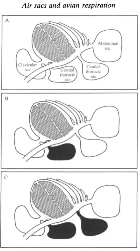
Fig. 1. Scheme of air sac blocking experiments. (A) controls; (B) CRT group; (C) CRT-CT group. The cervical air sac is not represented. The neopulmo is a small area of parabronchi connecting the dorsobronchi to the caudal air sacs and is not shown.
thoracic sac and 50-60 pieces to pack the caudal thoracic sac. The effectiveness of this technique for blocking the air sacs had been validated in preliminary experiments and was verified by post-mortem examination of the experimental birds (see Discussion).
230 J. H . BRACKENBURY, C. D A R B Y AND M . S. E L - S A Y E D
abdominal sac was cannulated immediately behind the base of the last rib and the .cannula was tied to the rib in front. Approximately 50 units of heparin was administered throughout the operation, and thereafter the arterial cannula was flushed regularly with heparinized saline.
l m l blood samples were removed anaerobically using a double-syringe tech-nique and analysed for POl and PQO2
o n a Radiometer BMS2 blood-gas analyser
thermostatted at 41-5°C. The electrodes were calibrated with a precision gas mixer after each measurement. The POz and PCoz of clavicular and abdominal air sac
gases were measured on Beckman OM-11 and LB-2 analysers, respectively, after drawing the samples through Drierite. The sampling flow rate was less than SOmlmin"1 and this was assumed to be small in comparison with the normal gas turnover in individual air sacs.
Oxygen consumption, VO2, was measured in four control birds to give an
indication of work load. The method has been described previously (Brackenbury
et al. 1981). Briefly, the subject wore a loose-fitting plastic mask into which air was
drawn from the atmosphere at a measured rate of 20-30 lmin"1 using a i m length of flexible tubing attached to a vacuum pump. A sample of the gas drawn through the mask was taken over Drierite into a Beckman OM-11 oxygen analyser. In individual experiments the flow rate of the gas collected from the mask was adjusted so that the analyser gave a displacement of approximately 1 % compared to atmospheric, thereby ensuring against loss of expirate from the mask. Measured values of Vch were corrected to STPD. The same mask was used to measure simultaneously respiratory evaporative water loss by drawing the collected gas through an airtight vessel containing a thermometer and a relative humidity detector (Vaisala, Finland) with an accuracy of ± 2 % . The response time of the detector to a step-wise increase in relative humidity was approximately 90s. The detector was calibrated with both dry and saturated air at known temperatures, and total respiratory water loss was calculated from the known gas flow rate, relative humidity and temperature. This value was converted to the corresponding value for expired minute ventilation, Vz, assuming that the expired gas had a temperature of 38°C. This indirect method of measuring ventilation has been validated by comparing values for estimated and directly measured ventilation in resting and exercising chickens (Brackenbury et al. 1982a). Respiratory rate was measured directly by drawing a sample of the collected gases directly into a Beckman LB-2 CO2 analyser which was capable of following breath-to-breath
variations in PQCH- Tidal volume was derived using the estimated VE and measured respiratory frequency values.
Air sac gas tensions, VOl ar>d the changes in relative humidity and PC O 2 of the
Air sacs and avian respiration Til
Experimental protocols
Each bird performed dual treadmill runs in two separate series of experiments. Except for the fact that different parameters were measured, the two series were carried out in identical conditions at a treadmill speed of 4-5 km h"1 (zero gradient), for a period of lOmin. In the first series, air sac gas tensions were monitored continuously before, during and for 10-15 min after the run. Arterial blood was sampled immediately after the cessation of the run and again approximately 10 min later when the air sac gas tension traces indicated a return to steady-state resting conditions. Preliminary trials had shown that measurements taken after a run generally gave a more stable indication of resting conditions than those taken before, when the birds tended to be more easily distracted by sounds and movements in the laboratory. The total delay between the end of a run and the final transfer of a blood sample into the blood gas analyser was less than 45 s. In the second series of runs, the birds were fitted with the mask and V"^, respiratory water loss and respiratory rate were measured simultaneously. Mean values of all the measured variables from both series were estimated over the last 2-3 min of exercise, and 10-15 min after exercise. All data are quoted as mean values ± 1 S.E.M. Significance tests (Student's f-test) were carried out on paired values at the 5 % level.
Results
Minute ventilation increased by approximately 200 % in the control birds and this was mainly due to elevated respiratory rate, with little change in tidal volume. Exercise resulted in a slight fall in arterial PCOz and a slight rise in arterial POl in
the control birds, and similar changes were reflected in the composition of the clavicular and the abdominal sac gases. Rectal temperature increased only marginally during exercise in the control birds, from 41-4 ± 0-17 to 41-8 ± 0-01 °C. Both experimental groups showed perfect compensation for the lack of function in the pertinent air sacs and their ventilatory characteristics at rest and during exercise were virtually indistinguishable from those of the controls (Table 1). Taken together, the control and experimental birds increased minute ventilation by approximately 250% compared to the non-exercised state. The CRT group regulated blood and air sac gases normally. The CRT-CT group showed a mild but significant hypoxaemia/hypercapnaemia compared with controls and this was also observed in the abdominal, although not the clavicular, sac gas tensions. As with the controls and the CRT group, the CRT-CT group displayed a slight freshening of both air sac and blood gases during exercise.
Oxygen consumption in four of the control birds increased by 300% from 16-00 ±0-95 ml kg"1 min"1 in the non-exercised state to 66-97 ± 1-43 ml kg"1 min^1 during exercise.
Post-mortem examination of the experimental birds confirmed the effectiveness
232 J. H. BRACKENBURY, C. DARBY AND M. S. EL-SAYED
Table 1. Respiratory characteristics of domestic fowl at rest and during exercise
following occlusion of the cranial thoracic air sac alone (CRT) or the cranial and caudal thoracic air sacs together (CRT-CT)
Respiratory evaporation (mmol kg"1 min"1) Minute volume
(mlkg-'min-1)**
Tidal volume (ml kg"1) Respiratory frequency
(min"1)
Arterial PCO2 (kPa)
Arterial P^ (kPa)
Clavicular sac PQO^ (kPa)
Clavicular sac F02 (kPa)
Abdominal sac Pco? (kPa) Abdominal sac P^ (kPa)
Control Rest 0-93 (0-09) 319 (32) 13-3 24 (0) 4-11 (0-09) 11-01 (0-17) 5-36 (0-08) 13-25 (0-13) 2-68 (0-11) 16-82 (0-19)
(N = 6)
Exercise 2-83 (0-11) 976 (39) 16-0 61 (3) 3-88 (0-08) 11-34 (0-27) 4-95 (0-09) 14-38 (0-12) 2-37 (0-11) 17-52 (0-23)
C R T (
Rest 1-06 (007) 367 (24) 14-7 25 (1) 4-11 (0-05) 10-61 (0-2) 5-25 (0-11) 13-88* (0-16)
-TV = 5 )
Exercise 3-57* (0-12) 1231* (42) 18-6 66 (1-5) 4-03 (0-05) 10-58* (0-16) 4-84 (0-15) 14-%* (0-12)
-C R T - -C T
Rest 0-97 (0-05) 335 (16) 14-0 24 (1) 4-40* (0-09) 10-30* (0-15) 5-56 (0-07) 13-53 (0-13) 3-08* (0-12) 16-26 (0-33)
(TV = 5)
Exercise 3-22 (0-17) 1110 (59) 15-4 72 (5) 4-21* (0-08) 10-53* (0-15) 4-66 (009) 15-17* (0-12) 2-93 (0-29) 16-64 (0-4)
Mean values (1 S.E.M.), except for tidal volume which was calculated from respiratory rate and minute volume.
TV = number of birds.
•Significantly different from control values ( P < 0 0 5 ) .
• • Estimated from respiratory evaporation using the method described in the text. lkPa = 7-52mmHg.
resulting in the entire plug of cotton wool becoming invested with a thick layer of fibrous tissue which bound it to the surrounding walls. In a small minority of cases the lumen of the air sac had not been completely obliterated but the volume of the remaining air space was only a few percent of its original volume.
Discussion
Critique of experimental methods
Initial attempts to design a flowmeter for the direct measurement of ventilation based on the face-mask design used previously in running hens (Brackenbury et al.
1982b) proved impracticable, since the size and shape of the comb in the White
Air sacs and avian respiration 233
1985) since we felt that this would interfere with normal respiration in an exercising bird. For similar reasons, we discounted the idea of using an acute tracheostomy. The method finally adopted, although indirect, has been validated in a previous study in which it was shown that there was a linear relationship between respiratory evaporative water loss and directly measured ventilation over a range of respiratory flow rates up to 61min~1, well above the maximum ventilatory flow rates encountered in the present study (Brackenbury et al. 1982a). The method, however, is sensitive to changes in temperature of the expired air since this governs the slope of the water loss versus ventilatory flow rate relationship. The assumption of an expired gas temperature of 38°C is based on the previously quoted study, but variations from a linear relationship would be expected if there were any significant alterations of expired gas temperature between rest and exercise. However, since rectal temperature hardly altered in the present experiments, it seems unlikely that there would be significant changes in expired gas temperature. To check on the reliability of the method, we compared ventilatory data in the present study with data obtained by direct measurement in hens running at the same mass-specific work loads (Brackenbury et al. 19826) and found excellent agreement.
The technique used to block the air sacs was simple in design and crude in operation but had been arrived at only after fairly extensive exploration of alternatives, including the implantation of inflatable balloons and the injection of inert foams. None of these methods proved as reliable as packing the sac with cotton wool. The only pathological reaction to the cotton wool was the fibrocytic response which not only isolated the implant but also sealed any small pockets of space that may have existed at the time of the implantation. The foreign body reaction appeared to be complete within 2-3 weeks and none of the birds showed chronic side-effects from the procedure. Although there was partial infiltration of the implants with tissue fluid, the additional mass was estimated to be less than 50 g or approximately 2 % of the mean body mass. No correction for this additional mass was made in the calculation of respiratory variables.
The dorsal wall of the cranial thoracic air sac is effectively the ventral surface of the lung underlain by the pulmonary aponeurosis. It could therefore be argued that direct pressure from the cotton wool implant might compress the pulmonary bronchi or the blood vessels. Such direct pressure could affect both airflow and blood perfusion, potentially creating ventilation/perfusion mismatches and lead-ing to reduced O2 diffusion capacity (Powell & Wagner, 1982; Hempleman &
Powell, 1986). However, as well as possessing intrinsic tensile strength, the pulmonary aponeurosis is also tensioned by the pulmocostales muscles and both of these would resist compression of the lung. Post-mortem examination showed that although the cotton wool implant was firmly bound to the aponeurosis by fibrous tissue, there was no sign of haemostasis or necrotic tissue in the lung.
234 J. H . BRACKENBURY, C . D A R B Y AND M . S. E L - S A Y E D
pronounced deficiencies in the experimental birds, particularly the CRT-CT group. Evidently, at the work load used, the birds were able to compensate for the lack of respiratory capacity by increasing the tidal excursion of the rib-cage, thereby enhancing the turnover of gas in the available air sacs. A higher work load might demand an excursion much closer to the mechanical limits of the rib-cage and might also incur a significant increase in the energy cost of ventilation. In view of the various instrumental attachments to the birds, however, it would be much more difficult to obtain smooth runs at higher treadmill speeds.
Ventilation, gas tensions and intrapulmonary airflow
The measured ventilatory responses of the birds to exercise were essentially the same as those previously documented: increased minute volume was achieved primarily by alteration of respiratory rate, with little change in tidal volume (Brackenbury et al. 19826). The slight fall in arterial PQO^ and rise in arterial POl as
a result of exercise (Table 1) are in keeping with the idea that there was a small degree of parabronchial hyperventilation, and this is supported by the freshening of gases that occurred in the air sacs. Parabronchial hyperventilation in exercising chickens is due, amongst other things, to thermal state and, at very high work loads, metabolic acidosis (Brackenbury & Gleeson, 1983), although other un-identified non-thermal factors may also be involved in the control of pulmonary ventilation (Gleeson & Molony, 1989). In the present study, it seems unlikely that either hyperthermia or lactacidosis would have contributed significantly to parabronchial hyperventilation.
According to the unidirectional airflow scheme, gas tensions in the cranial air sacs (i.e. the cervical, clavicular and cranial thoracic sacs) should all be identical (Bouverot & Dejours, 1971), although this assumption holds true only if the so-called inspiratory and expiratory valves in the lung work perfectly. There is considerable evidence of the effectiveness of these valves during resting respir-ation; this comes from direct flow recordings (Scheid & Piiper, 1972; Bretz & Schmidt-Nielsen, 1971, 1972), from gas sampling at intrapulmonary sites (Powell
et al. 1981) and, more recently, from plotting the time of appearance of injected
tracer gases in the cranial air sacs (Banzett et al. 1987). Nothing is known about the effectiveness of the valves during exercise hyperpnea, although Banzett et al. (1987) demonstrated that the inspiratory valve was more efficient at high flow rates and this suggests that exercise should, if anything, increase the efficiency of the aerodynamic valves.
Air sacs and avian respiration 235
the front, and the abdominal to the rear, lie outside the rib-cage. According to figures presented by King (1975), the combined volumes of both cranial and both caudal thoracic air sacs in the male chicken account for 25-30 % of the total air-sac volume, and presumably their contribution to the tidal volume is at least a comparable fraction, and probably a greater fraction in view of their mechanical advantage. Despite this, the CRT-CT group of experimental birds showed little sign of respiratory distress, even under challenge from exercise, apart from a minor hypoxaemia/hypercapnaemia (Table 1).
Although respiratory movements were not measured directly, a simple calcu-lation indicates that to bring about an additional increase in volume of the body cavity by 30 %, experimental birds need only increase the linear excursion of the rib-cage by a further (0-3)1//3, or less than 10%, compared to controls. The measured increase in arterial Pcch O I t n e CRT-CT group (Table 1), although small, would probably have been sufficient to elicit this level of response. Respiratory pattern did not differ significantly between the control and exper-imental birds and this, in turn, may be related to the similarity in arterial and intrapulmonary Pcch, both of which are involved in the control of avian breathing pattern. Mechanoreceptors, capable of responding to changes in volume of the air sacs or the body cavity, may also play a role, but the absence of any experimental data on respiratory movements in the present study makes it impossible to comment further on this subject.
The interesting question remains: to what extent did the pattern of intrapulmon-ary airflow differ in the experimental and control groups? We have no direct evidence, but the fact that the differential distribution of gas tensions between the CTanial and caudal air sacs (Table 1) was the same in control and experimental birds suggests that the unidirectional pattern of intrapulmonary airflow remained intact even after effective removal of the thoracic air sacs. The clear implication is that a normal unidirectional airflow pattern can be generated without the participation of the thoracic air sacs. It is logical to enquire whether other air sacs can be experimentally inactivated without influencing intrapulmonary airflow. In a preliminary attempt to answer this problem we tried blocking first the abdominal, then the clavicular air sacs, in separate series of experiments. Unfortunately, the distensibility of the abdominal sac is practically unlimited, and it is not possible to block this sac fully without imposing considerable pressure on the abdominal viscera. In a few cases we succeeded in blocking the cranial 30% or so of the abdominal air sac, in the area of its connection to the lung, and post-mortem examination showed that the entire air sac wall had collapsed around and fused with the implant, completely preventing inflation. The respiratory characteristics of these birds, including blood gases, were normal, although the data were too fragmentary to justify formal inclusion in the results.
236 J. H . BRACKENBURY, C. DARBY AND M . S. E L - S A Y E D
caused more by the pathological reaction to the implant than the physiological consequences of removal of air sac function. Post-mortem examination revealed copious secretions in the clavicular air sac and secondary bronchi of the lung, resulting from irritation of the mucosal coverings of the lung and the nerves, blood vessels and pericardium contained within the air sac. Further, there was evidence of a direct compression of the syrinx by the cotton wool. An alternative technique to direct physical block will have to be designed before the role of this sac can be investigated.
Finally, it is possible that blockage of the caudal thoracic air sac may interfere with normal airflow through the neopulmo, which is said to account for a significant fraction of gas exchange in the resting bird (Duncker, 1971, 1972; Piiper, 1978; Fedde etal. 1986). It is noteworthy that the resting PCch in the
abdominal air sac of the experimental birds was slightly but significantly higher than in the controls (Table 1). This may indicate that air that normally flows through the neopulmo to both the caudal thoracic and abdominal air sacs is now passing entirely to the abdominal sac, with a resultant increase in Pc<v These speculations, however, must also be weighed against evidence that caudal air sac gas composition in the penguin, which does not have a neopulmo, is similar to that in birds, such as the chicken, which have a well-developed neopulmo (Powell & Hempleman, 1985).
References
BANZETT, R. B., BUTLER, J. P., NATIONS, C. S., BARNAS, E. M., LEHR, J. L. & JONES, J. H.
(1987). Inspiratory aerodynamic valving in goose lungs depends on gas density and velocity.
Respir. Physiol. 70, 287-300.
BETHE, A. (1925). Atmung: Allgemeines und Vergleichendes. In Handbuch der Normalen und
Pathologischen Physiologie, vol. 2 (ed. A. Bethe, G. V. Bergmann, G. Embden & A.
Ellinger). Berlin: Springer Verlag.
BOUVEROT, P. & DEJOURS, P. (1971). Pathway of respired gas in the air sacs-lung apparatus of fowl and ducks. Respir. Physiol. 13, 330-342.
BRACKENBURY, J. H. (1979). Corrections to the Hazelhoff model of airflow in the avian lung.
Respir. Physiol. 36, 143-154.
BRACKENBURY, J. H., AVERY, P. & GLEESON, M. (1981). Respiration in exercising fowl. I. Oxygen consumption, respiratory rate and respired gases. /. exp. Biol. 93, 317-325.
BRACKENBURY, J. H., AVERY, P. & GLEESON, M. (1982a). Measurement of water loss in exercising animals using an electronic humidity detector. Med. biol. Eng. Comput. 20, 433-436.
BRACKENBURY, J. H. & GLEESON, M. (1983). Effects of Pco^ on respiratory pattern during thermal and exercise hyperventilation in domestic fowl. Respir. Physiol. 54, 109-119.
BRACKENBURY, J. H., GLEESON, M. & AVERY, P. (1982b). Respiration in exercising fowl. LTI. Ventilation. /. exp. Biol. 96, 315-324.
BRANDES, G. (1923). Atmung der Vogel. Verh. dt. Zool. Ges. 28, 57-59.
BRANDES, G. (1924). Beobachtungen und Reflexionen tiber die Atmung der V6gel. Pfliigers
Arch. ges. Physiol. 203, 492-511.
BRETZ, W. L. & SCHMIDT-NIELSEN, K. (1971). Bird respiration: flow patterns in the duck lung. /. exp. Biol. 54, 103-118.
BRETZ, W. L. & SCHMIDT-NIELSEN, K. (1972). The movement of gas in the respiratory system of the duck. J. exp. Biol. 56, 57-65.
Air sacs and avian respiration 237
DUNCKER, H. R. (1971). The lung air sac system of birds. Ergebn. Anat. EntwGesch. 45,1-171.
DUNCKER, H. R. (1972). Structure of avian lungs. Respir. Physiol. 14, 44-63.
FEDDE, M. R. (1980). Structure and gas-flow pattern in the avian respiratory system. Poult. Sci. 59, 2642-2653.
FEDDE, M. R., BURGER, R. E., GEISER, J., GRATZ, R. K., ESTAVILLO, J. A. & SCHEID, P. (1986).
Effects of altering dead space volume on respiration and air sac gases in geese. Respir.
Physiol. 66,109-122.
GLEESON, M., BARNAS, G. M. & RAUTENBERG, W. (1985). Respiratory and cardiovascular responses of the exercising chicken to spinal cord cooling at different ambient temperatures. n. Respiratory responses. /. exp. Biol. 114, 427-441.
GLEESON, M. & MOLONY, V. (1989). Control of breathing. In Form and Function in Birds, vol. 4, chapter 10 (ed. A. S. King & J. McLelland). London: Academic Press.
HAZELHOFF, E. H. (1943). Bouw en Functie van de Vogellong. Verslag van de gewone Vergaderingen der Afdeeling Natuurkunde. Kon. Ned. Akad. Wet. 52, 391-400.
HEMPLEMAN, S. C. & POWELL, F. L. (1986). Influence of pulmonary blood flow and O2 flux on DO2 in avian lungs. Respir. Physiol. 63, 285-292.
JONES, J. H., EFFMAN, E. L. & SCHMIDT-NIELSEN, K. (1981). Control of air flow in bird lungs: radiographic studies. Respir. Physiol. 45, 121-131.
KING, A. S. (1975). Aves Respiratory System. In The Anatomy of the Domestic Animals, vol. 2, chapter 64 (ed. R. Getty). Philadelphia: W. B. Saunders Company.
KUETHE.D. O. (1988). Fluid mechanical valvingof air flow in bird lungs. 7. exp. Biol. 136,1-12.
PHPER, J. (1978). Origin of carbon dioxide in caudal air sacs of birds. In Respiratory Function in
Birds, Adult and Embryonic (ed. J. Piiper), pp. 148-153. Berlin: Springer-Verlag.
POWELL, F., GEISER, J., GRATZ, R. K. & SCHEID, P. (1981). Airflow in the avian respiratory tract: variations of O2 and CO2 concentrations in the bronchi of the duck. Respir. Physiol. 44, 195-213.
POWELL, F. L. & HEMPLEMAN, S. C. (1985). Sources of carbon dioxide in penguin air sacs. Am.
J. Physiol. 248, R748-R752.
POWELL, F. L. & WAGNER, P. D. (1982). Ventilation-perfusion inequality in avian lungs. Respir.
Physiol. 48, 233-241.
SCHEID, P. (1979). Mechanisms of gas exchange in bird lungs. Rev. Physiol. Biochem. Pharmac.
86, 137-186.
SCHEID, P. & PIIPER, J. (1972). Mechanisms of unidirectional flow in the parabronchi of avian lungs: measurements in duck lung preparations. Respir. Physiol. 14, 83-95.
SCHEID, P. & PIIPER, J. (1989). Respiratory mechanics and air flow in birds. In Form and
Function in Birds, vol. 4, chapter 8 (ed. A. S. King & J. McLelland). London: Academic
Press.