Vijay K. Sadhu',2 Stanley F. Handel' Richard S. Pinto ',3 T. Franklin Glass,,4
Received June 21, 1979; accepted after r evi-sion September 14, 1979.
I Department of Diagnostic Radiology, Univer-sity of Texas Medical School at Houston, 6431 Fannin, Houston, TX 77030. Address reprint re-quests to S. F. Handel.
2 Present address: Department of Radiology, Boone County Hospital, Columbia, MO 65201.
3 Present address: Department of Radiology, New York University Medical Center, New York, NY 10016.
4 Present address: Department of Radiology, Medical Center of Central Georgia, 777 Hemlock, Macon, GA 31201.
AJNR 1 :39-44, January/February 1980 0195-6108/80/0011-0039 $00.00 © American Roentgen Ray Society
39
Neuroradiologic Diagnos
i
s of
Subdural Empyema an
d CT
Limitations
Five cases of subdural empyema are described. Two of the cases eluded a definitive computed tomography (CT) diagnosis despite classical clinical background. Extracer-ebral collection w.ith definitive border enhancement was seen in the other three cases.
Mass effect, present in all five cases, was related to the extracerebral collection in three cases and diffuse cerebral edema and/or infarction in two. Angiography in four cases initially demonstrated an extracerebral collection in three and inflammatory angiospasm in two. Repeat angiography demonstrated an extracerebral collection in the fourth case. In the proper clinical setting subdural empyema should be considered even in the absence of an extracerebral collection when mass effect or an infarction pattern is seen on CT. Angiography may be diagnostic in such cases. Hopefully, newer techniques will further the diagnostic efficacy of CT in this disease.
Computed tomography (CT) is essential for diagnosis and management of cerebral infections. Although extensive literature [1 -8] is available for CT diag -nosis of intracranial abscesses, subdural empyema specifically has not received adequate attention in recent reports. We describe our experience with five cases of subdural empyema, two of which had nonspecific CT findings, and the use of angiography in the appropriate clinical setting.
Case Reports
Case 1
A 21-year-old woman was seen with marked deterioration in sensorium, fever, and
nuchal rigidity 3 weeks after a gunshot wound to the left orbit that had resulted in a dural
laceration, Cerebrospinal fluid examination revealed leukocytosis with polymorphonuclear
predominance. CT revealed a low density subdural collection with border enhancement on the left and midline shift to the right (fig. 1 A). Angiography immediately after CT demo
n-strated a frontoparietal extracerebral collection without changes in the arterial caliber or
occlusions of vessels (fig. 1 B). At surgery a subdural empyema was drained.
Case 2
A 7 -month-old girl had an upper respiratory tract infection that was treated with antibiotics 3 weeks before admission. Fever, vomiting, and lethargy developed 7 days before admis-sion, and 5 days thereafter became obtunded with a stiff neck. At this time, provisional diagnosis of meningitis was made. On admission to Hermann Hospital in Houston, Tex. she was opisthotonic with deviation of head and eyes to the left and had rapid shallow breathing.
Examination demonstrated sustained clonus on the right side and a possible mild right
hemiparesis. Lumbar puncture revealed turbid, yellowish cerebrospinal fluid with a white blood cell count of 4,300/mm3
40 SADHU ET AL. AJNR: 1 , January/February 1980
A
B
g/dl, and glucose of 4 g/dl. The patient was put on a combination of antibiotics.
CT 1 day after admission showed a subdural collection of fluid with medial rim enhancement (fig. 2). Mass effect in the form of diminution of sulci on the periphery of the left hemisphere was seen.
A diagnosis of subdural empyema was suggested. After CT a left
parietal craniotomy revealed yellow mucoid pus along with a yellow membrane covering the brain surface. H. influenzae type B was
cultured from the pus. Follow-up CT 5 days after admission showed
a low density zone in the left frontal area suggestive of edema or
infarction. Angiography 12 days after admission revealed a residual
•
Fig. 1.- Case 1. A, Contrast-en -hanced scan. Low density extracerebral
collection with border enhancement on
lell (arrowheads) and midline shift of frontal horns to right. B, Midarterial
phase of left internal carotid angiogram.
Extracerebral crescentic collection ( ar-rows) without any definite caliber
changes of cerebral vasculature .
Fig. 2.-Case 2. A, Low density
ex-tracerebral collection on left. B, border
enhancement after contrast injection.
left extracerebral collection. At 20 days after admission, the pa-tient's neurologic status was gradually improving.
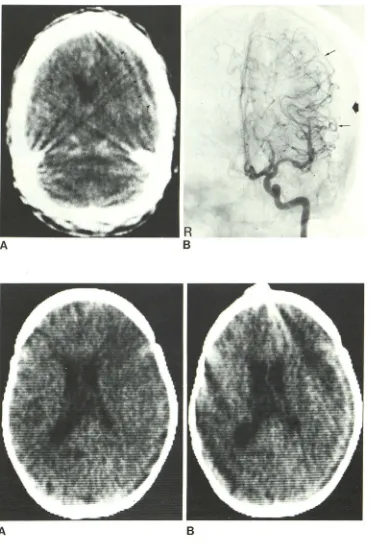
Case 3
An 11-year-old boy was admitted with a diagnosis of meningitis. He had complained of headaches 4 days before admission. During
the next 24 hr he also developed malaise, fever, and neck pain. At
this time the patient was treated with oral antibiotics, but 1 day
before admission he developed staggering gait, nuchal rigidity,
[image:2.613.54.424.76.621.2]gener-AJNR: 1 , January/February 1980
SUBDURAL EMPYEMA
41-L
c
alized seizure. On admission, the cerebrospinal fluid revealed a
white blood cell count of 5,200/mm3
, glucose of 6.9 g/dl, and
protein of 91 g/dl. Gram stain was negative.
CT scan 1 day after admission was interpreted as showing right
cerebral edema with displacement of the midline structures to the
left (figs. 3A and 38). In retrospect, the contrast-enhanced scan
showed an equivocal low density crescentic zone with slight border
enhancement paralleling the inner table of the skull. The patient deteriorated neurologically 2 days after admission, developing a right third, a right sixth, and a left seventh nerve palsy. A left hemiparesis was also found. Clinical diagnosis of encephalitis was made and the patient was taken to surgery for a brain biopsy. A large amount of subdural pus was encountered on a right tempo-roparietal craniotomy. Unidentified Gram-negative anaerobic rods were grown on culture. After operation, angiography was performed
o
Fig. 3.- Case 3. A and B, Contrast -enhanced scans. Right hemispheric
mass effect secondary to difluse cer
e-bral edema with midline shift to left and obliteration of right lateral ventricle.
Small extracerebrallow density crescen -tic zone suggested (B, arrowheads). C,
Anteroposterior view. Arterial phase of right internal carotid injection. Parafal -cine (thin arrows) and right hemispheric
(short arrows) extracerebral collection. Inner table of skull (longer thick arrow). D, Caliber changes in cerebral arteries (arrowheads). Prominent anterior falcine arteries (arrows).
on two separate occasions to localize other sites of subdural
em-pyema. Extracerebral collections were observed on both cerebral
convexities and with the interhemispheric fissure. Attenuation in the caliber of peripheral vessels as well as vessels at the base of the
brain was noted. Prominent anterior falcine arteries were observed
(figs. 3C and 3D). Pansinusitis was diagnosed on subsequent sinus
and skull radiographs. After a protracted course, the patient was
discharged. When last seen 17 months after his initial admission,
he was neurologically normal, seizure-free, and doing well in school.
Case 4
A 31-year-old man was seen at another hospital with a 2 week
history of severe headache, elevated temperature, and numbness
[image:3.615.51.558.67.564.2]42 SADHU ET AL. AJNR:1, January/February 1980
A
c
to a right hemiplegia. After a focal seizure, the patient was
trans-ferred to Hermann Hospital. At this time he was found to have
nuchal rigidity, slurred speech, and depressed mentation. Lumbar
puncture revealed an opening pressure of 230 mm water, protein
of 16 g/dl, and white blood cell count of 1 ,000/mm3
with 35%
lymphocytes. Treatment was continued with chloramphenicol,
pen-icillin, and streptomycin, which was begun at the other hospital.
Admission CT showed a midline shift from left to right with a low
density zone of edema in the left frontal lobe (fig. 4A). No
extracer-ebral collection was noted on either the pre-or postcontrast scans,
Sinus series showed clouding of the left frontal sinus with loss of
mucoperiosteal line consistent with sinusitis. Angiography revealed
a prolonged circulation time and several poorly opacifying middle
cerebral artery branches. A round shift fo the left anterior cerebral
artery was also observed (figs. 48 and 4C). CT scans over the next
18 days demonstrated a gyral enhancement pattern with
improve-ment in mass effect (fig. 40). Repeat angiography 18 days after the
initial study revealed an extracerebral collection and improvement
in the diffuse arterial changes (fig, 4E). A subdural empyema was
evacuated through a left frontoparietal craniotomy. Follow-up
al-most 1 year after initial admission revealed a residual right
hemi-paresis. A low density zone indicating infarction of the left frontal
lobe was seen on CT.
Case 5
An 18-year-old man was transferred from another hospital with
a 6 day history of headache, fever, chills, and photophobia. Lumbar
B
Fig. 4.- Case 4. A, Contrast-enhanced scan. Midline shift to right with left hemispheric edema pattern. No evidence of extracerebral collection. Band C, Late films of left internal carotid angiogram. Slow circulation, multi-ple attenuated basal, and hemispheric arteries. Round
shift of anterior cerebral artery noted on frontal projec -tions. Difficulty in demonstrating extracerebral collection from lack of convexity branches seen distally over parietal
lobe. D, Enhanced scan 18 days after initial scan (A).
Evolving infarction pattern with gyral enhancement. No extracerebral collection. E, Repeat left internal carotid
angiogram. Definite extracerebral collection (arrows), Im
-provement in inflammatory angiospasm vasculature and midline shift.
puncture at the other hospital showed a white blood cell count of
1 ,000/mm3 with polymorphonuclear predominance; glucose, 4.5
g/dl; and protein, 12.2 g/dl. Peripheral white blood cell count was
19,200/mma Subsequently, the patient became stuporous and
developed right leg weakness and bilateral papilledema. Paranasal
sinus radiographs showed frontal sinusitis, The patient was then
transferred to St. Luke's Episcopal Hospital in Houston, Tex. where
the frontal sinuses were drained. Despite antibiotic treatment the
patient deteriorated more the next day. CT showed what was
interpreted as a right frontal abscess and associated
interhemi-spheric empyema (fig. 5). This was confirmed at surgery where a brain abscess and interhemispheric empyema were drained. After
repeat drainage of the interhemispheric empyema, the patient had
a satisfactory hospital course and was subsequently discharged.
Discussion
Subdural empyema, a neurosurgical emergency [9],
should be considered when a patient has fever and focal neurologic deficits. It should also be suspected in a patient with meningitis who is refractory to medical therapy.
Con-comitant or previous history of paranasal sinusitis, osteo-myelitis of the calvarium, otitis media, or postcraniotomy infection should alert the clinician to the possibility of sub-dural empyema [10]. Symptoms are related to an ex
pres-AJNR: 1 , January/February 1980 SUBDURAL EMPYEMA 43
Fig. S.-Case 5. A, Low density in-terhemispheric collection. B, Peripheral
enhancement after contrast iniection.
Subdural empyema proven at surgery.
A
sure. Venous thromboses and infarction may ensue and lead to further complications such as seizures, hemiparesis, hemianopsia, and aphasia [11]. The course is often cata-strophic and mortality may range from 25% to 40% [12].
Computed tomography has become essential for diagno-sis and management of cerebral infection [8]. However, subdural empyema has not received specific attention in recent literature. Zimmerman et al. [8] reported that subdural empyema may appear as an extracerebral low density col-lection with mass effect. Medial or border enhancement has not been described in subdural empyema on contrast-en-hanced scans.
We found that subdural empyema may be seen with nonspecific CT findings. Three of our five cases revealed a
definite low density extracerebral collection with medial or border enhancement (figs. 1 A, 1 B, 2A, 2B, 5A, and 5B). Case 3 revealed a subtle low density extracerebral collec-tion without definite border enhancement (fig. 3). Case 4 was initially seen as diffuse edema of a cerebral hemisphere that evolved to an infarction pattern with peripheral gyral enhancement (figs. 4A and 40). Our only constant CT finding was mass effect. The mass effect in cases 1 and 2
may be totally explained on the basis of an extracerebral collection. The explanation for inadequate detection of low density extracerebral collections in cases 3 and 4 may be that these collections were small in total volume and/or were isodense in relation to the surrounding cerebral tissue.
We believe that the focal neurologic deficit is due more to the response of the cerebral vasculature and brain to the inflammatory process and less to the mass effect of the extracerebral collection. Therefore, even small
extracere-bral collections of pus, relatively unresolved on current CT scans, may produce severe neurologic changes. The
ab-sence of medial or border enhancement in some cases may be related to a short time interval from onset of the infectious process, with its effect on cerebral vessels and brain, and the CT scan. It is presumed that the border enhancement
B
we observed in three cases is due to development of a
granulation tissue wall over a period of 3 weeks. Analo-gously, this generalization is supported by the fact that the
enhanced fibrous tissue wall of intracerebral abscess may not be observed until the third week of its natural history [7]. Conceivably, sequential CT scans might demonstrate
the evolution of subdural empyema and appearance of
border enhancement. A definite cerebral edema pattern, as
visualized in cases 3 and 4, probably is responsible for the
major part of the observed mass effect. This edema pattern is probably related to the cerebral ischemia secondary to
an inflammatory angiospasm shown on subsequent
angio-grams. Concomitant venous thromobsis or cerebritis may
also contribute to the development of cerebral edema.
Kim et al. [2] described these specific angiographic signs
in subdural empyema: (1) an irregular border of the
extra-cerebral collection; (2) a thickened vascular wall of the dura;
and (3) a semilunar avascular zone in the lateral view. In
addition, the following angiographic findings of meningeal inflammation may be present: (4) spasm of large arteries at
the base of the brain with or without segmental arterial
dilatation; (5) multiple peripheral arterial stenoses; and (6)
enlargement of the meningeal arteries.
Angiography in cases 1 -4 revealed a subdural collection
in each case, although temporal relation of CT to
angiogra-phy varied from case to case (see Case Reports). Cases 3
and 4 showed definite spasm of large arteries at the base of
the brain (figs. 3C and 4B). Multiple peripheral arterial
stenoses with reduced flow to the hemispheres were also
observed in case 3. The presence of angiospasm and a
prolonged circulation time in case 4 prevented the detection of an extracerebral collection on the first angiogram (figs. 4B and 4C). Dilatation of the anterior falcine artery was
noted in case 1 (fig. 3D).
The neuroradiologic diagnosis of subdural empyema may
be elusive if CT findings alone are considered. We believe
[image:5.612.192.554.77.315.2]44 SADHU ET AL. AJNR: 1, January /February 1980
clinical setting when mass effect, diffuse cerebral edema,
or an infarction pattern is seen on CT without a definite
extracerebral collecton. In these cases arteriography should
be promptly performed although, as case 4 illustrates, a
n-giography may not always provide an answer. Moreover,
difficulty may occur in demonstrating interhemispheric, base
of brain, and posterior fossa extracerebral collections by
angiography. The subdural empyema seen in figure 5 may
only have been demonstrated by CT. On the other hand
newer CT techniques including high dose contrast infusion,
delayed scanning, and coronal imaging (direct or reco
n-structed) may further the diagnostic efficacy of CT for s
ub-dural empyema. It is hoped refinements of CT technique will
lessen the need for angiography in this surgical emergency.
ACKNOWLEDGMENT
We thank Paul Gerson of S1. Luke's Episcopal Hospital, Houston
for the use of case 5.
REFERENCES
1. Claveria CE, du Boulay GA, Moseley IF. Intracranial infections:
investigations by computerized axial tomography. Neurora-diology 1976; 12: 59-71
2. Kim KS, Weinberg PE, Magidson M. Angiographic features of subdural empyema. Radiology 1976;118: 621-625
3. Lott T, EI Gammal T, Dasilva R, Hanks D, Reynolds J. Evalu
a-tion of brain and epidural abscesses by computed tomography.
Radiology 1977;122:371-376
4. New PF, Davis KR, Ballantine HT. Computed tomography in
cerebral abscesses. Radiology 1976; 121 : 641 -646
5. Nielsen H, Gyldensted C. Computed tomography in the diag-nosis of cerebral abscesses. Neuroradiology 1977; 12: 207-217
6. Stevens EA, Norman D, Kramer RA, Messina AB, Newton TH. Computed tomographic brain scanning in intraparenchymal pyogenic abscesses. AJR 1978; 130: 111-114
7. Whalen MA, Hilal SK: The computed tomography of brain
abscesses. Presented at the annual meeting of the American Society of Neuroradiology, New Orleans, March 1978
8. Zimmerman RA, Patel S, Bilaniuk L T. Demonstration of puru-lent bacterial intracranial infection by computed tomography.
AJR 1976;127: 155-165
9. Bhandari YS, Sarkari NBS. Subdural empyema. J Neurosurg
1970;32: 35-39
10. Butler I, Johnson RT. Central nervous system infections. Pe
-diatr C/in North Am 1974;21 : 649-663
11. Hirchcock E, Andreadis A. Subdural empyema a review of 21
cases. J Neurol Neurosurg Psychiatry 1964;27: 422 12. LeBleau J, Creissard P, Harispe L, Redondo A. Surgical
treat-ment of brain abscess and subdural empyema. J Neurosurg