and External Auditory Canal
T. E. Mayer, H. Brueckmann, R. Siegert, A. Witt, and H. Weerda
PURPOSE: To determine CT findings in the external, middle, and inner ear of patients with microtia and external auditory canal dysplasia. METHODS: We used high-resolution CT, with multiplanar or axial 1-mm continuous sections, coronal or sagittal reformations, or low-dose spiral acquisitions, to examine 184 temporal bones of children with microtia. RESULTS: In cases of minor microtia, auditory canal stenosis was the most common associated abnormality; in those with major microtia, atresia was predominant. Middle ear malformations depended on the severity of the auricular anomalies. Inner ear changes could also be noted. Ossicle dysplasias occurred in 98% of patients (stapes, 72%), absence of the oval window in 36%, labyrinthine malformations in 13%, closed round window in 6%, facial canal displacement in up to 75%, and aberrations of the vascular canal in 38% of patients with third-grade auricular deformity. CONCLUSION: A variety of external, middle, and, less frequently, inner ear changes were detected in connection with microtia.
Index terms: Ear, abnormalities and anomalies; Temporal bone, computed tomography
AJNR Am J Neuroradiol18:53–65, January 1997
Congenital aural dysplasias occur in one of 3300 to 10 000 births (1, 2), except in the era of thalidomide embryopathy (1959 –1962), when it was diagnosed in one of 900 neonates born in Germany (3). Microtia has been described even on Babylonian tablets (4) and has been seen in prehistoric skulls (5). The classification of au-ricular deformities was proposed by Marx (6) and described by Rogers (7). Microtia is often associated with abnormalities of other organs as the result of genetic disorders, chromosomal defects, intrauterine infections, or environmen-tal teratogens.
Surgery for congenital aural atresia is one of the most difficult ear operations, as it requires reconstruction of the auricle, often done by us-ing grafts of skin and rib cartilage (8, 9), and, if necessary, restoration of hearing by drilling a
new auditory canal and reconstructing the os-sicular chain (10 –12). Surgery is also per-formed for congenital cholesteatoma, labyrin-thine fistula, infection, or facial nerve palsy from previous surgery (13). Even if there is labyrin-thine involvement by the malformation, inner ear function is preserved in most cases. Endo-scopic viewing is not possible in stenotic or atretic auditory canals. Staging by computed tomography (CT) is necessary to avoid the risk of facial nerve lesions, worsening of hearing, and bleeding, and to obtain a valid prognosis of success.
We present a review of the CT findings in 92 patients with microtia and external auditory ca-nal dysplasia.
Subjects and Methods
All 92 patients had high-resolution CT, 89 of them with 1- mm-thick sections and three with 2-mm-thick sections. Fifty-five patients only had axial CT, sometimes with ref-ormations of the coronal, oblique, or sagittal planes; 37 patients were scanned in axial and coronal planes, and five of these were additionally scanned in the sagittal orienta-tion with low-dose (85 mAs) spiral CT. In these cases, Received March 21, 1995; accepted after revision July 8, 1996.
From the Institute of Radiology and Neuroradiology (T.E.M., H.B., A.W.) and the Clinic of Ear, Nose, Throat, and Plastic Surgery (R.S., H.W.), Medical University, Lu¨beck, Germany.
Address reprint requests to Dr Thomas Mayer, Abteilung Neuroradiolo-gie, Klinikum Grosshadern, 81377 Munchen, Germany.
AJNR 18:53–65, Jan 1997 0195-6108/97/1801–0053 ©American Society of Neuroradiology
three-dimensional surface reconstructions and sagittal ref-ormations were also made, if necessary. Spiral CT allowed a reduction of motion artifacts in examinations of children. Four patients had magnetic resonance (MR) imaging.
The following structures were evaluated in 184 tempo-ral bones: 1) The external auditory canal: stenosis or atre-sia of the cartilaginous part of the auditory canal, stenosis or atresia of the bony part, thickness of the atresia plate (smallest dimension at the level of the hypotympanum). 2) Dysplasia or defects of the temporal bone, zygomatic bone, and mandibular condyle; if possible, the whole skull and the cervical spine (scout view) were analyzed for anomalies. 3) Vascular structures such as the carotid ca-nal, sigmoid sinus, and jugular bulb were looked at for dehiscence, aberrant course, or remnant embryological vessels. 4) The auditory tube, including tensor tympani muscle and grade of pneumatization. 5) Congenital or secondary cholesteatoma or epidermoid. 6) The extent of the middle ear cavity, form of tegmen. 7) Diminution, dysplasia, rotation, fusion, ectopia, tympanic wall adher-ence, and absence of the ossicles. 8) Labyrinthine win-dows open or closed; fistula. 9) Cochlear turns, vestibule, semicircular canals, aqueducts. 10) The internal auditory canal and facial nerve canal: aberration, dehiscence, hy-poplasia, thickening, splitting.
Classification criteria of auricular deformities used in this study were those described by Weerda (14), as fol-lows. First-degree dysplasia: macrotia, prominent ear, pocket ear, absence of the upper helix (Fig 1A), absence of the tragus, clefts, lobular deformities, and cup ear de-formities type 1 and 2. Second-degree dysplasia: cup ear deformity type 3 (Fig 1B) and mini-ear. Third-degree dys-plasia: absence of normal auricular structure (Fig 1C) (unilateral or bilateral) and anotia; severely dysplastic ears might be displaced downward owing to incomplete ascen-sion from the neck (15).
Results
Auricular Dysplasia
One hundred thirty-four of 184 ears exam-ined were microtic. Consequently, 23% of the patients had bilateral microtia. Auricular defor-mities were often visible on CT scans. Some patients had undergone several reconstructive operations on the external ear.
CT findings were compared with the clinical appearance of the auricle. The results are pre-sented as percentages of two groups: three quarters of the microtic ears were classified as third-degree dysplasia (including one case of anotia) and are referred to as major microtia (84 ears); one quarter of the dysplastic auricles had first-degree (16 ears) or second-degree (13 ears) dysplasia. The two groups are combined and referred to as minor microtia in the follow-ing sections (see Table).
External Auditory Canal
[image:2.587.49.549.85.265.2]Microtia and external auditory canal dyspla-sia were highly correlated (P , 10230). Three quarters of our patients with major microtia had an atresia; none had a normal external auditory canal. In three quarters of the minor microtic patients, the bony or cartilaginous part of the external auditory canal was stenotic (P,1027). The patients with a normal external ear (con-tralateral to a microtic ear) rarely had external auditory canal stenosis.
Fig 1. Pathologic anatomy of the auricle, classification after Weerda (14).
A, First-degree auricular dysplasia: small auricle with hypoplasia of the upper helix.
Petrous bone malformations correlated to the degree of auricular dysplasia (relative rate in each group of microtia)
Auricular Dysplasia, n5184
Normal (Contralateral) Ear,
n571
Minor Microtia (First and Second Grade),
n516113
Major Microtia (Third Grade),
n584
External auditory canal
Stenosis 5.6 72.4 21.4
Atresia 0 10.3 78.6
Malformations of:
Temporal bone 9.9 55.2 89.3
Mandible* 1.4 20.7 64.3
Zygomatic arch† 0 13.8 44.0
Others 14.1 17.2 29.8
Carotid canal
Slight aberrations 1.4 17.2 15.5
Hypoplasia 1.4 3.4 0
Sigmoid sinus, jugular bulb aberration, highriging 1.4 17.2 38.1 Eustachian tube, bony dysplasia 0 20.7 44.0 Pneumatization
Reduced‡ 7.0 10.3 19.0
Little or none‡ 0 17.2 47.6
Middle ear space epitympanum
Reduced§ 1.4 24.1 20.2
Minimal§ 0 3.4 41.7
Mesotympanum
Reduced§ 0 37.9 46.4
Minimal§ 0 6.9 41.7
Hypotympanum
Reduced§ 1.4 24.1 17.9
Minimal§ 0 13.8 73.8
Malleoincudal joint
Dysplastic, adherent 7.0 51.7 63.1
Absent 0 3.4 34.5
Incudostapedial joint, dysplastic/absent 4.2 68.9 89.3 Stapes
Dyplastic 1.4 17.2 15.5
Absent 0 34.5 56.0
Oval window, occluded 0 41.4 35.7 Fallopian canal abnormalities
Hypoplasia of the labyrinthine segment 0 10.3 9.5 Aberration of the labyrinthine segment 0 3.4 4.8
Tympanic dehiscence 0 24.1 26.2
Severe anomalies of the tympanic segment 0 0 2.4 Mastoid segment anterior dislocation 0 55.2 61.9 Severe aberration of the mastoid segment 0 3.4 11.9 Internal auditory canal, inclined/widened/agenesis 5.6 13.8 16.7 Round window, occluded 0 3.4 6.0 Inner ear dysplasia
Cochlea 0 0 3.6
Vestibulum 0 3.4 4.8
Lateral semicircular canal 0 6.9 13.1
Note.—Values given in percentages. * n5137.
† n5122.
‡ Reduced indicates 75% less than contralateral side or only periantral cells bilaterally; little or none, complete loss of mastoid pneumatisation; tympanum may or may not be air containing.
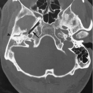
Stenotic auditory canals often showed a dis-tinctly more angulated course than normal. The canals sloped from caudolateral to craniome-dial. External auditory canal atresia varied from a fibrous closure to thick atretic plates (Fig 2) up to 31 mm in diameter (median, 14 mm).
Among patients with external auditory canal stenosis and atresia, 24% had a tympanic mass of solid tissue or fat density. Some patients had bone erosion, suggesting an epidermoid cyst or a congenital or secondary cholesteatoma (Fig 3A). One patient had fistulas to the skin of the side of the face combined with mandibular dys-plasia. In another patient, an epidermoid of the cerebellopontine angle had to be removed sur-gically (Fig 3B).
Other Skull Abnormalities
Microtia and external auditory canal atresia were most often associated with aplasia or hypoplasia of the tympanic part or mastoid process of the temporal bone (P , 10215), which correlated with the degree of auricular
dysplasia (P , .001). The second most
[image:4.587.212.546.83.528.2]fre-quent abnormality was dysplasia of the man-dibular condyle (P , 1029), followed by de-fects of the zygomatic arch (P , 1024) (Fig 4). In genetic disorders, such as Franceschetti (Treacher Collins) syndrome or Crouzon dis-ease, the whole skull was abnormal; and in Klippel-Feil syndrome, the cervical spine was dysplastic.
Fig 2. Axial CT scans of external au-ditory canal abnormalities.
A, Stenosis (arrow) is seen on the right.
B, Atresia and formation of an atretic plate (arrowhead) are on the right; a nor-mal external auditory canal is on the left (arrow).
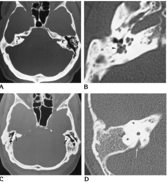
Fig 3. Cholesteatoma and dermoid. A, Axial CT scan shows masses of solid tissue in the middle ear cavity and mastoid, with bony erosion (arrowhead) in external auditory canal stenosis.
B, Coronal T1-weighted (360/15/4 [repetition time/echo time/excitations]) spin-echo MR image shows normal ex-ternal auditory canal on left (arrow) and external auditory canal missing and an epidermoid in the cerebellopontine angle on right (arrowheads).
Fig 4. Associated anomalies. A, Coronal CT scan shows dysplasia of the mandibular condyle and defect of the joint socket with a nearly obliterated middle ear in Franceschetti syndrome.
Temporal Bone Vessels
The carotid canal was asymmetric in one fifth of the patients with unilateral aural atresia, slightly rotated or elevated in relation to the hypoplastic base of the skull (P , .05). In two patients, the internal carotid artery was severely hypoplastic or aplastic, but these severe anom-alies did not correspond with the degree of mi-crotia (both had first-degree auricular dyspla-sia, and one was affected on the contralateral side). We did not find dehiscence into the mid-dle ear or a remnant stapedial artery. The sig-moid sinus and jugular vein showed normal asymmetries and emissaries and often a high-riding jugular bulb. In cases of severe hypopla-sia of the tympanic part of the temporal bone, the sigmoid was positioned somewhat ventrally (P,1024). Only three patients had an erosion of the squamous part of the temporal bone by venous structures, but none showed dehiscence into the middle ear (Fig 5).
Pneumatization of the Temporal Bone
The air content of the middle ear and mastoid was partially or completely reduced in one quarter of the minor and in two thirds of the major microtic ears; only a few normal con-tralateral ears had reduced mastoid pneumati-zation (P , 1027). The severity and frequency of decreased pneumatization correlated with the degree of anomalies of the external ear (P , .05).
The bony eustachian tube had either a
dys-plastic appearance (narrowing) or the tensor tympani muscle was hypoplastic (P , 1026) in about 20% of minor and 40% of major mi-crotic ears. In two patients, the tube was ex-cessively enlarged (Fig 6). The cartilaginous part of the tube was visible only in cases of enlargement.
The Tympanic Space
The width of the middle ear was frequently diminished (P , 10219), correlating with the degree of microtia (P , 1027). The epitympa-num was affected least often, but could be to-tally obliterated. In the mesotympanum, at least a small residual cleft was usually left in
other-Fig 5. Vessel canal aberrations.
A, Axial CT scan at the level of the foramen lacerum and basal turn of the cochlea shows hypoplastic carotid canal (arrow) in first-degree auricular dysplasia.
B, Axial CT scan shows dehiscent sigmoid sinus (arrow) and an emissary (arrowhead) in a severely microtic ear with hypoplastic temporal bone.
[image:5.587.53.551.84.243.2]C, Sagittal CT scan in the plane of the round window shows high-riding jugular bulb.
[image:5.587.346.508.316.477.2]wise nearly obliterated middle ears. The hypo-tympanum was reduced most often (Fig 7).
The Ossicular Chain
Changes of the ossicles were frequently present (P,10218) and consisted of a dysplas-tic shape, diminution, thickening (rarely), axis rotation, arthrodesis, bony or ligamentous ad-herence to the attic wall, ectopia, or complete absence (Figs 8 and 9A).
We subdivided the complex dysplasias ac-cording to embryological origin:
Malleus-incus-complex.—Few of the normal contralateral ears showed dysplastic changes of the malleus or incus. In minor microtia, half the patients had dysplastic ossicles; rarely, the os-sicles were absent. In severe microtia, only 2% of the ears were normal, almost two thirds were dysplastic, and one third were absent (P,1027 for the dependency of the degree of microtia).
Incus-stapes joint.—The incus-stapes com-plex was often also dysplastic if the malleoincu-dal articulation was affected. In cases of an ab-sent stapes, the remnant incus was always malformed and the joint counted as dysplastic or absent (P , .05 for the dependency of the degree of microtia).
Stapes.—The stapes were not involved as fre-quently as the rest of the ossicular chain. Even in severe microtia, about 30% of the stapes re-mained normal. In minor microtia, the results were similar, so the stapes anomalies had no significant correlation with the degree of
auric-ular anomalies. The most often observed abnor-mality was a missing stapes. Dysplasia was dif-ficult to see and was identified less often.
Oval Window
The oval window was always open in normal ears. It was absent in more than one third of minor and major microtic ears (P,1024), with no significant correlation with the degree of ex-ternal ear deformity (Fig 9).
Facial Nerve Canal
The course of the fallopian canal was often abnormal in patients with microtia (P,10213). Anomalies of the labyrinthine segment oc-curred rarely—three slight aberrations were lo-cated caudally, two medially—four of them in patients with severe microtia. In 11 patients, the labyrinthine segment was hypoplastic, eight of these had major microtia.
The tympanic segment was affected more frequently. Typically, it was displaced caudally, extending as inferiorly as the round window and was often dehiscent (Fig 10A and B). Dehis-cence of the tympanic part could be difficult to identify if the tympanum was filled with soft tissue. In one case, the course of the facial nerve was shown intraoperatively between the crura of the stapes (combined with closure of the oval and round window). In two patients, the facial nerve canal had an extremely anterior course, straight down from the geniculate ganglion.
[image:6.587.52.549.85.266.2]The mastoid segment was ventrally displaced
Fig 7. Axial CT scans of tympanic space reduction.
A, Obliterated epitympanum and loss of ossicles are on the right.
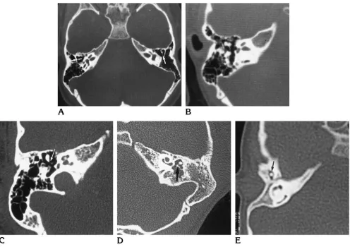
Fig 8. Axial CT scans of ossicle dysplasia.
A, Axis rotation and arthrodesis of the incudomalleal joint are on the right; the left side is normal (arrow). B, Fusion and adherence of the incudomalleal joint.
C, Dysplasia of the incudostapedial joint (arrow).
D, Rudimentary ossicles (arrow) in an opacified middle ear.
E, Ectopic (anterior) ossicle (arrow) in an obliterated temporal bone.
Fig 9. Oval window.
A, Coronal CT scan shows closed oval window (arrow) in a pneumatized middle ear, with loss of all ossicles and external auditory canal atresia.
[image:7.587.50.380.517.696.2]in half the patients with minor microtia and in three quarters of those with major microtia. Mostly, the dorsal attic wall was displaced ven-trally, reaching the level of the round window, leading to a deep tympanic sinus (Fig 10C and E). In eight of these cases, the facial nerve left the mastoid cranially to the stylomastoid fora-men in a ventral direction to the temporoman-dibular joint; in three cases, the exit was in a lateral direction. All but one of these patients had major microtia. A posterior course of the seventh nerve was found in two microtic and two normal ears. In one case of first-degree
auricular dysplasia, the mastoid facial nerve coursed medial to the tympanic sinus. The CT appearance could suggest splitting of the facial nerve, but it was indistinguishable from vascular canals leading to the nerve. In one case, dupli-cation was proved intraoperatively (combined with dehiscence, closure of the oval window, and loss of the stapes).
Internal Auditory Canal
[image:8.587.51.549.82.483.2]The internal auditory canal was dysplastic in one sixth of all patients with microtia. Typical
Fig 10. Axial CT scans of facial nerve canal aberration.
A, Caudal course of the second segment (arrow) and opacified narrowed tympanum (arrowhead). B, Dehiscence in the caudally displaced tympanic segment (arrowhead).
C, External auditory canal atresia and ventral displacement of the mastoid segment (black arrow), deepened sinus tympany (white arrow), and closed round window (lacking “air bubble”) on the right; normal round window, sinus tympani, and mastoid facial nerve (arrowheads) on the left, from ventral to dorsal.
D, Sagittal CT reformation of the normal mastoid facial nerve canal (arrow).
was an inclined course from craniomedial to caudolateral and widening of the porus (Fig 11A). In one patient, instead of the internal au-ditory canal, only a thin canal of the seventh and eighth nerve was preserved (Fig 11B), combined with a hypoplastic labyrinthine seg-ment of the facial nerve canal.
Round Window
The round window was absent in only 6% of the severely microtic ears (Fig 12). Closure of the round window was not significantly corre-lated with severe microtia or other dysplasias. It occurred also in two patients with well-pneuma-tized middle ear cavities (Fig 10C).
Labyrinth
The inner ear appeared slightly dysplastic in 13% of patients with microtia (P,.02); there was only one severe anomaly of this structure. In 13 cases, the lateral semicircular canal was hypo-plastic (Fig 13A); in six cases, the vestibule was abnormal (Fig 13B). Minor labyrinthine anoma-lies were especially frequent in patients with Franceschetti syndrome. In one case of thalido-mide embryopathy, only rudiments of the whole labyrinth, including the cochlea, were present bilaterally (Fig 13C). One case showed marked widening of the vestibular aqueduct (Fig 13D).
Discussion
To aid in understanding the complex anom-alies of the external, middle, and inner ear, we present a short synopsis of its embryology.
Development of the inner ear begins in the third week of gestation with formation of the otic placode, an ectodermal thickening in the neigh-borhood of the myelencephalon. Invagination of the otic placode leads to an otic pit and to fusion of their external lips to the otic vesicle. In the eighth week, the otic capsule is formed, provid-ing the stapes footplate and the ligament of the oval window. By the 12th week, the labyrinth is well differentiated. The facial nerve grows to reach its destinations between the fourth and fifth week.
The middle and external ear develops from the mesodermal first and second branchial arch and the endodermal first pharyngeal pouch be-tween the fourth and 30th weeks. Developmen-tal anomalies of the first pharyngeal pouch lead to disturbances of the eustachian tube and of the tympanic and mastoid pneumatization. Fail-ure of differentiation of the first branchial arch leads to malformations of the incudomalleal joint, tensor tympani muscle, and mandible. Failure of differentiation of the second branchial arch affects the facial nerve canal, the stapedius muscle, the lower part of the ossicular chain, and the styloid process. Disorders of the first and second branchial arches also result in dys-plasia of the auricular cartilage (leading to mi-crotia in the seventh to eighth week, the earlier the more severe, or to anotia in the seventh week).
The external auditory canal arises from deep-ening of the first branchial groove in the ninth week. Opening of the bony part of the external auditory canal starts only in the 30th week, after complete differentiation of the inner, middle,
Fig 11. Abnormality of the internal auditory canal.
A, Coronal CT reformation of an abnormal craniomedial to caudolateral course and widening of the internal auditory canal (arrowhead) on the left; the right side shows a normally horizontal direction.
[image:9.587.52.546.85.236.2]B, Axial CT scan of hypoplastic internal auditory canal (arrow).
and outer ear. Failure of the epithelial cells of the first branchial groove to split causes steno-sis or atresia of the external auditory canal, which might be isolated in an otherwise normal temporal bone (16 –19). Anomalies of the inter-nal carotid artery are thought to be caused by maldevelopment of the third branchial arch dur-ing the fourth week (20).
A variety of pathologic changes associated with microtia can be detected by high-resolu-tion CT (17, 21–23). We found that the extent of the auricular anomalies corresponded to the se-verity of changes in the external auditory canal (ie, stenosis or atresia). Excessive inclination of the external and internal auditory canals may be explained by incomplete ascension of the auri-cle from the neck (15).
The degree of auricular anomaly was also highly correlated with dysplasias of the middle ear cavity, mastoid cells, malleus, and incus, presumably because of a common embryolog-ical origin. The connection between
[image:10.587.217.546.84.443.2]malforma-tion of the auricule/external auditory canal and development of the middle ear as a whole was recently characterized (24) by means of Jahrs-doerfer’s score (25). Just as frequent were ab-errations of the facial nerve canal that devel-oped from the second branchial arch. Ventral dislocation of the mastoid segment might be caused directly by hypoplasia of Reichert’s car-tilage and the ventrally migrated mastoid, as described by Curtin (23). Ventral placement of the dorsal tympanic wall leads to a deep tym-panic sinus. The chorda tympani then courses lateral to the atresia plate (17). These changes corresponded to the severity of microtia and atresia. In some cases of anterior displacement of the mastoid segment and severe microtia, the facial nerve canal turned anteriorly or, less commonly, laterally, leaving the temporal bone to the temporomandibular articulation instead of continuing downward toward the stylomas-toid foramen. This finding corresponds to the observations of Proctor (26). The more frequent
Fig 13. Axial CT scans of labyrinthine dysplasia.
A, Diminutive lateral semicircular ca-nal (arrow) and hypoplasia of the tym-panic part of the temporal bone on the microtic side (right); the left side is nor-mal.
B, Dysplastic vestibulum (arrowhead) in Franceschetti syndrome.
C, Severe bilateral dysplasia of the lab-yrinth, including the cochlea (arrows) and the middle and external ear in thalid-omide embryopathy.
variation of the mastoid segment in humans with normal auricles is a posterior course (26, 27). We found this posterior “hump” less often, and not in connection with microtia. Caudal dis-placement and dehiscence of the tympanic seg-ment were also frequent CT findings, corre-sponding to the cases reported by Swartz (28). In two of our patients, the seventh nerve on the microtic side went straight down from the genic-ulate ganglion ventral to the tympanum, in ac-cordance with the description of a case of mi-crotia by Fowler (27). Another interesting finding was a facial nerve medial to the tym-panic sinus. A detailed classification of the fa-cial nerve aberrations and splitting based on anatomic specimens and surgical findings has been provided by Proctor (26), Fowler (27), and Mu¨ndnich and Terrahe (29). CT findings some-times suggest a separation of the facial nerve, but additional canals converging with the mas-toid segment (or chorda tympani) are mostly vascular (30). A true nerve separation might be confirmed only by MR imaging (31–35). In opacified temporal bones, visualization of the fallopian canal by CT is difficult, and
T1-weighted MR imaging with a 5123 512 matrix
and thin sections may have an advantage. Fa-cial (and abducens) nerve palsy is more com-mon in embryopathies. We did not find a corre-lation between facial nerve canal aberration and facial palsy; a bony facial nerve canal might be “empty” (26).
Abnormalities of structures representing the border between the branchial arches and the otic capsule, such as the stapes, oval window, and tympanic segment of the facial nerve canal, were less frequent and not significantly associ-ated with the severity of auricular malforma-tions. The derivates of the otocyst, such as the round window and labyrinth, were rarely af-fected. Absence of the round window was ex-ceptional (6%) and was occasionally found in otherwise minor malformations of temporal bone. The cochlea was involved (bilaterally) in only one of our cases, and that was due to
thalidomide embryopathy. Embryopathies,
whether toxic (eg, thalidomide) or viral (eg, ru-bella), generally carry a high risk of inner ear malformation and deafness (18). With regard to genetic disorders, a controversial discussion in the literature of the past concerned the issue of whether the inner ear is involved in malforma-tions of the external and middle ear. Since the advent of polytomography, changes of
labyrin-thine structures in microtia have most often been described at the lateral semicircular canal (2, 36, 37), which is in keeping with our find-ings. In our cases, lateral semicircular canal
hypoplasias occurred relatively frequently
(13%), particularly in patients with severe in-volvement of the middle ear. The relationship between these anomalies is unclear. The lateral semicircular canal has a spatial relationship to the tympanic cavity, but these structures have separate embryologic derivations. Although the otic capsule begins its development before the middle ear is formed, complete differentiation of the labyrinth might be influenced by branchial structures. In cases of inner ear malformation without microtia, lateral semicircular canal dys-pasia appears in 40% (17). A detailed description of inner ear malformations was published by Mu¨ndnich and Terrahe (29) and radiologic find-ings by Curtin (23). Even changes of the vestib-ular organ seem to be a sign of inner ear dysfunc-tion (38), but audiometry is decisive in this instance. We did not find a CSF fistula, common in labyrinthine dysplasias (but not microtia).
As in the report by Tanghe (17), we found few changes in the labyrinthine segment of the fa-cial nerve canal. Severe dysplasias of the inter-nal auditory cainter-nal are also rare. Only a few reports concern internal auditory canal abnor-malities in the setting of microtia (29), and a few cases of aberrations of the canalicular segment of the facial nerve canal (without microtia) have also been reported (26, 29, 39).
Epidermoids or cholesteatomas, congenital or secondary, can involve the facial nerve, inner ear, and brain, and they require surgery (40). Congenital cholesteatomas account for 2% of all cholesteatomas and are located at varying places in the temporal bone. In some of our cases there was a typical pattern. Epidermoids also occur intracranially, most often in the cer-ebellopontine angle, as in our patients, or in the suprasellar cistern (41).
the middle ear (embryological inferior tympanic artery) and may result in enlargement of the tympanic segment of the facial nerve canal. This is caused by the persistent stapedial artery, which leads to the medial meningeal artery (42). Hypoplasia or agenesis of the carotid ca-nal is uncommon and is more often unilateral (as in our two cases) than bilateral (43). It may be associated with intracranial aneurysms (44, 45) and neurovascular compressive symptoms due to compensatory vertebral artery ectasia (46).
In conclusion, we found a decrease in the rate of microtic anomalies from the external auditory canal to the middle ear, the boarder of the mid-dle and inner ear, the labyrinth, the internal auditory canal, and the cerebellopontine angle. The current most important and validated pre-operative criteria for hearing repair surgery (12), proposed by Jahrsdoerfer (25) and re-cently substantiated (47), include the clinical appearance of the external ear and the findings at CT examination. The ear surgeon is inter-ested in multiplanar, especially sagittal, views when planning restoration of the external audi-tory canal. Therefore, direct scanning in these planes (at a low dose) or reconstruction of a three-dimensional data set, acquired by spiral CT (to prevent motion artifacts when imaging children), is necessary.
References
1. Melnick M, Myrianthopoulos NC, Paul NW. External ear malfor-mations: epidemiology, genetics and narural history. In: Birth Defects: Original Article Series.New York, NY: Alan Liss; 1979; 15:1–140
2. Mu¨ndnich K. Ho¨rverbessernde und Plastische Operationen bei Ohrmissbildungen. In: Berendes J, Heilkunde RL, and Zo¨llner F, eds. Hals-Nasen-Ohren Heilkunde. Stuttgart, Germany: Georg Thieme; 1966;3(part 1):637– 668
3. Lenz W. Das Thalidomid-Syndrom.Fortschr Med1963;81:148 – 158
4. Ballantyne GW. The teratologic records of Chaldea.Teratologia
1894;1:127–132
5. Hrdlicka A. Seven prehistoric American skulls with complete ab-sence of external auditory meatus.Am J Phys Anthropol1933; 17:355–360
6. Marx H. Die Missbildungen des Ohres. In: Denker A, Kahler O, eds.Handbuch der Hals-Nasen-Ohrenheilkunde.Berlin, Germa-ny: Springer; 1926;6:565–583
7. Rogers B. Anatomy, embryology and classification of auricular deformities. In: Tanzer R, Edgerton M, eds.Symposium on Recon-struction of the Auricle.St Louis, Mo: Mosby; 1974;10:3–11 8. Weerda H. Reconstructive surgery of the auricle.Fac Plast Surg
1988;5:399 – 410
9. Weerda H. Plastic surgery of the ear. In: Booth J, ed.Otology. 5th ed. London, England: Butterworth; 1987;3:185–202
10. Weerda H, Bockenheimer S, Truebi M. Middle ear surgery in congenital malformations of the auricle with atresia: experience with 89 operated ears.HNO1985;33:449 – 452
11. Lambert PR. Major congenital ear malformations: surgical man-agement and results.Ann Otol Rhinol Laryngol1988;97:641– 649
12. Aguilar EA, Jahrsdoerfer RA. The surgical repair of congenital microtia and atresia.Otolaryngol Head Neck Surg1988;98:600 – 606
13. Jahrsdoerfer RA. Clinical aspects of temporal bone anomalies.
AJNR Am J Neuroradiol1992;13:821– 825
14. Weerda H. Classification of congenital deformities of the auricle.
Fac Plast Surg1988;5:385–388
15. Schumacher GH, Christ BEA.Embryonale Entwicklung des Men-schen. Anatomie und Klinik.10th ed. Berlin, Germany: Ullstein Mosby; 1993:373–374
16. Siegert R, Weerda H, Remmert S. Embryology and surgical anat-omy of the auricle.Fac Plast Surg1994;232–243
17. Tanghe H. Congenital malformations and anatomic variations of the ear.Riv Neuroradiol1994;7:341–348
18. Mattox DE, Nager GT, Levin LS. Congenital aural atresia: embry-ology, pathembry-ology, classification, genetics and surgical manage-ment. In: Paparella MM, Shumrick DA, eds.Otolaryngology.3rd ed. Philadelphia, Pa: Saunders; 1991;20:1191–1225
19. Nager GT, Levin LS. Congenital aural atresia: embryology, pa-thology, classification, genetics and surgical management. In: Paparella MM, Skumrick DA, eds.Otolaryngology.Philadelphia, Pa: Saunders; 1980:1303–1344
20. Lie TA. Congenital anomalies of the carotid arteries.Excerpta Medica.Amsterdam, Netherlands: 1968;35–51
21. Bockenheimer S, Weerda H, Hartenstein V. Das hochaufloesende Computertomogramm des Felsenbeines bei Ohrmuschelmissbil-dungen.Arch Otolaryngol1984;(suppl II):211–213
22. Swartz JD, Wolfson RJ, Popky GL, et al. External auditory canal dysplasia: CT evaluation.Laryngoscope1985;95:841– 845 23. Curtin HD. Congenital malformations of the ear.Otolaryngol Clin
North Am1988;21:317–336
24. Kountakis SE, Helidonis E, Jahrsdoerfer RA. Microtia grade as an indicator of middle ear development in aural atresia.Arch Otolar-yngol Head Neck Surg1995;121:885– 886
25. Jahrsdoerfer RA, Yeakley JW, Aguilar EA, Cole RR, Gray LC. Grading system for the selection of patients with congenital aural atresia.Am J Otol1992;13:6 –12
26. Proctor B.Surgical Anatomy of the Ear and Temporal Bone.New York, NY: Thieme; 1989:100 –111
27. Fowler EP. Variations in the temporal bone course of the facial nerve.Laryngoscope1961;71:937–946
28. Swartz JD. The facial nerve canal: CT analysis of the protruding tympanic segment.Radiology1984;153:443– 447
29. Mu¨ndnich K, Terrahe K. Mißbildungen des Ohres. In: Berendes J, Link RL, Zo¨llner F, eds. Hals-Nasen-Ohrenheilkunde. 2nd ed. Stuttgart, Germany: Thieme; 1979;18:1– 49
30. Dimiopoulos PA, Muren C, Smedby Oe, Wadin K. Anatomical variations of the tympanic and mastoid portions of the facial nerve canal: a radioanatomical investigation.Acta Radiol(in press) 31. Dohringer E, Albegger K, Sinzinger G, Schmoller HJ.
Idiopa-thische Fazialisparese und Magnetresonanztomographie. HNO
1991;39:362–366
Meeus L. Constructive interference in steady state-3DFT MR im-aging of the inner ear and cerebellopontine angle.AJNR Am J Neuroradiol1993;14:47–57
34. Casselman JW, Kuhweide R, Ampe W, Meeus L, Steyaert L. Pathology of the membranous labyrinth: comparison of T1- and T2-weighted and gadolinium-enhanced spin-echo and 3DFT-CISS imaging.AJNR Am J Neuroradiol1993;14:59 – 69 35. Roos EH, Lufkin RB. Magnetic resonance imaging of the temporal
bone. In: Swartz JD, ed.Imaging of the Temporal Bone. Philadel-phia, Pa: Saunders; 1986;9:203–211
36. Franceschetti A, Klein D. Mandibulofacial dysostosis: a new he-reditary syndrome.Acta Ophthalmol1949;27:143–148 37. Hutchinson JC, Caldarelli DD, Valvassori GE, Pruzansky S, Parris
PJ. The otologic manifestation of mandibulofacial dysostosis.
Trans Am Acad Ophthalmol Otolaryngol1977;84:520 –528 38. Murata K, Isono M, Ohta F, Yoshida A, Ishida O. Quantitative
analysis of inner ear anomalies by high resolution CT scanning of the temporal bone.J Otolaryngol1987;16:133–136
39. Weissman JL, Arriga M, Curtin C, Hirsch B. Duplication anomaly of the internal auditory canal.AJNR Am J Neuroradiol1991;12: 867– 869
40. Mehra YN, Dubey SP, Mann SBS, Suri S. Correlation between high-resolution computed tomography and surgical findings in
congenital aural atresia.Arch Otolaryngol Head Neck Surg1988; 114:137–141
41. Swartz JD. Middle ear and mastoid. In: Swartz DJ, ed.Imaging of the Temporal Bone.Philadelphia, Pa: Saunders; 1992:33– 87 42. Harnsberger HR. Vascular aberrations. In: Swartz DJ, ed.Imaging
of the Temporal Bone.Philadelphia, Pa: Saunders; 1992:154 –191 43. Grand CM, Louryan S, Bank WO, Baleriaux D, Brotchi J, Raybaud C. Agenesis of the internal carotid artery and cavernous sinus hypoplasia with contralateral cavernous sinus meningioma. Neu-roradiology1993;35:588 –590
44. Waga S, Okada M, Kojima T. Saccular aneurysm associated with absence of the left cervical carotid arteries.Neurosurgery1978; 3:208 –212
45. Kunishio K, Sunami N, Yamamoto Y, Asari S. Agenesis of the left internal carotid artery, common carotid artery and the main trunk of the external carotid artery associated with multiple cerebral aneurysms: case report.Neurol Surg(Tokyo) 1986;14:873– 879 46. Kiuchi K, Kowada M, Kojima H. Hypoplasia of the internal carotid artery associated with spasmodic torticollis: the possible role of altered vertebrobasilar hemodynamics.Neuroradiology1995;37: 362–364