THE USE OF
SURFACE ELECTROMYOGRAPHY
WITHIN
EQUINE PERFORMANCE ANALYSIS
JANE MICHELLE WILLIAMS
A thesis submitted in partial fulfilment of the requirements of the University of the West of England, Bristol for the degree of Doctor of Philosophy by Publication
Faculty of Health and Life Sciences, University of the West of England, Bristol
i CONTENTS
Chapter Page
number
Contents page i
List of tables vi
List of figures viii
List of plates viii
List of appendices ix
Evidence sources presented in the thesis xi
Acknowledgements xii
Statement of authorship xiii
Statement of work xiv
Statement of training xv
Abstract xvi
1 THE RESEARCH JOURNEY: AN INTRODUCTION
1.1What is surface electromyography? 1.2sEMG as a performance analysis tool 1.3Performance analysis in equestrian sport 1.4Hypotheses
1.5Research aims 1.6Research objectives 1.7Structure of the thesis
1 1 2 3 6 7 7 8
2 EQUINE PERFORMANCE
2.1 The equine athlete
ii
2.2 Equine performance: a multifactorial concept 2.3 Defining success
2.4 Why investigate equine performance? 2.5 Evidence source 1
12 14 18 20
3 AN INTRODUCTION TO SURFACE
ELECTROMYOGRAPHY
3.1 Introduction to electromyography 3.2 Interpretation of the electrical signal
3.3Indwelling versus surface electromyography 3.4sEMG versus indwelling EMG in the horse 3.5The Delsys® Trigno ™ sEMG system 3.6Data collection
3.7Physiological influences on the EMG signal 3.8 Data processing
3.8.1 Filters
3.8.2 Full wave rectification 3.8.3 Linear enveloping 3.8.4 Integrated EMG
3.9 Interpretation of the processed EMG signal 3.9.1 Muscle fibre profile
3.9.2 Contraction type 3.9.3 Comparing events 3.9.4 Assessment of fatigue
21 21 22 24 29 29 32 34 35 37 41 41 43 44 44 44 45 46
4 SURFACE ELECTROMYOGRAPHY AND THE EQUINE
ATHLETE
iii
4.1 An introduction to muscle physiology 4.1.1 Muscle contraction
4.2 Muscle supporting performance
4.2.1 The influence of muscle fibre profiles 4.2.2 Muscle fibre recruitment during exercise 4.3 Principles of training to promote performance
4.3.1 Evaluation of training regimens 4.4 Training equine muscle
4.5 Training the ‘whole’ horse
4.6 sEMG and the horse
4.7 Application of sEMG to training the equine athlete 4.7.1 Muscle recruitment
4.7.2 A balanced athlete 4.7.3 Fitness and fatigue
4.7.4 Training versus competition 4.7.5 Injury
4.8 Challenges in equine sEMG research 4.8.1 Preparation
4.8.2 Dynamic evaluation 4.8.3 Speed
4.8.4 Individuality 4.9 Evidence source 2
4.9.1 Rationale
4.9.2 Research methods and limitations
4.9.3 Contribution to the field of equine
iv
performance
4.9.4 Implications and questions generated 4.10 Evidence source 3
4.10.1 Rationale
4.10.2 Research methods and limitations
4.10.3 Contribution to the field of equine performance
4.10.4 Implications and questions generated 4.11 Evidence source 4
4.11.1 Rationale
4.11.2 Research methods and limitations
4.11.3 Contribution to the field of equine performance
4.11.4 Implications and questions generated
105 107 108 108 112 113 114 114 116 118 119
5 DISCUSSION
5.1 Field assessment of sEMG in the equine athlete 5.2 Muscle recruitment
5.3Muscle activity
5.3.1 Measures of muscle activity
5.4 Comparison to previous equine sEMG studies 5.5 Laboratory versus field assessment
5.6 Individuals versus defined samples
5.7 sEMG: a relevant performance analysis tool? 5.8 Limitations and challenges within sEMG research 5.9 The future of sEMG research
v
5.10 Applied equine performance research
5.10.1 The future of applied equine research 5.11 Spreading the message
5.12 Final thoughts
141 144 145 145
6 CONCLUSIONS 147
References 149
Appendices 184
A1 Evidence sources presented in the thesis 185
A2 Definition of authorship 196
A3 Collaborative relationships 197
A4 Attainment of Doctoral learning criteria 201
A5 Training and Continuing Professional Development 204
A6 Glossary of terms and list of abbreviations 205
A7 Curriculum Vitae 208
A8 Reflection on the research journey 229
A9 Practical sEMG demonstration ISES 2012 233
vi
LIST OF TABLES
Table Page
number
1 Examples of equestrian measures of success 16
2 Electromyography measures of muscle performance 23
3 Comparison of fine-wire and needle EMG 26
4 Advantages and disadvantages of surface and indwelling EMG
27
5 Technical specifications of the Delsys® Trigno™ Wireless EMG system
31
6 Factors which can influence the EMG signal 32
7 The three common applications of sEMG 36
8 Examples of EMG filtering protocols utilised in human dynamic studies
42
9 Types of contraction in skeletal muscle 49
10 Key features of motor units which can impact force production 51
11 Characteristics of equine muscle fibre types 55
12 Factors which can influence equine muscle fibre Profiles
56
13 Distribution of fibre type in horses trained for various disciplines
57
14 Key objectives when training the performance horse 59 15 Intrinsic and extrinsic factors that can influence equestrian
training and performance
60
vii
training in the horse and their relationship to sEMG
17 Categories of training for the equine athlete 68 18 Summary of equine muscular adaptations to training 69
19 Equine electromyography research 74
20 Muscle fibre recruitment during exercise in the horse 86 21 Variables which can influence the reliability or interpretation of
sEMG data during equine research
91
22 Sources of noise in the sEMG signal 92
23 Inclusion criteria St George and Williams (2013) 109 24 Key areas for recommended future sEMG research areas 142
APPENDICES:
A2 Overview of methodologies and author contribution within the evidence sources
196
A3 Overview of collaboration for the research presented in the thesis
200
viii
LIST OF FIGURES
Figure Page
number 1 An overview of equine sporting disciplines in Great Britain 10
2 Overview of British equestrian competition 11
3 EMG electrode pickup zone 22
4 EMG features which can be an measured within a motor unit action potential
24
5 Application of different types of EMG 25
6 The four basic filter types 39
7 Equine muscle fibre recruitment during exercise 54 APPENDICES:
A10.1 Muscle hierarchy 239
LIST OF PLATES
Plate Page
number
ix
LIST OF APPENDICES
Appendix Page
number A1 Evidence sources presented in the thesis:
A1.1 Evidence source 1
A1.1A Reflection on evidence source 1 A1.2 Evidence source 2
A1.2B Erratum A1.3 Evidence source 3 A1.4 Evidence source 4
185 187 188 192 193 194 195
A2 Definition of authorship 196
A3 Collaborative relationships 197
A4 Attainment of Doctoral learning criteria 201
A5 Training and Continuing Professional Development 204
A6 Glossary of terms and list of abbreviations 205
A7 Curriculum Vitae 208
A8 Reflection on the research journey A8.1 Developing a research philosophy
A8.2 Reflection on personal development during the research journey
229 229 231
A9 Practical sEMG demonstration ISES 2012 A9.1 Rationale
A9.2 Research methodologies and limitations A9.3 Summary of demonstration and results
x
A9.3.1 Visual assessment of muscle activity A9.4 Contribution to the field of equine performance A9.5 Implications and questions generated
236 236 237 A10 Skeletal Muscle
A10.1 Anatomical hierarchy A10.2 Functionality
A10.3 Muscle fibre characteristics A10.3.1 The sarcomere A10.3.2 Force-length curves A10.4 Muscle twitch
A10.5 Excitation - contraction coupling A10.6 Energy requirements of contraction
xi Evidence sources presented in the thesis
Evidence source 1: Williams, J.M. (2013) Performance analysis in equestrian sport Comparative Exercise Physiology. 9(2), pp. 67-77.
Evidence source 2: Williams, J.M., Gundry, P., Richards, J. and Protheroe, L. (2013) A preliminary evaluation of surface electromyography as a tool to measure muscle fatigue in the National Hunt racehorse. The Veterinary Nurse. 4(9), pp. 566-572.
Erratum: Williams, J.M., Gundry, P., Richards, J. and Protheroe, L. (2013) A preliminary evaluation of surface electromyography as a tool to measure muscle fatigue in the National Hunt racehorse. The Veterinary Nurse. 5(6), pp. 319.
Evidence source 3: St George, L. and Williams, J.M. (2013) Electromyographic evaluation of approach stride, jump stride and intermediate stride in selected superficial muscles of the jumping horse: A preliminary study. Comparative Exercise Physiology. 9(1), pp. 23-32.
xii Acknowledgements
I would like to take the opportunity to thank my husband, Karl, for his support and ability to make me laugh throughout the research process. Thanks are also extended to my supervisory team: Dr Jon Pollock, Dr Stephen Waite and in particular Dr Hayley Randle who initiated this journey many years ago. Recognition is also required for the input of Delsys® and my colleagues throughout the research projects undertaken and heartfelt gratitude is extended to Dr Tim Parkin and Professor Jim Richards for their support and continued encouragement. Thanks are also extended to the undergraduate and postgraduate students from Hartpury College for their enthusiasm in my research ideas and for their help in collecting the data presented in the evidence sources.
xiii Statement of authorship
I can confirm that the research presented in the critical commentary is the product of my own work. The research projects incorporated have been performed, interpreted and prepared for publication by myself in conjunction with colleagues; Table A2 (Appendix 2) explicitly identifies my contribution to the work presented.
xiv Statement of work
xv Statement of training
xvi Abstract
Equine athletes participate in a wide range of equestrian disciplines. Performance analysis in sport is the collection and subsequent analysis of data, or key information sets, related to facets of training and / or competition, to accelerate and improve athletic performance. Equine performance analysis research aims to optimise the potential competition success of the horse whilst concurrently promoting health and welfare and increasing career longevity. Despite the benefits associated with performance analysis, its application is limited in equine sport.
Surface electromyography (sEMG) is a non-invasive technique which illustrates recruitment patterns of superficial skeletal muscle and can provide quantitative data on the activity within muscle during dynamic motion. sEMG has the potential to contribute to equine performance analysis particularly via assessment of muscle recruitment, activity and adaptation within training regimens and during competition. The critical commentary demonstrates the potential of surface electromyography (sEMG) as an effective performance analysis tool that could be used to assess the physiological response of muscle during field-based exercise in the horse and provides examples of how sEMG data obtained could guide improvements in the efficacy of training regimens for the equine athlete.
xvii
interval training. Opportunities also exist to determine the efficacy of muscle-related clinical and therapeutic interventions such as prophylactic dentistry or physiotherapy. The preliminary research presented suggests the use of equine sEMG as a performance analysis tool has most value to assess and compare muscle performance during exercise within individual horses. However further research is required to substantiate this. Future studies integrating larger sample sizes, horses selected from specific equestrian disciplines and breeds, and further exploration of the impact of coat length and sEMG sensor placement on data obtained would be worthwhile to standardise and validate the protocols employed here.
Equine performance is a complex area; future work needs to focus on the individual characteristics that contribute to desired performance goals, but should also evaluate performance as a holistic entity. It is essential for progression in the performance field that research undertaken is shared with the equine industry to enable practical implementation. The use of sEMG in the equine athlete has the potential to increase understanding of how muscle responds to exercise and could help create an evidence-base to inform individual and discipline-specific training regimens. Increased efficacy in training should promote success, enhancing performance and extending career longevity for the equine athlete, whilst indirectly benefiting the horse’s health and welfare through improved management practices and injury reduction.
1
CHAPTER ONE
THE RESEARCH JOURNEY: AN INTRODUCTION
This commentary focuses on the use of surface electromyography (sEMG) in the analysis of the muscle performance of the equine athlete. The evidence sources (Appendices 1.1 to 1.4) demonstrate the potential of sEMG as a quantitative tool for equine performance analysis, which could inform training and management practices within the equine industry.
1.1 What is surface electromyography?
non-2
invasive nature and ability to utilise sEMG in the field presents opportunities for the technology to be used within performance analysis.
1.2 sEMG as a performance analysis tool
Electromyography has been used as a performance analysis tool to assess the efficacy of training and rehabilitation regimens in humans (Richards et al., 2008; Hanon, Thepaut-Matieu and Vanderwalle, 2005). sEMG can identify muscle recruitment through the onset and offset of motor unit action potentials (MUAP) (Winter, 2009) and measure muscle activity-levels via analysis of EMG signal amplitude and frequency (Hanon, Thepaut-Matieu and Vanderwalle, 2005; Hanon et al., 1998). sEMG data provide an objective measure of (muscle) fitness (Duc, Betik and Grappe, 2005) and fatigue (Hanon, Thepaut-Matieu and Vanderwalle, 2005) through EMG frequency analysis. Some equine EMG research has been conducted, but the majority of studies have been restricted to the laboratory environment (Zsoldos et al., 2010a, b; Licka, Frey and Peham, 2009; Peham et al., 2001).
3 1.3 Performance analysis in equestrian sport
Performance analysis in sport relies on the objective assessment of data or key information related to training and / or competition, to improve athletic performance (Hughes and Bartlett, 2002). An individual human’s or Equid’s performance capacity is underpinned by the preparation or training regimen implemented for the targeted competition test. The reliability of the existing anecdotal evidence-base for the training practices employed across equestrian sport has been questioned (van Weeren and Back, 2014; Ely et al., 2010; McLean and McGreevy 2010a; McGreevy and McLean, 2007). Training regimens typically aim to prepare the equine athlete for specific competitive tests. Several years of physiological conditioning and technical training (the acquisition of discipline-specific motor skills) are required for the equine athlete to reach their full potential (Leisson, Uaakma and Seene, 2008). However, despite scientific advances and the financial rewards associated with success in most disciplines (Thiruvenkadan, Kandasamy and Panneerselvam, 2009), training in equestrianism remains largely based upon tradition which itself is based on anecdotal evidence of prior achievements (McLean and McGreevy 2010a; Powers and Harrison, 1999; Smith et al., 1999).
4
which muscles are recruited during golfing-swings and demonstrated differences between the modern and traditional golf-swing are not influenced by expertise (handicap) (Ashish, Shweta and Singh, 2008; Aggarwal, Shenoy and Sandhu, 2008).
5
understanding of factors which contribute to muscle fatigue and how to condition muscles to sustain and optimise physical activity to improve performance.
6
A better understanding of muscle physiology during specific exercise combined with examination of how muscle responds to cumulative exercise sessions over time (training regimens) is needed (Ferrari et al., 2009). Using sEMG to assess muscle exercise physiology, using actively competing equine athletes (Felici, 2006), could increase the evidence-base against which current equestrian training regimens can be assessed and, if needed, inform modifications to make regimens more fit for purpose (Hughes and Bartlett, 2002). As a result of the lack of an evidence-base in equine training, it was decided to investigate if sEMG could be used as a valid tool to analyse equine performance through the examination of muscle recruitment and activity in proven equine athletes in the field.
1.4 Hypotheses
7 1.5 Research aims:
The overarching aims of the research were to demonstrate:
1. That telemetric sEMG is a relevant technology allowing the evaluation of muscle activity and recruitment during ‘real-life’ assessment of training and competition, and,
2. The potential that muscle activity records obtained via sEMG could be used to guide improvements in training regimens for the equine athlete.
1.6 Research objectives
The work presented aimed to achieve the following research objectives:
1. To analyse the potential of sEMG as a tool to analyse muscle recruitment during defined activities, specifically cantering and when jumping a fence, in the horse,
2. To assess the potential of using sEMG as a comparative tool to enable quantitative measurement of muscle adaptation in the equine athlete:
a) to determine progress within training regimens via interval training, and,
b) to measure how muscle activity varies over time after routine dental-treatment (rasping), and,
8 1.7 Structure of the thesis
9
CHAPTER TWO
EQUINE PERFORMANCE
Measuring performance is complex across all sports due to the multiple factors which contribute to the targeted event undertaken. In equestrianism, the presence of a second, inarticulate athlete, the horse, complicates performance analysis further. Chapter Two introduces the equine athlete alongside the concept of equine performance to identify opportunities for increased analysis, via the contribution of field-based technologies such as sEMG.
2.1 The equine athlete
10
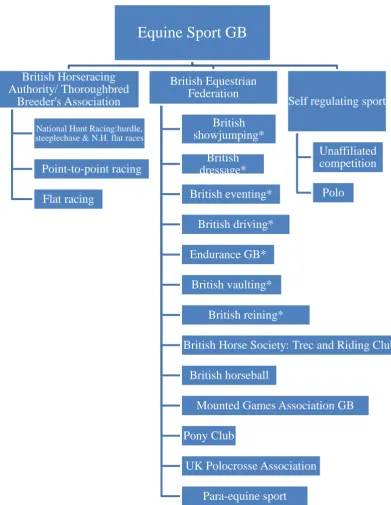
Figure 1: An overview of equine sporting disciplines in Great Britain; *FEI regulated disciplines.
Equestrian sport was reviewed in Great Britain to identify the range of equine sports that occur and the Governing Bodies, where appropriate, which regulate these sports.
Equine Sport GB
British Horseracing Authority/ Thoroughbred
Breeder's Association
National Hunt Racing:hurdle, steeplechase & N.H. flat races
Point-to-point racing
Flat racing
British Equestrian Federation
British showjumping*
British dressage*
British eventing*
British driving*
Endurance GB*
British vaulting*
British reining*
British Horse Society: Trec and Riding Clubs
British horseball
Mounted Games Association GB
Pony Club
UK Polocrosse Association
Para-equine sport
Self regulating sport
Unaffiliated competition
[image:29.595.136.528.68.574.2]11
[image:30.595.157.488.214.582.2]Equestrian competitions take place at affiliated and unaffiliated levels (Figure 2). Within competition, individuals are often assigned a status relative to competition experience and success of the rider, horse or horse and rider as a team. The unaffiliated sector mirrors the competitive disciplines; therefore work considering factors related to performance is applicable to both.
Figure 2: Overview of British equestrian competition. BHA: British Horseracing Authority (FEI, 2014; BHA, 2013); FEI: Federation Equéstré International; BD: British Dressage; BS: British
Showjumping; BE: British Eventing; BHS: British Horse Society; RC: Riding Clubs, PC: Pony Clubs.
12
2.2 Equine performance: a multifactorial concept
Equine performance is a complex phenomenon and many contributing factors interrelate to produce the tangible output observed. Epidemiological methodologies can be employed to review performance related variables such as precursory risk factors to injury or non-completion (Mata, Williams and Marks, 2012; Parkin et al., 2004; Stover, 2003; Williams et al., 2001) or to predict factors related to success (Williams, Heath and Da Mata, 2013; Marlin, Williams and Parkin, 2014). The potential also exists to engage in systematic review of relevant research related to injury, physiological or biomechanical parameters that are intrinsically or extrinsically associated with performance.
Knowledge of the critical components of performance can be utilised to inform training and management protocols for the equine athlete (Williams, 2013). However the challenge that exists in all forms of performance analysis is how to effectively evaluate the degree of interaction between contributing variables in order to analyse their cumulative impact on the final result (Stover, 2003).
13
invalidating direct appraisal between events, as observed in eventing (Williams, Marlin and Marks, 2012).
Defining performance within any sport is difficult as it requires analysis of a multifactorial output often without free access to all the influential factors that contribute to a specific performance (Hughes and Bartlett, 2002). The concept of sports performance analysis is well established in human sport (McGarry, 2009). Performance analysis depends on detailed review of physiological, biomechanical and psychological performance related variables and how these change in different environments, contextualised to the sporting discipline or test, and how they can be managed to optimise performance (McGarry, 2009; Hughes and Bartlett, 2002).
14
with competitive tests for example: speed (Pinchbeck et al., 2002), drop fences (Singer et al., 2008) and surfaces (Murray et al., 2010), and identified a genetic link with predisposition to tendon injury (Tully et al., 2013). Research has also evaluated emerging treatments which promote an improved prognosis and return to competition such as platelet rich plasma (Bazzano et al., 2013; Bosch et al., 2010) and stem cells (Godwin et al., 2012). Yet despite the advances made, tendon injury remains a leading cause of days lost from training or competition in the horse (Dyson et al., 2008). The reasons for a lack of progression in equine performance remain unknown but could be associated with the limited integration of scientific methods within training regimens or the poor uptake of performance analysis in equine sport in general (van Weeren and Crevier-Denoix, 2006).
2.3 Defining success
15
races or events at the same level due to environmental differences, could be linked to individual or team performance and can also be dependent on the stage of training of the athlete/s (McGarry, 2009).
16
valid and reliable data in the field which could identify muscle recruitment and activity-levels enabling usable conclusion related to equine performance to be drawn.
Table 1: Examples of equestrian measures of success
A keyword search of equestrian peer reviewed databases identified measures of success in equestrian sport and breeding. Additional discipline and competition success measures outlined by FEI and BHA were analysed to formulate a summary of commonly employed measures of success in the equine athlete.
Competitive performance
1. Genetic analysis of populations and individuals can identify polymorphic genetic markers which can indicate predilection for success related to specific performance variables for example a given race distance.
2. Individual performance in horseracing can be rated by prize money won, winning or cumulative wins and/or places in races, race times, a horse’s handicap rating or lifetime earnings.
3. The Olympic disciplines of showjumping, dressage and eventing rank horses (and riders) according to prize money won and competitive success, often via a cumulative points system and accumulated prize money.
4. Olympic, World Equestrian Games or Championship medals. Breeding
performance
1. Winning / placing in specific races of high renown to attain enhanced breeding value (known as attaining black type).
2. Breeding values can be based upon breed society’s grading systems were variables are assigned to individual horses’ combining an individual’s results with that of their offspring e.g. BLUP: best linear unbiased prediction.
3. Offspring competitive success.
4. Reproductive fecundity of stallions and mares.
5. Heritability indices, population measures which quantitatively assess phenotypic expression of performance for set biomechanical or gait-related criteria for selection of superior athletic potential or breeding value in the horse.
17
18
as a performance analysis tool if reliable data can be obtained to measure and assess muscle responses during exercise.
2.4 Why investigate equine performance?
Analysis of performance can be advantageous to sporting achievement. Obvious benefits include promoting competitive success in the combinations evaluated. At elite level, success can include winning medals within international competition leading to increased funding, which has the potential to cascade positively through all levels of the sporting discipline involved (Bosscher et al., 2009). Additional benefits for the wider population observing and participating in sport have also been recorded. For example, success in elite sport has been documented to benefit society through a positive association with national pride, happiness and socio-economic benefits (Hallman, Bruer and Kuhnreich, 2013). Within a discipline, funding may support work to enhance the knowledge of factors which contribute to success or to injury. Therefore increased understanding of competitive demands has the potential to improve health and welfare in all participants via improved preparation for competition (Hughes and Bartlett, 2002).
19
20 2.5EVIDENCE SOURCE 1
Williams, J.M. (2013) Performance analysis in equestrian sport. Comparative Exercise Physiology, 9(2), pp. 67-77.
21
CHAPTER THREE
AN INTRODUCTION TO
SURFACE ELECTROMYOGRAPHY
Muscles contribute the power to enable dynamic movement during exercise in the horse. Therefore evaluation of their contribution to performance is essential during equine performance analysis. Electromyography is the study of MUAPs, the electrical signals that occur in muscles during contraction, using variable types of electrode (Back and Clayton, 2001; Clayton and Schamhardt, 2001). Chapter Three introduces EMG and compares the different methodologies available for research: indwelling and surface EMG. Key concepts in data acquisition and processing are explored to enable reasoned judgements on the validity of sEMG to investigate muscle recruitment and activity.
3.1 Introduction to electromyography
22
depolarisation occurs and the distance from the source of the current to the electrode (Morris and Lawson, 2009).
Individual electrode overlap pickup zone electrode pickup zone (signal from same motor units)
Figure 3: EMG electrode pickup zone.
EMG evaluation of muscle requires the placement of two electrodes at a set distance from each other to record data effectively. Incorrect placement where the two electrodes are too close to each other can result in data anomalies related to the overlapping pickup zone. Each electrode will record motor unit action potentials (MUAP) within its own individual range but there will also be an overlap pickup zone representing where both electrodes are recording the same MUAP.
3.2 Interpretation of the electrical signal
23
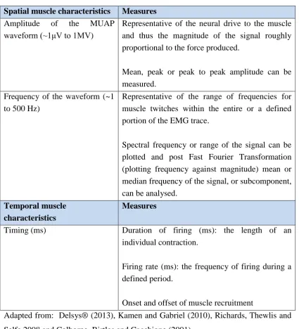
Table 2: Electromyography measures of muscle performance
Previous EMG research in humans and horses was reviewed to identify common characteristics associated with muscle contraction which can be measured by EMG. Characteristics were divided into temporal or timing related measures and spatial or workload related measures.
Spatial muscle characteristics Measures Amplitude of the MUAP
waveform (~1μV to 1MV)
Representative of the neural drive to the muscle and thus the magnitude of the signal roughly proportional to the force produced.
Mean, peak or peak to peak amplitude can be measured.
Frequency of the waveform (~1 to 500 Hz)
Representative of the range of frequencies for muscle twitches within the entire or a defined portion of the EMG trace.
Spectral frequency or range of the signal can be plotted and post Fast Fourier Transformation (plotting frequency against magnitude) mean or median frequency of the signal, or subcomponent, can be analysed.
Temporal muscle characteristics
Measures
Timing (ms) Duration of firing (ms): the length of an individual contraction.
Firing rate (ms): the frequency of firing during a defined period.
Onset and offset of muscle recruitment
24
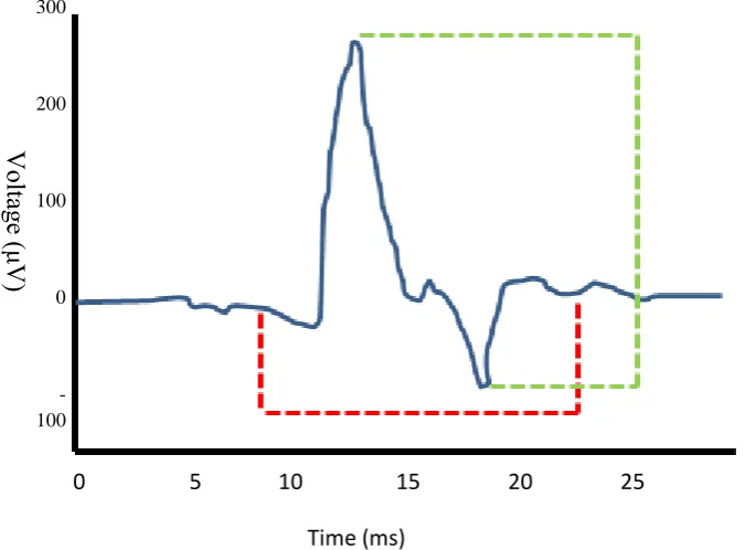
Figure 4: EMG features which can be measured within a motor unit action potential (MUAP): duration of MUAP; peak to peak amplitude of MUAP (adapted from Yousefi and Hamilton-Wright, 2014)
Surface electromyography records motor unit action potentials (MUAP). Analysis of EMG profiles obtained for the MUAP associated to muscle contraction for a defined activity can identify the duration of the MUAP i.e. how long a contraction is. The strength of contraction can also be estimated by measuring the peak to peal amplitude of MUAP within the EMG signal.
3.3 Indwelling versus surface electromyography
There are two methods of kinesiological EMG: indwelling EMG where electrodes are inserted into muscles of interest, and surface EMG where electrodes are applied to the extracellular skin surface above muscles of interest (Winter, 2009; Lamb and Hobart, 1992). Research has concluded that the results from each method are comparative; therefore experimental objectives and conditions should determine the choice of EMG employed (Chapman et al., 2010; Jacobsen, Gabel and Brand, 1995) as their scope varies (Drost et al., 2006) (Figure 5).
V
oltag
e (
μV)
300
200
100
0
-100
0 5 10 15 20 25
25
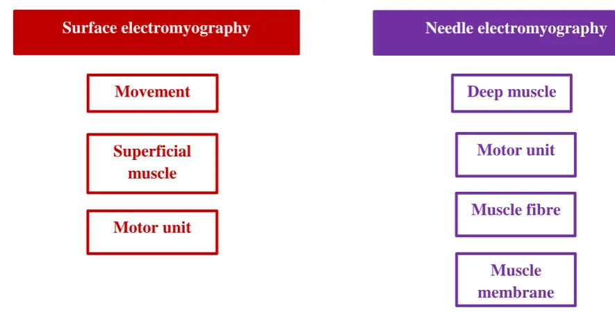
Figure 5: Application of different types of EMG (adapted from Drost et al., 2006)
Two types of electromyography (EMG) exist: surface and indwelling EMG. Surface EMG is non-invasive with sensors attached to the skin whilst indwelling EMG inserts fine-wire or needle electrodes into muscle. Surface EMG and needle EMG were selected to what each technique could be used to measure based upon reviewing EMG research in humans and the horse.
sEMG is a non-invasive technique which illustrates recruitment patterns of superficial skeletal muscle (Drost et al., 2006; Hanon, Thepaut-Matieu and Vanderwalle, 2005; Back and Clayton, 2001). There are two types of surface electrode: active and passive (Kamen and Gabriel, 2010). Active electrodes contain integral amplifiers and do not require the presence of electro- conductive gels and extensive skin preparation of passive electrodes (Drost et al., 2006). Indwelling EMG electrodes are smaller than surface electrodes and two types occur: fine-wire and needle EMG (Winter, 2009; Drost et al., 2006). Both techniques have been used in equine EMG studies (Wijnberg et al., 2003; Roberts et al., 2001; Colborne, Birtles and Cacchione, 2001), however the non-invasive nature of sEMG facilitates access
Movement
Superficial muscle
Deep muscle
Motor unit
Muscle fibre
Muscle membrane Motor unit
26
[image:45.595.108.525.245.647.2]to competitive equine athletes and is ethically more acceptable for use. Needle and fine-wire EMG are compared in Tables 3 and 4.
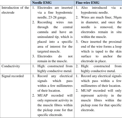
Table 3: Comparison of fine-wire and needle EMG
Two forms of indwelling electromyography (EMG) exist: needle and fine-wire EMG. Each method has unique and shared advantages and disadvantages. Previous reviews appraising the use of indwelling EMG were examined to summarise electrode insertion, outline conductivity and the signal each type of electrode records.
Needle EMG Fine-wire EMG
Introduction of the electrode
1. Electrodes are inserted via a fine hypodermic needle, 23-28 gauge. 2. Recording wires run
through the central cannula and have an uninsulated tip, which is placed into a specific area of interest for the targeted muscle.
3. Electrodes do not remain in the muscle.
1. Also introduced via a hypodermic needle.
2. Wires are much finer, 50μm in diameter, and once the needle is removed, the electrodes remain in situ within the muscle.
3. Once inserted the proximal end of the wire forms a loop which is taped to the skin surface to anchor the electrode in place.
Conductivity
1. High: constructed from highly conductive metal.
2. High: constructed from highly conductive metal. Signal recorded 1. Record any electrical
signals which pass within a few millimetres of their location.
2. MUAP recorded will only represent activity in the muscle fibres within the pickup zone for that specific electrode.
1. Record any electrical signals which pass within a few millimetres of their location. 2. MUAP recorded will only represent activity in the muscle fibres within the pickup zone for that specific electrode.
27
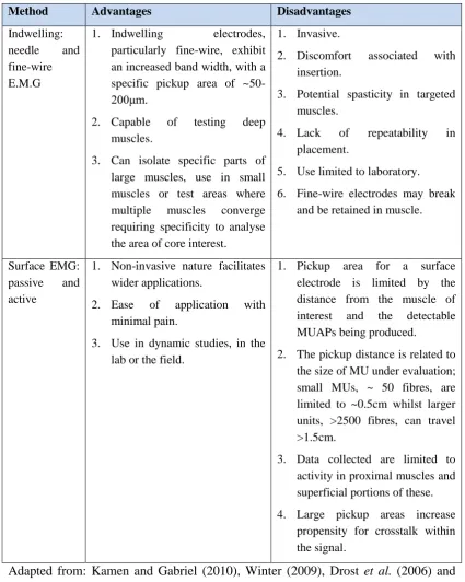
Table 4: Advantages and disadvantages of surface and indwelling EMG
Two types of electromyography (EMG) exist: surface and indwelling EMG. Previous reviews comparing the two methodologies were examined to summarise the advantages and disadvantages of surface EMG and indwelling EMG.
Method Advantages Disadvantages
Indwelling: needle and fine-wire E.M.G
1. Indwelling electrodes, particularly fine-wire, exhibit an increased band width, with a specific pickup area of ~50-200μm.
2. Capable of testing deep muscles.
3. Can isolate specific parts of large muscles, use in small muscles or test areas where multiple muscles converge requiring specificity to analyse the area of core interest.
1. Invasive.
2. Discomfort associated with insertion.
3. Potential spasticity in targeted muscles.
4. Lack of repeatability in placement.
5. Use limited to laboratory.
6. Fine-wire electrodes may break and be retained in muscle.
Surface EMG: passive and active
1. Non-invasive nature facilitates wider applications.
2. Ease of application with minimal pain.
3. Use in dynamic studies, in the lab or the field.
1. Pickup area for a surface electrode is limited by the distance from the muscle of interest and the detectable MUAPs being produced.
2. The pickup distance is related to the size of MU under evaluation; small MUs, ~ 50 fibres, are limited to ~0.5cm whilst larger units, >2500 fibres, can travel >1.5cm.
3. Data collected are limited to activity in proximal muscles and superficial portions of these.
4. Large pickup areas increase propensity for crosstalk within the signal.
28
It is normal practice, regardless of EMG type, for two electrodes to be used to reliably assess the muscle under investigation (Richards, Thewlis and Selfe, 2008). The electrodes are placed a set distance apart from each other. Each electrode will detect the MUAP but at a defined point in time, which enables the difference in potential between the electrodes to be recorded (Kamen and Gabriel, 2010). In reality, the signal should be virtually identical at each electrode but slightly shifted in time. It is important to recognise that timing-phasing exists during analysis of the EMG signal to enable accurate interpretation through appropriate selection of filter order (De Luca, 2003).
29 3.4 sEMG versus indwelling EMG in the horse
Choice of electrode will largely be dependent on the research objectives of individual studies (Rash, 1999). sEMG may exert an advantage in equine research over fine-wire and needle EMG, as sEMG electrodes have the ability to sample larger muscle volumes (more MUs per electrode) ,which are better suited to the large muscles of the horse and as sEMG is a more ethically acceptable methodology (Winter, 2009). However scope is limited to the surface musculature since examining muscles further away from the skin surface reduces the reliability of the source of the sEMG signal detected (Lowery, Stoykov and Kuiken, 2003). Therefore indwelling electrodes may be the methodology of choice to assess finessed movement, deep musculature or defined MUs (Rash, 1999). The majority of equine EMG work undertaken to date has occurred in laboratory environments attributable to limitations in the technology, for example use of fine-needle systems, fixed electrodes or restricted range in sEMG telemetric systems. Laboratory research can benefit from standardising extrinsic variables such as surface via treadmill use (Crook, Wilson and Hodson-Tole, 2010) but does not mimic the training or competition environment. Evolving technology provides opportunities for increased applied and field-based research using sEMG to analyse equine performance where indwelling systems would prove defunct due to the research environment or because riders and trainers would not sanction using indwelling electrodes in their horses.
3.5The Delsys® Trigno ™ sEMG system
30
the horse. The Delsys® Trigno™ Wireless EMG system (Table 5; Plate 1) (Delsys®; Boston, USA) was selected for use in the sEMG work presented after pilot studies performed in conjunction with Delsys® representatives confirmed its suitability for use in dynamic equine research (Delsys®, 2014).
31
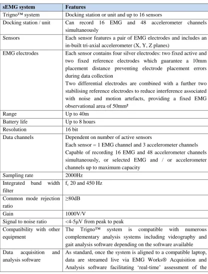
Table 5: Technical specifications of the Delsys® Trigno™ Wireless EMG system
The Delsys® Trigno™ Wireless EMG system was used to collect and analyse sEMG data for Evidence Sources 1.2 to 1.4 in the thesis. The technical specification for key aspects of the unit related to sEMG data collection are summarised from the unit’s handbook in the table.
sEMG system Features
Trigno™ system Docking station or unit and up to 16 sensors
Docking station / unit Can record 16 EMG and 48 accelerometer channels simultaneously
Sensors Each sensor features a pair of EMG electrodes and includes an in-built tri-axial accelerometer (X, Y, Z planes)
EMG electrodes Each sensor contains four silver electrodes: two fixed active and two fixed reference electrodes which guarantee a 10mm placement distance preventing electrode placement errors during data collection
Two differential electrodes are combined with a further two stabilising reference electrodes to reduce interference associated with noise and motion artefacts, providing a fixed EMG observational area of 50mm²
Range Up to 40m
Battery life Up to 8 hours Resolution 16 bit
Data channels Dependent on number of active sensors
Each sensor = 1 EMG channel and 3 accelerometer channels Capable of recording 16 EMG and 48 accelerometer channels simultaneously, or selected EMG and / or accelerometer channels up to maximum capacity
Sampling rate 2000Hz Integrated band width
filter
fc 20 and 450 Hz
Common mode rejection ratio
≥80dB
Gain 1000V/V
Signal to noise ratio <4-5μV from peak to peak Compatibility with other
equipment
The Trigno™ system is compatible with numerous complementary analysis systems including videography and gait analysis software depending on the software available Data acquisition and
analysis software
32
signal and subsequent data analysis Adapted from Delsys® (2014)
3.6 Data collection
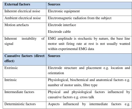
The ideal sEMG study should aim for consistency in the acquired EMG signal (Smoliga et al., 2010). Numerous factors affect the reliability of sEMG data collected or may potentially influence interpretation of results (Reaz, Hussain and Mohd-Yasin, 2006) (Table 6).
Table 6: Factors which influence the EMG signal
[image:51.595.99.525.415.764.2]The electromyography (EMG) signal will record all electrical activity within its defined pickup zone. The quality of the EMG signal received will also be affected by the location of the sensor and how well it is attached to the subject. Review of previous research identified core factors which can result in interference to the EMG signal. The sources of interference and their impact are provided in the table.
External factors Sources
Inherent electrical noise Electronic equipment
Ambient electrical noise Electromagnetic radiation from the subject Motion artefacts Electrode interface
Electrode cable Inherent instability of
signal
EMG amplitude is stochastic by nature, the base line motor unit firing rate at rest is not usually wanted within experimental EMG data
Causative factors (direct effect)
Sources
Extrinsic Electrode structure and placement e.g. location and orientation
Intrinsic Physiological, biochemical and anatomical factors e.g. number of motor units, fibre type
Intermediate factors Physical and physiological factors influenced by causative factors e.g. cross talk
33
amplitude, firing rates Adapted from: Reaz, Hussain and Mohd-Yasin (2006)
The optimal EMG signal represents the total of all MUAPs under investigation and should contain no distortion, noise or artefacts. Prior to data collection, four areas should be considered to optimise the quality of the signal:
1. Amplifier gain and its dynamic range, 2. Input impedance (Z),
3. Frequency response, and, 4. Common mode rejection.
34
frequency response should be set to enable all the frequencies present within an EMG signal to be collected (Winter, 2009). The EMG spectrum ranges between 5 and 2000Hz, which includes the physiological MUAP amplitudes and non-relevant electrical signals (Kamen and Gabriel, 2010). sEMG data collection units commonly apply frequency ranges between 10 and 1000Hz (De Luca, 2003; Delsys, 2014). Mammals are good conductors of electromagnetic radiation and will absorb signals from nearby power sources potentially introducing anomalies into the EMG signal being recorded (De Luca et al., 2010). The common mode rejection represents the differential amplification required to eliminate these extraneous sources from the functional EMG signal (Winter, 2009).
3.7 Physiological influences on the EMG signal
35
Other factors may influence muscle fibre conductivity. Temperature has been shown to change the velocity of APs (Kamen and Gabriel, 2010). Cold temperatures depress fibre excitability resulting in a reduced contraction speed, comprising lower spectral frequencies, whilst warmer temperatures increase contraction velocity (Kimura, 2001; Winkel and Jorgensen, 1991). Muscle and muscle fibre length will also influence the frequency characteristics of MUAPs, usually EMG frequency decreases as muscle length increases, whilst shorter fibres produce a higher spectral frequency (Kamen and Gabriel, 2010). The quantity and type of tissue between the electrode and MU will also influence the amplitude and frequency characteristics of the sEMG signal received (Kamen and Gabriel, 2010; Winter, 2009). The dermal layers, in particular subcutaneous adipose tissue, act as a low-pass filter on the EMG signal, effectively dampening it (Konrad, 2005). The dampening effect increases with tissue depth, therefore resultant EMG data are biased (reduced signal transfer) with increased representation of the MUAP from superficial fibres (Kamen and Gabriel, 2010).
3.8 Data processing
36
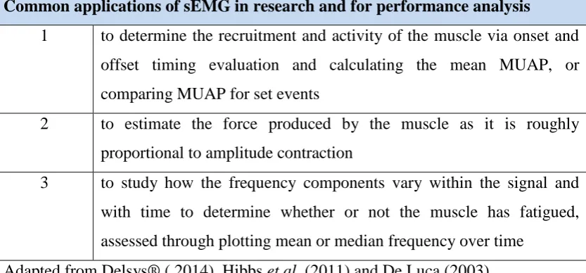
Table 7: The three common applications of sEMG; MUAP: motor unit action potential
Surface electromyography has evaluated muscle recruitment, activity levels, force production, fatigue and muscle relationships to motor skills within clinical studies to assess myoneuralgia and motor disease, during rehabilitation regimens, and for performance analysis during exercise and training. Brief details of how sEMG is used to assess these areas are provided in the table.
Common applications of sEMG in research and for performance analysis 1 to determine the recruitment and activity of the muscle via onset and
offset timing evaluation and calculating the mean MUAP, or comparing MUAP for set events
2 to estimate the force produced by the muscle as it is roughly proportional to amplitude contraction
3 to study how the frequency components vary within the signal and with time to determine whether or not the muscle has fatigued, assessed through plotting mean or median frequency over time
Adapted from Delsys® ( 2014), Hibbs et al. (2011) and De Luca (2003)
Methodologies commonly employed to facilitate further examination for the determination of amplitude, timing and spectral, or frequency, analyses include:
Categorisation of the frequencies within the signal via the application of filters, Half or full wave rectification of the absolute value of the EMG data,
Application of a linear envelope (after rectification and combined with a low
pass filter),
Integration of the full wave rectified signal (integrated or i.EMG):
[image:55.595.111.528.305.499.2]37 o For a fixed time period, or,
o Compared to a pre-set (baseline) level
(Delsys®, 2013; Hug, 2011; Kamen and Gabriel, 2010; Winter, 2009). 3.8.1 Filters
During muscle activity, the amplitude of the EMG signal at any given instant is stochastic and will contain multiple frequencies contributing to the force produced (Hug, 2011). Within the signal, the initial MU input is a high frequency signal which ‘fires’ the subsequent muscle twitch, however the muscle fibre acts like a capacitor, therefore contractions produce lower frequency outputs (Winter, 2009). Likewise, external sources of noise could contribute to the frequency domain recorded (De Luca et al., 2010). Therefore filters are applied to ensure only relevant frequencies that contribute to the event being assessed are analysed (Kamen and Gabriel, 2010; Winter, 2009).
38
and phase response (timing) within their specification to accurately filter the data obtained (De Luca, 2003; Kamen and Gabriel, 2010). All filters delay the timing of the EMG signal to some extent which would not be a problem if the delay was linear and consistent across all frequencies within the signal, however delays may only exist in certain frequencies distorting the signal (Winter, 2009). Delays in timing between two sinusoids of the same frequency results in each passing through the zero point at different times causing signals to be out of phase creating a phase-lag (Kamen and Gabriel, 2010). Therefore consideration of phase response within the signal is another important aspect that needs to be contemplated when applying filters.
The behaviour of different filter models is shown in Figure 6; practically a filter does not transition from pass-band to stop-band regions (or vice versa) immediately as can be seen by the lack of a 90º angle within each graph. In reality each filter type will have a transition zone where the signal transmission changes from pass-band to stop-band regions or vice versa (DeLuca, 2003).
39
[image:58.595.178.449.158.374.2]attenuate at -40dB / decade and will reduce the amplitude of the input signal by 1/20th for every decade (Delsys, 2013). Therefore with higher filter orders a lower value for fc can be used.
Figure 6: The four basic filter types: where filter response amplitude is 1 are defined as pass-band regions; while frequencies where the filter response amplitude is 0 are defined as stop-band regions. The cut-off frequency is denoted by fc. (a) Low-pass filter, (b) High-pass filter, (c) Band-pass filter and (d) Band-stop filter (DeLuca, 2003). Reproduced with kind permission from Delsys®.
Filters are applied to electromyography data to remove erroneous data related to movement artefacts, noise or other forms of electrical interference prior to analysis and to remove the impact of time-lag from the EMG signal. Filters utilise the frequency component of the EMG signal to apply one or more cut-off frequencies to isolate the desirable data within the signal. Four basic filter types exist, how each works is outlined in figure 6.
40
The Butterworth filter is the filter of choice for application of a linear envelope in gait studies (De Luca et al., 2010; Kamen and Gabriel, 2010; Winter, 2009). Butterworth filters are used as their roll rate is maximally flat in the pass-band which minimises the pass-band ripple in the attenuated signal resulting in the production of a smooth wave; they are best suited to applications requiring preservation of amplitude linearity in the pass-band region i.e. linear enveloping in kinesiological EMG (Kamen and Gabriel, 2010). The magnitude and phase responses of Butterworth filters are quantified by the maximum band-pass gain, fc and the filter order selected for use. Butterworth filters demonstrate a linear phase lag which is only beneficial if the phasing of all frequencies in a signal are consistent (Kamen and Gabriel, 2010).
41
3.8.2 Full wave rectification
Full wave rectification is often the initial building block for analysis of the filtered EMG data (Kamen and Gabriel, 2010; Winter, 2009). The result is a positive signal, which does not cross zero and fluctuates according to the strength of MUAPs facilitating further quantitative analysis. Visual examination of the signal should occur after rectification and will provide functional information on muscle contraction through assessment of signal amplitudes and their duration (Richards, Thewlis and Selfe, 2008).
3.8.3 Linear enveloping
42
[image:61.595.106.532.245.745.2]literature, ranging from 3 to 60Hz, but the majority of human gait studies utilise a fc <20Hz (Kamen and Gabriel, 2010) (Table 8).
Table 8: Examples of EMG filtering protocols utilised in human dynamic studies.
A range of human surface electromyography (EMG) studies examining dynamic movement were reviewed to identify the filtering protocols applied to the EMG data to facilitate subsequent comparison.
Sport Filtering protocol applied to EMG data Study Downhill
running
High-pass filter with fc of 20Hz Full wave rectification
Linear envelope: low-pass filter, zero-lag, fc of 5Hz
Sheenan and Gotschall, 2013
Rowing Band-pass filter with lower and higher fc of 20 and 400Hz, respectively
Full wave rectification
Linear envelope: low-pass Butterworth filter, 4th Order, fc of 8Hz
Turpin et al., 2011
Pole vaulting Band-pass filter with lower and higher fc of 19 and 395Hz, respectively
Full wave rectification
Linear envelope: low-pass Butterworth filter, 4th Order, fc of 5Hz
Frere et al., 2012
Muscle
synergy during cycling
High-pass filter fc of 20Hz Full wave rectification
Linear envelope: low-pass filter, zero lag and fc of 5Hz
Hug et al., 2010
Cross-country skiing
Band-pass filter with lower and higher fc of 20 and 450Hz, respectively
Full wave rectification
Linear envelope: low-pass Butterworth filter, 4th Order, fc of 50Hz
Zoppirolli et al., 2013
Landing from a drop jump
Band-pass filter with lower and higher fc of 10 and 500Hz, respectively
Full wave rectification
Linear envelope: low-pass Butterworth filter, 2nd Order, fc of 6Hz
43
There is no general rule that should be applied to give the most appropriate low pass fc (Kamen and Gabriel, 2010). The selection of fc can be derived from muscle twitch times providing a biological basis for the value applied (Winter, 2009), from calculation of -3dB point of the signal (De Luca, 2003) or set at the value which represents 95% of the Total Power of the signal (Kamen and Gabriel, 2010). In humans, twitch times have been extrapolated from isometric EMG analysis for a number of muscle groups (Winter, 2009), however isometric analysis is not achievable in the horse. For dynamic studies, fc may be defined as the value (Hz) which represents 95% of the Total Power of the movement signal under consideration. Fc selection should be sufficient to reduce EMG variation related to phasing during movement whilst minimising distortion within the signal but will be dependent on the filtering protocol applied (Kamen and Gabriel, 2010; Shiavi et al., 1998). In the absence of a standardised protocol, Vint et al. (2001) suggest that a range of fc combined with low pass filtering are applied to data and subsequent EMG traces overlaid to evaluate variability within EMG profiles to determine the effect of, and to select a suitable value for fc. The majority of previous equine EMG research has utilised an fc of 10Hz combined with a Butterworth filter (for example: Zsoldos et al., 2010a, b; Licka, Frey and Peham, 2009).
3.8.4 Integrated EMG
44 3.9 Interpretation of the processed EMG signal
Interpretation of sEMG data is acknowledged as challenging as a number of factors can influence the resultant signal (Reaz, Hussain and Mohd-Yasin, 2006). Variation in signal amplitude may be attributed to increased numbers of active MUs being recruited or a change in the frequency of activation i.e. the firing rate has increased but the same number of MUs are being recruited (Konrad, 2005; Stegeman et al., 2000).
3.9.1 Muscle fibre profile
Muscle fibre profile will influence the EMG trace produced (Kamen and Gabriel, 2010). An elevated frequency for amplitude of the sEMG signal could be related to an increase in fast-twitch fibre recruitment or a higher firing rate in slow-twitch fibres (both equalling more muscle effort) or could be the result of decreased synchronisation between MUs that are firing (muscle starting to fatigue) (Staudenmann et al., 2010; Winter, 2009; Rahnama, Lees and Reilly, 2006). In contrast, a decreased frequency may be attributed to a reduced firing rate (fatigue) or increased MU synchronisation (coordinated muscle activity; no fatigue present) (Staudenmann et al., 2010; Winter, 2009; Rahnama, Lees and Reilly, 2006).
45
The category of muscle contraction will also impact the MUAPrecorded (Richards, Thewlis and Selfe, 2008). During dynamic evaluation, muscles perform anisometric contractions dependent upon their function. In concentric contractions, tension is reduced as length shortens during muscle contraction. Eccentric muscles display the opposite pattern, with tension increasing as the muscle lengthens during activity. Therefore concentric muscles have to work harder to function effectively than eccentric ones. Although contraction magnitude is individualised, broad characteristics are observed in the resultant EMG trace (Staudenmann et al., 2010). Contractions in concentric muscles will exhibit a larger magnitude than eccentric ones representing their greater workload (Richards, Thewlis and Selfe, 2008).
3.9.3 Comparing events
46
3.9.4 Assessment of fatigue
47
CHAPTER FOUR
SURFACE ELECTROMYOGRAPHY AND THE EQUINE ATHLETE
Limited research has been conducted to analyse muscle performance during exercise in the horse (Ferrari et al., 2009). Factors which relate to muscle performance are commonly assessed observationally, for example fatigue, or via analysis of allied physiological variables, such as heart rate (for example: Williams and Fiander, 2014). However these techniques cannot quantify recruitment, activity-levels or adaptation in muscle tissue, or account for individual or breed variation; factors which could directly influence performance. Chapter Four reviews the capacity of sEMG as an objective and quantifiable measure of muscle performance related to the core principles of equestrian training and muscle physiology.
4.1 An introduction to muscle physiology
48
During activity, muscles produce ‘power’: the product of the force generated multiplied by velocity associated changes in the muscle length (Kearns, McKeever and Abe, 2002). Movement is not uniform in its mechanics; different categories of contraction exist and are identifiable in sEMG-traces (Table 9) (Section 4.8.2). Maximal force production is related to the physiological cross-sectional area (CSA) and the muscle fibre profile of an individual muscle (Rivero, 2014; Rietbroek et al., 2007). Force and velocity are dependent on the functional biochemical characteristics of muscle fibres, combined with the configuration of fibres in respect of the muscle in its entirety (Rivero, 2014; Rietbroek et al., 2007; Kearns, McKeever and Abe, 2002). The potential force which can be generated is directly proportional to the number of sarcomeres that are parallel and in series within active muscle fibres (Rivero, 2014; Kearns, McKeever and Abe, 2002). The duration of the force produced, or the contraction, will be reliant upon the ability of the muscle not to fatigue (Rivero, 2014; Rivero and Piercy, 2008). Therefore skeletal muscle also requires an adequate blood supply to support its function, which is achieved via a highly organised integral capillary network (Marlin and Nankervis, 2002).
4.1.1 Muscle contraction
49
Table 9: Types of contraction in skeletal muscle
Different types of muscle contraction occur in mammals. The key features of each type of contraction were examined and are summarised in the table. Practical examples are provided to enable their identification during movement.
Contraction type:
Key features:
Isometric Resting length of the muscle body remains of constant length during contractions
Muscle force equals load and no movement occurs until fatigued
Example: human holding a weight still at arm’s length, isometric contraction of the Triceps brachii and Biceps brachii Isotonic Muscle tension remains constant, but muscle length varies
Muscle force exceeds load on the muscle Examples: eccentric and concentric contractions
Concentric Resting length of the muscle body shortens during the contraction
Tension reduces as muscle shortens: attributed to cross-bridges in the contractile element breaking and reforming in a shortened condition
Muscle force exceeds resistance
Example: flexion of the human arm, concentric contraction of the Biceps brachii
Eccentric Resting length of the muscle body lengthens during contraction Tension increases as muscle length increases due to increased loading: greater force required to break cross-bridges and lengthen, than to maintain isometric length
External load exceeds muscle force; often assisted by gravity Example: extension of the human arm, eccentric contraction of the Biceps brachii
Isokinetic Muscle contraction velocity is constant but force varies
Analysis method employed on individual muscle within in vitro experiments
Adapted from: Winter (2009) and Rivero and Piercy (2008)
50
fibres (Yousefi and Hamilton-Wright, 2014). The arrival of the AP at the MEP stimulates changes in the ionic properties of the sarcolemma (Konrad, 2005). At rest the intrinsic polarity of the muscle fibre is negative; once an AP arrives acetylcholine is released increasing conductivity within the sarcolemma. The result is an increase in Na+ ions entering the fibre which produce a change in polarity (positive) creating a MUAP when the stimulus is of sufficient size. The process is known as polarisation. Polarisation is followed by depolarisation when the K+ channels open and the polarity reverts to its resting status (Konrad, 2005). sEMG records the changes in polarity as a MUAP.
51
Table 10: Key features of motor units which can impact force production
Activation of motor units by a propagating action potential stimulated sequential contraction in the sarcomeres of muscle fibres, ultimately resulting in muscle contraction. Knowledge of the types of motor unit and understanding their characteristics aids interpretation of sEMG data. The three types of motor unit and key features are outlined in the table.
Feature Description
Motor unit type 3 types are recognised in mammalian tissue:
slow (S) found in Type I muscle fibres: associated with long duration-low intensity exercise e.g. postural support
fast resistant to fatigue (FR) found in Type IIA muscle fibres: associated with long duration - medium intensity exercise e.g. sustained running
fast fatigable (FF) found in Type IIX muscle fibres: associated with short duration – high intensity exercise e.g. sprinting or jumping
Activation threshold Related to MU type and size
The size of the AP required to recruit MU increases sequentially within S > FR > FF
Firing rates S: slow
FR: intermediate FF: fast
MUAP magnitude Related to MU type and size, magnitude increases sequentially: S>FR>FF
Sex distribution Males have increased FF than females
Age related changes Contraction times decrease in S and FR with aging
Force production increases in S units and decreases in mainly FF but also FR units with aging
52 4.2 Muscle supporting performance
The skeletal musculature of the horse has evolved to facilitate superior athletic performance in comparison to other mammals (Rivero, 2014). Skeletal muscle constitutes approximately 42% of an average horse’s bodyweight, which increases to ~55% in the mature, trained equine athlete (Hinchcliff and Geor, 2008). The biological importance of skeletal muscle is demonstrated by its structural and functional plasticity, which facilitates phenotypic adaptation in response to exercise and training (Rivero, 2014; McGivney et al., 2009; Serrano, Quiroz-Rothe and Rivero, 2000).
4.2.1 The influence of muscle fibre profiles
sEMG profiles are dependent on the MUAP production in muscles under investigation (Reaz, Hussain and Mohd-Yasin, 2006). Equine muscles contain a combination of fibre types, producing a muscle fibre profile which will influence the sEMG profile obtained during exercise. Fibre type varies between breeds (Lopez-Rivero et al., 1992), can differ according to function (Choi and Kim, 2009), sex (Ozawa et al., 2000; Choi and Kim, 2009), age (Smarsh and Williams, 2014; Holloszy and Larsson, 1995), and with hormones and fitness status (Bell et al., 2000; Rivero et al., 1995).