A second monoclinic polymorph of
N
-(pyrazin-2-yl)aniline
Zanariah Abdullah and Seik Weng Ng*
Department of Chemistry, University of Malaya, 50603 Kuala Lumpur, Malaysia Correspondence e-mail: [email protected]
Received 23 September 2008; accepted 4 October 2008
Key indicators: single-crystal X-ray study;T= 100 K; mean(C–C) = 0.002 A˚;
Rfactor = 0.042;wRfactor = 0.113; data-to-parameter ratio = 15.8.
The two aromatic rings in the title compound, C10H9N3, are
aligned at 23.4 (1) and the bridging C—N—C angle is 128.9 (1). In the crystal structure, intermolecular N—H N hydrogen bonds result in a chain motif, the repeat distance of which is half thebaxial length of 8.8851 (3) A˚ .
Related literature
In the P21/c modification, the aromatic rings are aligned at
15.2 (1), and the repeat distance of the helical chain is half the b-axial length of 7.8423 (3) A˚ ; see: Wan Saffieeet al.(2008).
Experimental
Crystal data
C10H9N3 Mr= 171.20
Monoclinic, P21=n
a= 8.2194 (3) A˚
b= 8.8851 (3) A˚
c= 11.8395 (4) A˚
= 104.643 (2)
V= 836.56 (5) A˚3 Z= 4
MoKradiation
= 0.09 mm1 T= 100 (2) K 0.250.050.03 mm
Data collection
Bruker SMART APEX diffractometer
Absorption correction: none 7621 measured reflections
1922 independent reflections 1389 reflections withI> 2(I)
Rint= 0.045
Refinement
R[F2> 2(F2)] = 0.042 wR(F2) = 0.113 S= 1.03 1922 reflections 122 parameters
H atoms treated by a mixture of independent and constrained refinement
max= 0.20 e A˚
3
min=0.23 e A˚
3
Table 1
Hydrogen-bond geometry (A˚ ,).
D—H A D—H H A D A D—H A
N1—H1 N3i
0.90 (2) 2.17 (2) 3.062 (2) 175 (2)
Symmetry code: (i)xþ5 2;y
1 2;zþ
3 2.
Data collection:APEX2(Bruker, 2007); cell refinement:SAINT (Bruker, 2007); data reduction:SAINT; program(s) used to solve structure:SHELXS97(Sheldrick, 2008); program(s) used to refine structure: SHELXL97 (Sheldrick, 2008); molecular graphics: X-SEED (Barbour, 2001); software used to prepare material for publication:publCIF(Westrip, 2008).
The authors thank the University of Malaya for supporting this study (grant Nos. FS 358/2008A and FA 067/2006A).
Supplementary data and figures for this paper are available from the IUCr electronic archives (Reference: PK2124).
References
Barbour, L. J. (2001).J. Supramol. Chem.1, 189–191.
Bruker (2007).APEX2andSAINT. Bruker AXS Inc., Madison, Wisconsin, USA.
Sheldrick, G. M. (2008).Acta Cryst.A64, 112–122.
Wan Saffiee, W. A. M., Idris, A., Abdullah, Z., Aiyub, Z. & Ng, S. W. (2008).
Acta Cryst.E64, o2105.
Westrip, S. P. (2008).publCIF. In preparation. Acta Crystallographica Section E
Structure Reports
Online
supporting information
Acta Cryst. (2008). E64, o2106 [doi:10.1107/S1600536808031954]
A second monoclinic polymorph of
N
-(pyrazin-2-yl)aniline
Zanariah Abdullah and Seik Weng Ng
S1. Comment
The cell dimensions of the reported monoclinic P21/c modification are: a = 10.0644 (3), b = 7.8423 (3), c = 10.8907 (3)
Å; β = 116.439 (2)° (Wan Saffiee et al., 2008). The cell dimensions of the present modification (Scheme I, Fig. 1), after
transformation to the standard P21/c setting, are: a = 8.2194 (3), b = 8.8851 (3), c = 12.5909 (4) Å, β = 114.525 (2)°.
S2. Experimental
The P21/c modification of 2-pyrazinyl-N-aniline (0.10 g, 0.4 mmol), zinc acetate (0.09 g, 0.4 mmol) and water (18 ml)
were heated in a 23-ml Teflon-lined Parr bomb at 403 K for 2 days. The bomb was cooled to room temperature at 5 K
min-1. Several faint yellow prisms were picked out manually from the cool solution.
S3. Refinement
Carbon-bound H-atoms were placed in calculated positions (C—H 0.95 Å) and were included in the refinement in the
riding model approximation, with U(H) fixed at 1.2U(C). The amino H-atom was located in a difference Fourier map,
[image:2.610.128.481.416.637.2]and was freely refined.
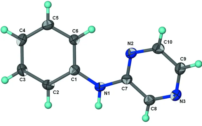
Figure 1
Thermal ellipsoid plot (Barbour, 2001) of C10H9N3 at the 70% probability level; hydrogen atoms are drawn as spheres of
N-(pyrazin-2-yl)aniline
Crystal data
C10H9N3
Mr = 171.20 Monoclinic, P21/n Hall symbol: -P 2yn
a = 8.2194 (3) Å
b = 8.8851 (3) Å
c = 11.8395 (4) Å
β = 104.643 (2)°
V = 836.56 (5) Å3
Z = 4
F(000) = 360
Dx = 1.359 Mg m−3
Mo Kα radiation, λ = 0.71073 Å Cell parameters from 1282 reflections
θ = 2.7–26.1°
µ = 0.09 mm−1
T = 100 K
Prism, pale yellow 0.25 × 0.05 × 0.03 mm
Data collection
Bruker SMART APEX diffractometer
Radiation source: fine-focus sealed tube Graphite monochromator
ω scans
7621 measured reflections 1922 independent reflections
1389 reflections with I > 2σ(I)
Rint = 0.045
θmax = 27.5°, θmin = 2.7°
h = −10→10
k = −11→11
l = −15→14
Refinement
Refinement on F2 Least-squares matrix: full
R[F2 > 2σ(F2)] = 0.042
wR(F2) = 0.113
S = 1.03 1922 reflections 122 parameters 0 restraints
Primary atom site location: structure-invariant direct methods
Secondary atom site location: difference Fourier map
Hydrogen site location: inferred from neighbouring sites
H atoms treated by a mixture of independent and constrained refinement
w = 1/[σ2(F
o2) + (0.0547P)2 + 0.1331P] where P = (Fo2 + 2Fc2)/3
(Δ/σ)max = 0.001 Δρmax = 0.20 e Å−3 Δρmin = −0.23 e Å−3
Fractional atomic coordinates and isotropic or equivalent isotropic displacement parameters (Å2)
x y z Uiso*/Ueq
N1 1.00115 (16) 0.42084 (14) 0.63143 (11) 0.0216 (3)
H1 1.103 (2) 0.377 (2) 0.6527 (15) 0.035 (5)*
N2 0.84988 (15) 0.63197 (14) 0.66840 (11) 0.0230 (3)
N3 1.15770 (16) 0.76478 (14) 0.78284 (11) 0.0228 (3)
C1 0.86787 (18) 0.33136 (16) 0.56827 (12) 0.0193 (3)
C2 0.89595 (19) 0.17682 (17) 0.56499 (13) 0.0254 (4)
H2 1.0021 0.1367 0.6049 0.030*
C3 0.7717 (2) 0.08147 (17) 0.50462 (14) 0.0264 (4)
H3 0.7933 −0.0235 0.5030 0.032*
C4 0.61582 (19) 0.13750 (17) 0.44633 (13) 0.0232 (3)
H4 0.5296 0.0717 0.4058 0.028*
C5 0.58777 (19) 0.29106 (17) 0.44814 (13) 0.0222 (3)
C6 0.71238 (18) 0.38836 (16) 0.50783 (13) 0.0211 (3)
H6 0.6916 0.4936 0.5074 0.025*
C7 0.99583 (18) 0.56191 (16) 0.67668 (13) 0.0192 (3)
C8 1.14967 (18) 0.63040 (16) 0.73478 (13) 0.0213 (3)
H8 1.2514 0.5772 0.7393 0.026*
C9 1.00948 (18) 0.83515 (18) 0.77382 (13) 0.0248 (4)
H9 1.0085 0.9325 0.8069 0.030*
C10 0.8604 (2) 0.76892 (17) 0.71788 (14) 0.0254 (4)
H10 0.7592 0.8226 0.7139 0.030*
Atomic displacement parameters (Å2)
U11 U22 U33 U12 U13 U23
N1 0.0144 (7) 0.0200 (7) 0.0276 (7) 0.0019 (5) 0.0003 (5) −0.0014 (5)
N2 0.0173 (6) 0.0230 (7) 0.0272 (7) 0.0004 (5) 0.0027 (5) −0.0036 (5)
N3 0.0195 (7) 0.0228 (7) 0.0251 (7) −0.0033 (5) 0.0037 (5) −0.0011 (5)
C1 0.0180 (7) 0.0205 (7) 0.0193 (7) −0.0010 (6) 0.0048 (6) −0.0009 (6)
C2 0.0217 (8) 0.0227 (8) 0.0289 (8) 0.0040 (6) 0.0012 (6) 0.0000 (6)
C3 0.0293 (9) 0.0176 (8) 0.0307 (9) 0.0005 (6) 0.0043 (7) −0.0019 (6)
C4 0.0217 (8) 0.0233 (8) 0.0244 (8) −0.0059 (6) 0.0053 (6) −0.0035 (6)
C5 0.0173 (7) 0.0251 (8) 0.0229 (8) 0.0005 (6) 0.0026 (6) −0.0006 (6)
C6 0.0195 (8) 0.0190 (7) 0.0237 (8) 0.0005 (6) 0.0031 (6) −0.0011 (6)
C7 0.0175 (7) 0.0198 (7) 0.0194 (7) 0.0004 (6) 0.0030 (6) 0.0020 (6)
C8 0.0177 (7) 0.0224 (8) 0.0233 (8) −0.0001 (6) 0.0041 (6) 0.0015 (6)
C9 0.0228 (8) 0.0220 (8) 0.0284 (8) −0.0016 (6) 0.0047 (6) −0.0049 (6)
C10 0.0202 (8) 0.0241 (8) 0.0307 (9) 0.0032 (6) 0.0043 (6) −0.0041 (7)
Geometric parameters (Å, º)
N1—C7 1.3681 (19) C3—H3 0.9500
N1—C1 1.4061 (19) C4—C5 1.385 (2)
N1—H1 0.897 (18) C4—H4 0.9500
N2—C7 1.3330 (18) C5—C6 1.389 (2)
N2—C10 1.3438 (19) C5—H5 0.9500
N3—C8 1.3171 (19) C6—H6 0.9500
N3—C9 1.3494 (19) C7—C8 1.415 (2)
C1—C6 1.393 (2) C8—H8 0.9500
C1—C2 1.395 (2) C9—C10 1.370 (2)
C2—C3 1.379 (2) C9—H9 0.9500
C2—H2 0.9500 C10—H10 0.9500
C3—C4 1.385 (2)
C7—N1—C1 128.94 (13) C4—C5—H5 119.4
C7—N1—H1 114.0 (12) C6—C5—H5 119.4
C1—N1—H1 116.7 (11) C5—C6—C1 119.86 (14)
C7—N2—C10 115.67 (13) C5—C6—H6 120.1
C8—N3—C9 116.12 (13) C1—C6—H6 120.1
C6—C1—N1 123.92 (13) N2—C7—C8 120.79 (13)
C2—C1—N1 117.35 (13) N1—C7—C8 118.11 (13)
C3—C2—C1 120.88 (14) N3—C8—C7 122.74 (13)
C3—C2—H2 119.6 N3—C8—H8 118.6
C1—C2—H2 119.6 C7—C8—H8 118.6
C2—C3—C4 120.52 (14) N3—C9—C10 121.23 (14)
C2—C3—H3 119.7 N3—C9—H9 119.4
C4—C3—H3 119.7 C10—C9—H9 119.4
C5—C4—C3 118.88 (14) N2—C10—C9 123.44 (14)
C5—C4—H4 120.6 N2—C10—H10 118.3
C3—C4—H4 120.6 C9—C10—H10 118.3
C4—C5—C6 121.11 (14)
C7—N1—C1—C6 21.9 (2) C10—N2—C7—N1 178.88 (14)
C7—N1—C1—C2 −159.27 (15) C10—N2—C7—C8 0.2 (2)
C6—C1—C2—C3 −1.0 (2) C1—N1—C7—N2 2.9 (2)
N1—C1—C2—C3 −179.81 (14) C1—N1—C7—C8 −178.36 (14)
C1—C2—C3—C4 −0.3 (2) C9—N3—C8—C7 0.0 (2)
C2—C3—C4—C5 1.0 (2) N2—C7—C8—N3 −0.2 (2)
C3—C4—C5—C6 −0.5 (2) N1—C7—C8—N3 −178.92 (13)
C4—C5—C6—C1 −0.8 (2) C8—N3—C9—C10 0.1 (2)
C2—C1—C6—C5 1.5 (2) C7—N2—C10—C9 0.0 (2)
N1—C1—C6—C5 −179.75 (13) N3—C9—C10—N2 −0.1 (2)
Hydrogen-bond geometry (Å, º)
D—H···A D—H H···A D···A D—H···A
N1—H1···N3i 0.90 (2) 2.17 (2) 3.062 (2) 175 (2)