(2
E
)-2-[(3
E
)-4-Phenylbut-3-en-2-ylidene]-hydrazinecarboxamide
S. Samshuddin,aRay J. Butcher,bSema Ozturk Yıldırım,c Mehmet Akkurt,d* B. Narayanaaand H. S. Yathirajane
aDepartment of Studies in Chemistry, Mangalore University, Mangalagangotri, Mangalore 574 199, India,bDepartment of Chemistry, Howard University, 525 College Street NW, Washington, DC 20059, USA,cDepartment of Chemistry, Howard University, 525 College Street NW, Washington, DC 20059, USA, and, Department of Physics, Faculty of Sciences, Erciyes University, 38039 Kayseri, Turkey,dDepartment of Physics, Faculty of Sciences, Erciyes University, 38039 Kayseri, Turkey, andeDepartment of Studies in Chemistry, University of Mysore, Manasagangotri, Mysore 570 006, India
Correspondence e-mail: [email protected]
Received 5 December 2011; accepted 6 December 2011
Key indicators: single-crystal X-ray study;T= 123 K; mean(C–C) = 0.002 A˚; disorder in main residue;Rfactor = 0.055;wRfactor = 0.176; data-to-parameter ratio = 21.0.
In the title compound, C11H13N3O, the phenyl ring is disordered over two sites, with occupancy factors in a 0.520 (17):0.480 (17) ratio. The dihedral angle between the ring planes of the major and minor components of the disordered ring is 12.9 (2). In the crystal, molecules are linked
by N—H O hydrogen bonds, formingR2 2
(8) ring motifs. C— H O, C—H N and C—H interactions also occur.
Related literature
For background to the biological activity of semicarbazones, see: Beraldo et al. (2002); Teixeira et al. (2003); Du et al.
(2004); Kucukguzelet al.(2006); Beraldo & Gambino (2004). For related structures, see: Naik & Palenik (1974); Wanget al.
(2004); Yathirajanet al.(2006); Sarojiniet al.(2007).
Experimental
Crystal data
C11H13N3O
Mr= 203.24 Monoclinic,C2=c a= 15.1094 (8) A˚
b= 24.4445 (11) A˚
c= 7.0368 (4) A˚
= 109.908 (6)
V= 2443.7 (2) A˚3
Z= 8
MoKradiation
= 0.07 mm1
T= 123 K
0.400.300.18 mm
Data collection
Oxford Diffraction Xcalibur Ruby Gemini diffractometer Absorption correction: multi-scan
(CrysAlis RED; Oxford Diffraction, 2007)
Tmin= 0.987,Tmax= 1.000
12712 measured reflections 3528 independent reflections 2748 reflections withI> 2(I)
Rint= 0.026
Refinement
R[F2> 2(F2)] = 0.055
wR(F2) = 0.176
S= 1.05 3528 reflections
168 parameters
H-atom parameters constrained max= 0.26 e A˚
3
min=0.22 e A˚
[image:1.610.315.564.276.377.2]3
Table 1
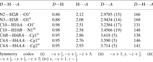
Hydrogen-bond geometry (A˚ ,).
Cg1 andCg2 are the centroids of the disordered benzene rings C1A–C6Aand C1B–C6B, respectively.
D—H A D—H H A D A D—H A
N2—H2B O1i
0.88 2.12 2.9785 (15) 166 N3—H3B O1ii 0.88 2.08 2.9434 (14) 168 C10—H10A O1i
0.98 2.51 3.2384 (17) 131 C10—H10B N1iii
0.98 2.58 3.4566 (19) 148 C4B—H4BA Cg1iv 0.95 2.86 3.618 (5) 138 C4A—H4AA Cg1iv
0.95 2.76 3.590 (5) 146 C4A—H4AA Cg2iv
0.95 2.93 3.714 (5) 141
Symmetry codes: (i) xþ1 2;yþ
1
2;zþ3; (ii) xþ1;y;zþ 7 2; (iii)
xþ1 2;yþ
1
2;zþ2; (iv)x;yþ1;z 1 2.
Data collection: CrysAlis PRO(Oxford Diffraction, 2007); cell refinement: CrysAlis PRO; data reduction: CrysAlis RED (Oxford Diffraction, 2007); program(s) used to solve structure:SHELXS97 (Sheldrick, 2008); program(s) used to refine structure:SHELXL97 (Sheldrick, 2008); molecular graphics: ORTEP-3 (Farrugia, 1997); software used to prepare material for publication:WinGX(Farrugia, 1999) andPLATON(Spek, 2009).
BN thanks the UGC for financial assistance through SAP and BSR one-time grants for the purchase of chemicals. HSY thanks the University of Mysore for research facilities. RJB wishes to acknowledge the NSF–MRI program (grant CHE-0619278) for funds to purchase the diffractometer.
Supplementary data and figures for this paper are available from the IUCr electronic archives (Reference: TK5032).
References
Beraldo, H. & Gambino, D. (2004).Mini Rev. Med. Chem.4, 31–39. Beraldo, H., Sinisterra, R. D., Teixeira, L. R., Vieira, R. P. & Doretto, M. C.
(2002).Biochem. Biophys. Res. Commun.296, 241–246.
Du, C. X., Guo, C., Hansell, E., Doyle, P. S., Caffrey, C. R., Holler, T. P., McKerrow, J. H. & Cohen, F. E. (2004).J. Med. Chem.47, 3212–3219. Farrugia, L. J. (1997).J. Appl. Cryst.30, 565.
Farrugia, L. J. (1999).J. Appl. Cryst.32, 837–838.
Kucukguzel, G., Kocatepe, A., De Clercq, E., Sahin, F. & Gulluce, M. (2006).
Eur. J. Med. Chem.41, 353–359.
Naik, D. V. & Palenik, G. J. (1974).Acta Cryst.B30, 2396–2401.
Oxford Diffraction (2007). CrysAlis PRO and CrysAlis RED. Oxford Diffraction Ltd, Abingdon, England.
Sarojini, B. K., Narayana, B., Bindya, S., Yathirajan, H. S. & Bolte, M. (2007).
Acta Cryst.E63, o2946.
Sheldrick, G. M. (2008).Acta Cryst.A64, 112–122. Spek, A. L. (2009).Acta Cryst.D65, 148–155.
organic compounds
o76
Samshuddinet al. doi:10.1107/S160053681105255X Acta Cryst.(2012). E68, o76–o77Acta Crystallographica Section E Structure Reports
Online
Teixeira, L. R., Sinisterra, R. D., Vieira, R. P., Doretto, M. C. & Beraldo, H. (2003).J. Incl. Phenom. Macro. Chem.47, 77–82.
Wang, J.-L., Jia, Y.-J. & Yu, M. (2004).Acta Cryst.E60, o662–o663.
Yathirajan, H. S., Bindya, S., Narayana, B., Sarojini, B. K. & Bolte, M. (2006).
supporting information
sup-1
Acta Cryst. (2012). E68, o76–o77supporting information
Acta Cryst. (2012). E68, o76–o77 [doi:10.1107/S160053681105255X]
(2
E
)-2-[(3
E
)-4-Phenylbut-3-en-2-ylidene]hydrazinecarboxamide
S. Samshuddin, Ray J. Butcher, Sema Ozturk Y
ı
ld
ı
r
ı
m, Mehmet Akkurt, B. Narayana and H. S.
Yathirajan
S1. Comment
Semicarbazones presents a wide range of biological applications such as antitumoral, anticonvulsant, anti-trypanosomal, herbicidal and biocidal activities (Beraldo & Gambino, 2004; Beraldo et al., 2002; Teixeira et al., 2003). They can also be used as important intermediates in organic synthesis, mainly for obtaining heterocycle rings, such as thiazolidones, oxa-diazoles, pyrazolidones, and thiadiazoles (Du et al., 2004; Kucukguzel et al., 2006)
Crystal structures of some semicarbazone derivatives, viz., acetone semicarbazone and benzaldehyde semicarbazone (Naik & Palenik, 1974); 3,4- methylenedioxybenzaldehyde semicarbazone (Wang et al.,2004);
4-(methyl-sulfanyl)benzaldehyde thiosemicarbazone (Yathirajan et al., 2006) and 4-(Methylsulfanyl)benzaldehyde semicarbazone (Sarojini et al., 2007) have been reported. In view of the importance of semicarbazones, the title compound (I) was prepared and its crystal structure is reported.
Fig. 1 shows the molecular structure of the title compound (I) with the disordered phenyl ring. The dihedral angle between the major and minor disorder components of the phenyl ring is 12.9 (2)°. The C7—C8—C9—C10, C7—C8— C9—N1, C10—C9—N1—N2, C8—C9—N1—N2, N1—N2—C11—N3 and N1—N2—C11—O1 torsion angles are -2.7 (2), 178.13 (13), -1.37 (19), 179.53 (10), -1.40 (17) and 179.22 (11)°, respectively, and indicate planarity in the molecule.
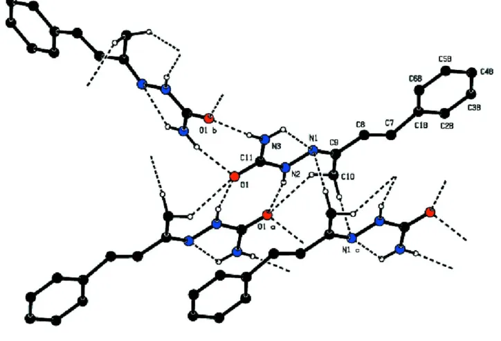
In the crystal, the molecules form centrosymmetric dimers with an R22(8) ring motif through a pair of N—H···O
hydrogen bonds. These dimers are further connected into a three-dimensional network by intermolecular C—H···O and C —H···N hydrogen bonds (Table 1, Fig. 2). Weak intermolecular C—H···π interactions further stabilize the crystal structure.
S2. Experimental
To a mixture of a benzylidene acetone (1.46 g, 0.01 mol) and semicarbazide hydrochloride (1.12 g, 0.01 mol) in 50 ml ethanol was added a sodium acetate solution (2 g in 5 ml water) which was then refluxed for 4 h. The resultant solution was concentrated to half of its volume and poured into 50 ml ice-cold water. The precipitate thus formed was collected by filtration and purified by recrystallization from ethanol. The single crystal was grown from its absolute alcohol solution by slow evaporation. The yield was 74%. (M.pt. 455–459 K).
S3. Refinement
The phenyl ring is disordered over two positions with refined site occupancies of 0.520 (17) and 0.480 (17). All H atoms were placed in idealised positions and refined in the riding model approximation [N—H = 0.88 Å, aromatic C—H = 0.95 Å and methyl C—H = 0.98 Å, and with Uiso(H) = 1.2 or 1.5 Ueq(parent atom)]. In the crystal structure, there is an 206 Å3
void, but the low electron density (0.26 e.Å-3) in the difference Fourier map suggests no solvent molecule occupying this
Figure 1
The disordered molecule (I) showing the atom labeling scheme. Atoms of the minor disorder components are joined with dashed lines. Displacement ellipsoids for non-H atoms are drawn at the 50% probability level.
Figure 2
View of the N—H···O mediated dimers in (I) and their connections to other molecules by C—H···O and C—H···O hydrogen bonding.
(2E)-2-[(3E)-4-Phenylbut-3-en-2-ylidene]hydrazinecarboxamide
Crystal data
C11H13N3O
Mr = 203.24
Monoclinic, C2/c Hall symbol: -C 2yc a = 15.1094 (8) Å b = 24.4445 (11) Å c = 7.0368 (4) Å
β = 109.908 (6)° V = 2443.7 (2) Å3
Z = 8 F(000) = 864 Dx = 1.105 Mg m−3
[image:4.610.125.487.292.538.2]supporting information
sup-3
Acta Cryst. (2012). E68, o76–o77θ = 3.0–30.9° µ = 0.07 mm−1
T = 123 K
Prism, colourless 0.40 × 0.30 × 0.18 mm
Data collection
Oxford Diffraction Xcalibur Ruby Gemini diffractometer
Radiation source: Enhance (Mo) X-ray Source Graphite monochromator
Detector resolution: 10.5081 pixels mm-1
ω scans
Absorption correction: multi-scan
(CrysAlis RED; Oxford Diffraction, 2007) Tmin = 0.987, Tmax = 1.000
12712 measured reflections 3528 independent reflections 2748 reflections with I > 2σ(I) Rint = 0.026
θmax = 30.9°, θmin = 3.0°
h = −20→20 k = −34→26 l = −7→9
Refinement
Refinement on F2
Least-squares matrix: full R[F2 > 2σ(F2)] = 0.055
wR(F2) = 0.176
S = 1.05 3528 reflections 168 parameters 0 restraints
Primary atom site location: structure-invariant direct methods
Secondary atom site location: difference Fourier map
Hydrogen site location: inferred from neighbouring sites
H-atom parameters constrained w = 1/[σ2(F
o2) + (0.0981P)2 + 0.6768P]
where P = (Fo2 + 2Fc2)/3
(Δ/σ)max < 0.001
Δρmax = 0.26 e Å−3
Δρmin = −0.22 e Å−3
Special details
Geometry. Bond distances, angles etc. have been calculated using the rounded fractional coordinates. All su's are estimated from the variances of the (full) variance-covariance matrix. The cell e.s.d.'s are taken into account in the estimation of distances, angles and torsion angles
Refinement. Refinement on F2 for ALL reflections except those flagged by the user for potential systematic errors.
Weighted R-factors wR and all goodnesses of fit S are based on F2, conventional R-factors R are based on F, with F set to
zero for negative F2. The observed criterion of F2 > σ(F2) is used only for calculating -R-factor-obs etc. and is not
relevant to the choice of reflections for refinement. R-factors based on F2 are statistically about twice as large as those
based on F, and R-factors based on ALL data will be even larger.
Fractional atomic coordinates and isotropic or equivalent isotropic displacement parameters (Å2)
x y z Uiso*/Ueq Occ. (<1)
O1 0.36888 (6) 0.23438 (4) 1.62948 (12) 0.0323 (3) N1 0.34428 (7) 0.30220 (5) 1.17621 (15) 0.0337 (3) N2 0.31934 (7) 0.27704 (4) 1.32596 (14) 0.0310 (3) N3 0.47771 (7) 0.26174 (6) 1.49376 (16) 0.0458 (4)
C1B 0.3063 (5) 0.40506 (19) 0.5724 (7) 0.0301 (9) 0.520 (17) C2B 0.2428 (5) 0.4316 (2) 0.4074 (6) 0.0354 (10) 0.520 (17) C3B 0.2747 (6) 0.45824 (19) 0.2685 (6) 0.0405 (13) 0.520 (17) C4B 0.3701 (6) 0.45829 (14) 0.2947 (8) 0.0409 (13) 0.520 (17) C5B 0.4336 (6) 0.4317 (2) 0.4598 (11) 0.0469 (14) 0.520 (17) C6B 0.4017 (5) 0.4051 (2) 0.5986 (11) 0.0434 (11) 0.520 (17) C7 0.25908 (11) 0.37670 (5) 0.71144 (19) 0.0393 (4)
C10 0.17796 (10) 0.32953 (6) 1.0129 (2) 0.0395 (4) C11 0.38883 (8) 0.25686 (5) 1.49014 (17) 0.0304 (3)
C3A 0.2443 (5) 0.4630 (2) 0.2639 (7) 0.0458 (13) 0.480 (17) C4A 0.3345 (6) 0.45553 (15) 0.2576 (7) 0.0378 (13) 0.480 (17) C5A 0.3972 (6) 0.4208 (2) 0.3956 (10) 0.0408 (14) 0.480 (17) C6A 0.3697 (5) 0.39363 (19) 0.5398 (9) 0.0337 (11) 0.480 (17) C2A 0.2169 (4) 0.4358 (2) 0.4081 (7) 0.0402 (11) 0.480 (17) C1A 0.2795 (4) 0.40110 (19) 0.5461 (6) 0.0276 (10) 0.480 (17) H7A 0.19470 0.37930 0.69980 0.0470*
H3BA 0.23130 0.47640 0.15570 0.0490* 0.520 (17) H5BA 0.49890 0.43180 0.47770 0.0560* 0.520 (17) H6BA 0.44510 0.38700 0.71150 0.0520* 0.520 (17) H10A 0.17330 0.33360 1.14770 0.0590*
H10B 0.14520 0.29610 0.94940 0.0590* H10C 0.14890 0.36120 0.92980 0.0590*
H4BA 0.39200 0.47650 0.19980 0.0490* 0.520 (17) H8A 0.37790 0.34890 0.90100 0.0450*
H2B 0.25980 0.27410 1.31570 0.0370* H3B 0.52440 0.24890 1.59690 0.0550* H3C 0.48940 0.27780 1.39300 0.0550*
H2BA 0.17760 0.43160 0.38950 0.0420* 0.520 (17) H2AA 0.15520 0.44090 0.41240 0.0480* 0.480 (17) H3AA 0.20150 0.48670 0.16960 0.0550* 0.480 (17) H4AA 0.35330 0.47410 0.15910 0.0450* 0.480 (17) H5AA 0.45880 0.41570 0.39130 0.0490* 0.480 (17) H6AA 0.41250 0.36990 0.63410 0.0400* 0.480 (17)
Atomic displacement parameters (Å2)
U11 U22 U33 U12 U13 U23
supporting information
sup-5
Acta Cryst. (2012). E68, o76–o77C6A 0.038 (2) 0.0309 (16) 0.0338 (19) 0.0064 (14) 0.0144 (18) 0.0112 (13) C2A 0.053 (2) 0.0369 (19) 0.0353 (17) 0.0087 (17) 0.0212 (16) 0.0099 (13) C1A 0.038 (2) 0.0219 (14) 0.0268 (15) −0.0032 (14) 0.0161 (14) −0.0043 (12)
Geometric parameters (Å, º)
O1—C11 1.2472 (15) C5A—C6A 1.389 (10)
N1—N2 1.3788 (15) C5B—C6B 1.389 (10)
N1—C9 1.2915 (17) C7—C8 1.3344 (18)
N2—C11 1.3618 (15) C8—C9 1.4610 (19)
N3—C11 1.3398 (17) C9—C10 1.494 (2)
N2—H2B 0.8800 C2A—H2AA 0.9500
N3—H3B 0.8800 C2B—H2BA 0.9500
N3—H3C 0.8800 C3A—H3AA 0.9500
C1A—C7 1.432 (5) C3B—H3BA 0.9500
C1A—C6A 1.391 (10) C4A—H4AA 0.9500
C1A—C2A 1.390 (7) C4B—H4BA 0.9500
C1B—C2B 1.390 (7) C5A—H5AA 0.9500
C1B—C6B 1.390 (11) C5B—H5BA 0.9500
C1B—C7 1.556 (6) C6A—H6AA 0.9500
C2A—C3A 1.389 (8) C6B—H6BA 0.9500
C2B—C3B 1.390 (9) C7—H7A 0.9500
C3A—C4A 1.391 (12) C8—H8A 0.9500
C3B—C4B 1.390 (13) C10—H10B 0.9800
C4A—C5A 1.391 (9) C10—H10C 0.9800
C4B—C5B 1.391 (9) C10—H10A 0.9800
N2—N1—C9 118.31 (11) C3A—C2A—H2AA 120.00 N1—N2—C11 118.53 (11) C1A—C2A—H2AA 120.00
N1—N2—H2B 121.00 C3B—C2B—H2BA 120.00
C11—N2—H2B 121.00 C1B—C2B—H2BA 120.00
C11—N3—H3B 120.00 C4A—C3A—H3AA 120.00
H3B—N3—H3C 120.00 C2A—C3A—H3AA 120.00
C11—N3—H3C 120.00 C4B—C3B—H3BA 120.00
C1A—C6A—C5A 120.0 (6) C1B—C7—H7A 126.00 C1B—C6B—C5B 120.0 (6) C8—C7—H7A 114.00
C1B—C7—C8 119.7 (3) C9—C8—H8A 117.00
C1A—C7—C8 132.9 (3) C7—C8—H8A 117.00
C7—C8—C9 126.08 (15) C9—C10—H10C 110.00 N1—C9—C8 114.27 (13) C9—C10—H10B 109.00 C8—C9—C10 120.69 (11) H10B—C10—H10C 109.00 N1—C9—C10 125.04 (12) H10A—C10—H10B 110.00 N2—C11—N3 117.53 (11) H10A—C10—H10C 109.00 O1—C11—N3 122.23 (11) C9—C10—H10A 110.00 O1—C11—N2 120.24 (12)
C9—N1—N2—C11 −173.34 (11) C2B—C1B—C7—C8 −178.1 (3) N2—N1—C9—C8 −179.53 (10) C6B—C1B—C7—C8 2.8 (6) N2—N1—C9—C10 1.37 (19) C1B—C2B—C3B—C4B 0.0 (7) N1—N2—C11—O1 179.22 (11) C2B—C3B—C4B—C5B 0.0 (7) N1—N2—C11—N3 −1.40 (17) C3B—C4B—C5B—C6B 0.0 (8) C6B—C1B—C2B—C3B 0.0 (7) C4B—C5B—C6B—C1B 0.0 (8) C7—C1B—C2B—C3B −179.2 (4) C1B—C7—C8—C9 175.5 (2) C2B—C1B—C6B—C5B 0.0 (8) C7—C8—C9—N1 178.13 (13) C7—C1B—C6B—C5B 179.1 (4) C7—C8—C9—C10 −2.7 (2)
Hydrogen-bond geometry (Å, º)
Cg1 and Cg2 are the centroids of the disordered benzene rings C1A –C6A and C1B–C6B, respectively.
D—H···A D—H H···A D···A D—H···A
N2—H2B···O1i 0.88 2.12 2.9785 (15) 166
N3—H3B···O1ii 0.88 2.08 2.9434 (14) 168
N3—H3C···N1 0.88 2.28 2.6397 (16) 104 C10—H10A···O1i 0.98 2.51 3.2384 (17) 131
C10—H10B···N1iii 0.98 2.58 3.4566 (19) 148
C4B—H4BA···Cg1iv 0.95 2.86 3.618 (5) 138
C4A—H4AA···Cg1iv 0.95 2.76 3.590 (5) 146
C4A—H4AA···Cg2iv 0.95 2.93 3.714 (5) 141