(7
R
,8
S
,8a
S
)-8-Hydroxy-7-phenyl-perhydroindolizin-3-one
Lˇubomı´r Sˇvorc,aViktor Vra´bel,a* Sˇtefan Marchalı´n,bPeter Sˇafa´rˇb and Jozef Kozˇı´sˇekc
aInstitute of Analytical Chemistry, Faculty of Chemical and Food Technology, Slovak
Technical University, Radlinske´ho 9, SK-81237 Bratislava, Slovak Republic,
bInstitute of Organic Chemistry, Catalysis and Petrochemistry, Faculty of Chemical
and Food Technology, Slovak Technical University, Radlinske´ho 9, SK-81237 Bratislava, Slovak Republic, andcInstitute of Physical Chemistry and Chemical
Physics, Faculty of Chemical and Food Technology, Slovak Technical University, Radlinske´ho 9, SK-81237 Bratislava, Slovak Republic
Correspondence e-mail: viktor.vrabel@stuba.sk
Received 12 May 2009; accepted 15 May 2009
Key indicators: single-crystal X-ray study;T= 298 K; mean(C–C) = 0.003 A˚; Rfactor = 0.035;wRfactor = 0.101; data-to-parameter ratio = 10.4.
In the title compound, C14H17NO2, the six-membered ring of
the indolizine system adopts a chair conformation. In the crystal, molecules form chains parallel to the b axis via
intermolecular O—H O hydrogen bonds. The absolute molecular configuration was assigned from the synthesis.
Related literature
For industrial uses of indolizines, see: Jaung & Jung (2003); Rotaruet al.(2005); Delattreet al.(2005); Kelinet al.(2001). For biological uses, see: Nashet al.(1988); Molyneux & James (1982); Harrell (1970); Ruprechtet al.(1989); Liuet al.(2007); Smithet al.(2007); Guptaet al.(2003); Rosseelset al.(1982); Oslund et al. (2008); Ostby et al. (2000). For synthesis of indolizines, see: Chuprakov & Gevorgyan (2007); Yan & Liu (2007). For the synthesis methods used, see: Sˇafa´rˇet al.(2009). For structures related to the title compound, see: Sˇvorcet al.
(2009). For comparison of molecular parameters, see: Camus
et al. (2003); Lokajet al.(1999); Brown & Corbridge (1954); Pedersen (1967). For a general analysis of puckering, see: Cremer & Pople (1975).
Experimental
Crystal data
C14H17NO2 Mr= 231.29 Orthorhombic,Pca21 a= 11.4164 (3) A˚
b= 6.6372 (2) A˚
c= 15.5118 (4) A˚
V= 1175.38 (6) A˚3 Z= 4
MoKradiation
= 0.09 mm1 T= 298 K
0.600.560.13 mm
Data collection
Oxford Diffraction Gemini R CCD diffractometer
Absorption correction: analytical (Clark & Reid, 1995)
Tmin= 0.901,Tmax= 0.989
26298 measured reflections 1632 independent reflections 1128 reflections withI> 2(I)
Rint= 0.023
Refinement
R[F2> 2(F2)] = 0.035 wR(F2) = 0.101 S= 1.03 1632 reflections 157 parameters
1 restraint
H-atom parameters constrained max= 0.17 e A˚
3
min=0.12 e A˚
3
Table 1
Hydrogen-bond geometry (A˚ ,).
D—H A D—H H A D A D—H A
O2—H2 O1i
0.82 2.00 2.807 (2) 170
Symmetry code: (i)x;yþ1;z.
Data collection: CrysAlis CCD (Oxford Diffraction, 2006); cell refinement: CrysAlis RED (Oxford Diffraction, 2006); data reduc-tion:CrysAlis RED; program(s) used to solve structure:SHELXS97
(Sheldrick, 2008); program(s) used to refine structure:SHELXL97
(Sheldrick, 2008); molecular graphics: DIAMOND (Brandenburg, 2001); software used to prepare material for publication: enCIFer
(Allenet al., 2004).
The authors thank the Grant Agency of the Slovak Republic (grant Nos. 1/0161/08 and 1/0817/08) and Structural Funds, Interreg IIIA, for financial support in purchasing the diffractometer, and the Development Agency under contract No. APVV-0210-07.
Supplementary data and figures for this paper are available from the IUCr electronic archives (Reference: BG2260).
References
Allen, F. H., Johnson, O., Shields, G. P., Smith, B. R. & Towler, M. (2004).J. Appl. Cryst.37, 335–338.
Brandenburg, K. (2001).DIAMOND. Crystal Impact GbR, Bonn, Germany. Brown, C. J. & Corbridge, D. E. C. (1954).Acta Cryst.7, 711–715.
Camus, F., Norberg, B., Bourry, A., Akue´-Ge´du, R., Rigo, B. & Durant, F. (2003).Acta Cryst.E59, o1002–o1003.
Chuprakov, S. & Gevorgyan, V. (2007).Org. Lett.9, 4463–4466. Clark, R. C. & Reid, J. S. (1995).Acta Cryst.A51, 887–897. Cremer, D. & Pople, J. A. (1975).J. Am. Chem. Soc.97, 1354–1362. Delattre, F., Woisel, P., Surpateanu, G., Cazier, F. & Blach, P. (2005).
Tetrahedron,61, 3939–3945.
Gupta, S. P., Mathur, A. N., Nagappa, A. N., Kumar, D. & Kumaran, S. (2003).
Eur. J. Med. Chem.38, 867–873.
Harrell, W. B. (1970).J. Pharm. Sci.59, 275–277.
Jaung, J. Y. & Jung, Y. S. (2003).Bull. Korean Chem. Soc.24, 1565–1566.
organic compounds
o1368
Sˇvorcet al. doi:10.1107/S1600536809018455 Acta Cryst.(2009). E65, o1368–o1369Acta Crystallographica Section E
Structure Reports
Online
2074–2075.
Liu, Y., Song, Z. & Yan, B. (2007).Org. Lett.9, 409–412.
Lokaj, J., Kettmann, V. & Marchalin, S. (1999).Acta Cryst.C55, 1103–1105. Molyneux, R. J. & James, L. F. (1982).Science,216, 190–191.
Nash, R. J., Fellows, L. E., Dring, J. V., Stirton, C. H., Carter, D., Hegarty, M. P. & Bell, E. A. (1988).Phytochemistry,27, 1403–1406.
Oslund, R. C., Cermak, N. & Gelb, M. H. (2008).J. Med. Chem.51, 4708– 4714.
Ostby, O. B., Dalhus, B., Gundersen, L. L., Rise, F., Bast, A. & Haenen, G. R. M. M. (2000).Eur. J. Org. Chem.9, 3763–3770.
Oxford Diffraction (2006). CrysAlis CCD and CrysAlis RED. Oxford Diffraction Ltd, Abingdon, Oxfordshire, England.
Pedersen, B. F. (1967).Acta Chem. Scand.21, 1415–1424.
Bauthier, J., Richard, J., Tornay, C., Colot, M. & Claviere, M. (1982).Eur. J. Med. Chem.17, 581–584.
Rotaru, A. V., Druta, I. D., Oeser, T. & Mu¨ller, T. J. (2005).Helv. Chim. Acta,
88, 1798–1812.
Ruprecht, R. M., Mullaney, S., Anderson, J. & Bronson, R. (1989).J. Acquir. Immune Defic. Syndr.2, 149–157.
Sˇafa´rˇ, P., Zˇ u´zˇiova´, J., Marchalı´n, Sˇ., To´thova´, E., Pro´nayova´, N., Sˇvorc, Lˇ., Vra´bel, V. & Daich, A. (2009).Tetrahedron Asymmetry,20, 626–634. Sheldrick, G. M. (2008).Acta Cryst.A64, 112–122.
Smith, C. R., Bunnelle, E. M., Rhodes, A. J. & Sarpong, R. (2007).Org. Lett.9, 1169–1171.
Sˇvorc, Lˇ ., Vra´bel, V., Zˇu´zˇiova´, J., Bobosˇı´kova´, M. & Kozˇı´sˇek, J. (2009).Acta Cryst.E65, o895–o896.
supporting information
sup-1
Acta Cryst. (2009). E65, o1368–o1369
supporting information
Acta Cryst. (2009). E65, o1368–o1369 [doi:10.1107/S1600536809018455]
(7
R
,8
S
,8a
S
)-8-Hydroxy-7-phenylperhydroindolizin-3-one
Ľ
ubom
í
r
Š
vorc, Viktor Vr
á
bel,
Š
tefan Marchal
í
n, Peter
Š
af
ář
and Jozef Ko
žíš
ek
S1. Comment
Heterocycles are involved in a wide range of biologically important chemical reactions in living organisms, and therefore
they form one of the most important and well investigated classes of organic compounds. One group of heterocycles,
indolizines, has received much scientific attention during the recent years. They are known for their use as synthetic dyes
(Jaung & Jung, 2003), fluorescent materials (Rotaru et al., 2005; Delattre et al., 2005) and also as key intermediates for
the synthesis of indolizine based molecules (Kelin et al., 2001). Indolizines both synthetic and natural have also been
ascribed with a number of useful biological activities such as antibacterial, antiviral, antiinflammatory (Nash et al., 1988;
Molyneux & James, 1982), testosterone-3&-reductase inhibitors, 5-HT4 receptor antagonists, CNS depressants (Harrell
et al., 1970), anti-HIV (Ruprecht et al., 1989), anti-cancer (Liu et al., 2007; Smith et al., 2007) and have been used for
treating cardiovascular ailments (Gupta et al., 2003). For instance, aminoalkyloxybenzenesulfonylindolizine compounds
such as fantofarone and butoprozine have been used for the treatment of hypertension, arrhythmia and angina pectoris
(Rosseels et al., 1982). Several oxygenated indolizines have been shown to prevent, due to their strong anti-oxidative
effects, the initiation of oxidation processes that lead to DNA damage (Oslund et al., 2008; Ostby et al., 2000).
Consequently, synthesis of indolizines have attracted considerable attention and a number of synthetic methodologies
have been developed for a variety of indolizines, making use of in particular, transition metal catalyzed reactions
(Chuprakov & Gevorgyan, 2007; Yan & Liu, 2007). In addition, indolizines and their derivatives are important in the
field of material science owing to their unique photophysical properties.
Based on these facts and in continutation of our interest in developing simple and efficient routes for the synthesis of
novel indolizine derivatives, we report here the synthesis and molecular and crystal structure of the title compound, (I)
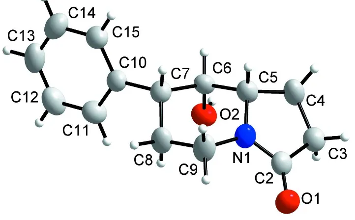
(Fig. 1). A similar analysis of its enantiomer (the stereochemistry of atom C6 was confirmed as R) has already been
published (Švorc et al., 2009). The absolute configuration of (I) was established by the synthesis and is depicted in the
scheme and Fig. 1. The expected stereochemistry of atoms C5, C6 and C7 was confirmed as S, S and R, respectively (Fig.
1). The central six-membered N-heterocyclic ring is not planar and adopts a chair conformation (Cremer & Pople, 1975).
A calculation of least-squares planes shows that this ring is puckered in such a manner that the four atoms C5, C6, C8 and
C9 are coplanar to within 0.010 (2) Å, while atoms N1 and C7 are displaced from this plane on opposite sides, with
out-of-plane displacements of -0.555 (2) and 0.711 (2) Å, respectively. The phenyl ring attached to the indolizine ring system
is planar (mean deviation is 0.009 (2) Å). The N1—C5 and N1—C9 bonds are approximately equivalent (See
supplementary material) and both are much longer than the N1—C2 bond. Moreover, the N1 atom is sp2 hybridized, as
evidenced by the sum of the valence angles around it [359.9 (2)°]. These data are consistent with conjugation of the
lone-pair electrons on N1 with the adjacent carbonyl and agree with literature values for simple amides (Brown & Corbridge,
1954; Pedersen, 1967). The bond length of the carbonyl group C2═O1 is 1.236 (2) Å, respectively, is somewhat longer
than typical carbonyl bonds. This may be due to the fact that atom O1 participates as acceptor in intermolecular hydrogen
and angles in the indolizine ring system are in good agreement with values from the literature (Camus et al., 2003; Lokaj
et al., 1999).
S2. Experimental
The title compound (7R,8S,8aS)-8-hydroxy-7-phenylhexahydroindolizin-3(5H)-one was prepared according literature
procedures of Šafář et al. (2009).
S3. Refinement
All H atoms were placed in geometrically idealized positions and constrained to ride on their parent atoms, with C—H
distances in the range 0.93–0.98 Å and O—H distance 0.85 Å and Uiso set at 1.2Ueq of the parent atom. The absolute
configuration could not be reliably determined for this compound using Mo radiation, and has been assigned according to
[image:4.610.126.487.269.493.2]the synthesis; Friedel pairs have been merged.
Figure 1
Molecular structure of (I) with the atomic numbering scheme. Displacement ellipsoids are drawn at the 50% probability
supporting information
sup-3
[image:5.610.128.485.72.391.2]Acta Cryst. (2009). E65, o1368–o1369
Figure 2
A packing of the molecule of (I), viewed along the b axis.
(7R,8S,8aS)-8-Hydroxy-7-phenylperhydroindolizin-3-one
Crystal data
C14H17NO2
Mr = 231.29
Orthorhombic, Pca21
Hall symbol: P 2c -2ac a = 11.4164 (3) Å b = 6.6372 (2) Å c = 15.5118 (4) Å V = 1175.38 (6) Å3
Z = 4
F(000) = 496 Dx = 1.307 Mg m−3
Mo Kα radiation, λ = 0.71073 Å Cell parameters from 13180 reflections θ = 3.3–29.4°
µ = 0.09 mm−1
T = 298 K Block, white
0.60 × 0.56 × 0.13 mm
Data collection
Oxford Diffraction Gemini R CCD diffractometer
Radiation source: fine-focus sealed tube Graphite monochromator
Detector resolution: 10.4340 pixels mm-1
Rotation method data acquisition using ω and φ scans
Absorption correction: analytical (Clark & Reid, 1995)
Tmin = 0.901, Tmax = 0.989
26298 measured reflections 1632 independent reflections 1128 reflections with I > 2σ(I) Rint = 0.023
θmax = 29.4°, θmin = 3.6°
Refinement on F2
Least-squares matrix: full R[F2 > 2σ(F2)] = 0.035
wR(F2) = 0.101
S = 1.03 1632 reflections 157 parameters 1 restraint
Primary atom site location: structure-invariant direct methods
Secondary atom site location: difference Fourier map
Hydrogen site location: inferred from neighbouring sites
H-atom parameters constrained w = 1/[σ2(F
o2) + (0.0644P)2 + 0.0334P]
where P = (Fo2 + 2Fc2)/3
(Δ/σ)max < 0.001
Δρmax = 0.17 e Å−3
Δρmin = −0.12 e Å−3
Extinction correction: SHELXL97 (Sheldrick, 2008), Fc*=kFc[1+0.001xFc2λ3/sin(2θ)]-1/4
Extinction coefficient: 0.014 (4)
Special details
Experimental. face-indexed (CrysAlis RED; Oxford Diffraction, 2006)
Geometry. All e.s.d.'s (except the e.s.d. in the dihedral angle between two l.s. planes) are estimated using the full covariance matrix. The cell e.s.d.'s are taken into account individually in the estimation of e.s.d.'s in distances, angles and torsion angles; correlations between e.s.d.'s in cell parameters are only used when they are defined by crystal symmetry. An approximate (isotropic) treatment of cell e.s.d.'s is used for estimating e.s.d.'s involving l.s. planes.
Refinement. Refinement of F2 against ALL reflections. The weighted R-factor wR and goodness of fit S are based on F2,
conventional R-factors R are based on F, with F set to zero for negative F2. The threshold expression of F2 > σ(F2) is used
only for calculating R-factors(gt) etc. and is not relevant to the choice of reflections for refinement. R-factors based on F2
are statistically about twice as large as those based on F, and R- factors based on ALL data will be even larger.
Fractional atomic coordinates and isotropic or equivalent isotropic displacement parameters (Å2)
x y z Uiso*/Ueq
C2 0.2640 (2) −0.3204 (3) 0.32882 (13) 0.0425 (5)
C3 0.21718 (19) −0.1773 (3) 0.26133 (17) 0.0490 (5)
H3A 0.1398 −0.1296 0.2773 0.059*
H3B 0.2121 −0.2438 0.2058 0.059*
C4 0.3027 (2) −0.0043 (4) 0.25750 (16) 0.0590 (6)
H4A 0.2626 0.1227 0.2670 0.071*
H4B 0.3409 0.0003 0.2017 0.071*
C5 0.3932 (2) −0.0428 (3) 0.32948 (15) 0.0466 (5)
H5 0.4708 −0.0578 0.3033 0.056*
C6 0.40010 (17) 0.1156 (3) 0.40092 (13) 0.0400 (4)
H6 0.4340 0.2393 0.3771 0.048*
C7 0.47970 (18) 0.0382 (3) 0.47386 (13) 0.0421 (5)
H7 0.5558 0.0075 0.4478 0.051*
C8 0.4309 (2) −0.1608 (3) 0.50890 (16) 0.0514 (6)
H8A 0.4814 −0.2100 0.5545 0.062*
H8B 0.3537 −0.1378 0.5332 0.062*
C9 0.4226 (2) −0.3184 (3) 0.43821 (15) 0.0557 (6)
H9A 0.5006 −0.3569 0.4197 0.067*
H9B 0.3832 −0.4375 0.4600 0.067*
C10 0.50152 (16) 0.1935 (3) 0.54318 (14) 0.0407 (5)
C11 0.42606 (19) 0.2235 (4) 0.61193 (16) 0.0507 (5)
supporting information
sup-5
Acta Cryst. (2009). E65, o1368–o1369
C12 0.4494 (2) 0.3654 (4) 0.67466 (16) 0.0580 (6)
H12 0.3976 0.3833 0.7203 0.070*
C13 0.5498 (2) 0.4808 (4) 0.66972 (17) 0.0599 (7)
H13 0.5662 0.5754 0.7122 0.072*
C14 0.6248 (2) 0.4553 (4) 0.60216 (17) 0.0623 (7)
H14 0.6919 0.5343 0.5982 0.075*
C15 0.60156 (19) 0.3120 (4) 0.53928 (16) 0.0501 (5)
H15 0.6538 0.2951 0.4938 0.060*
N1 0.35736 (17) −0.2359 (3) 0.36550 (12) 0.0473 (4)
O1 0.22237 (14) −0.4869 (2) 0.34724 (11) 0.0545 (5)
O2 0.28771 (12) 0.1598 (2) 0.43433 (11) 0.0496 (4)
H2 0.2647 0.2673 0.4146 0.074*
Atomic displacement parameters (Å2)
U11 U22 U33 U12 U13 U23
C2 0.0503 (12) 0.0381 (10) 0.0391 (11) 0.0064 (9) 0.0004 (9) −0.0066 (9)
C3 0.0507 (13) 0.0483 (11) 0.0480 (12) 0.0040 (9) −0.0032 (10) 0.0014 (10)
C4 0.0858 (18) 0.0477 (12) 0.0436 (12) −0.0088 (11) −0.0148 (13) 0.0042 (10)
C5 0.0585 (13) 0.0404 (10) 0.0408 (10) −0.0013 (9) −0.0015 (10) 0.0023 (9)
C6 0.0488 (11) 0.0331 (10) 0.0382 (10) −0.0015 (8) 0.0012 (9) 0.0038 (8)
C7 0.0395 (10) 0.0449 (12) 0.0419 (11) 0.0035 (8) −0.0023 (9) −0.0003 (9)
C8 0.0681 (15) 0.0384 (11) 0.0478 (11) 0.0032 (10) −0.0163 (11) 0.0054 (9)
C9 0.0736 (15) 0.0366 (11) 0.0570 (14) 0.0078 (9) −0.0211 (12) 0.0039 (10)
C10 0.0405 (10) 0.0417 (11) 0.0397 (10) 0.0017 (8) −0.0059 (9) 0.0024 (8)
C11 0.0523 (11) 0.0496 (12) 0.0501 (12) 0.0022 (9) 0.0080 (11) −0.0004 (10)
C12 0.0783 (16) 0.0509 (12) 0.0447 (12) 0.0132 (12) 0.0045 (12) −0.0045 (11)
C13 0.0818 (17) 0.0480 (12) 0.0500 (13) 0.0058 (11) −0.0209 (13) −0.0055 (11)
C14 0.0621 (14) 0.0556 (14) 0.0693 (16) −0.0098 (11) −0.0195 (14) 0.0004 (12)
C15 0.0453 (11) 0.0587 (13) 0.0464 (12) −0.0024 (10) −0.0031 (10) 0.0035 (10)
N1 0.0593 (11) 0.0359 (9) 0.0465 (9) 0.0005 (8) −0.0100 (8) 0.0014 (8)
O1 0.0640 (11) 0.0413 (8) 0.0582 (11) −0.0047 (6) −0.0065 (8) 0.0027 (7)
O2 0.0456 (8) 0.0486 (8) 0.0546 (9) 0.0083 (6) 0.0023 (7) 0.0085 (7)
Geometric parameters (Å, º)
C2—O1 1.237 (2) C8—C9 1.518 (3)
C2—N1 1.332 (3) C8—H8A 0.9700
C2—C3 1.511 (3) C8—H8B 0.9700
C3—C4 1.508 (3) C9—N1 1.458 (3)
C3—H3A 0.9700 C9—H9A 0.9700
C3—H3B 0.9700 C9—H9B 0.9700
C4—C5 1.542 (3) C10—C11 1.385 (3)
C4—H4A 0.9700 C10—C15 1.388 (3)
C4—H4B 0.9700 C11—C12 1.380 (3)
C5—N1 1.457 (3) C11—H11 0.9300
C5—C6 1.530 (3) C12—C13 1.380 (4)
C6—C7 1.540 (3) C13—H13 0.9300
C6—H6 0.9800 C14—C15 1.388 (3)
C7—C10 1.510 (3) C14—H14 0.9300
C7—C8 1.533 (3) C15—H15 0.9300
C7—H7 0.9800 O2—H2 0.8200
O1—C2—N1 125.78 (19) C9—C8—H8A 109.4
O1—C2—C3 125.9 (2) C7—C8—H8A 109.4
N1—C2—C3 108.33 (17) C9—C8—H8B 109.4
C2—C3—C4 106.09 (18) C7—C8—H8B 109.4
C2—C3—H3A 110.5 H8A—C8—H8B 108.0
C4—C3—H3A 110.5 N1—C9—C8 109.40 (16)
C2—C3—H3B 110.5 N1—C9—H9A 109.8
C4—C3—H3B 110.5 C8—C9—H9A 109.8
H3A—C3—H3B 108.7 N1—C9—H9B 109.8
C3—C4—C5 106.18 (18) C8—C9—H9B 109.8
C3—C4—H4A 110.5 H9A—C9—H9B 108.2
C5—C4—H4A 110.5 C11—C10—C15 117.7 (2)
C3—C4—H4B 110.5 C11—C10—C7 122.92 (19)
C5—C4—H4B 110.5 C15—C10—C7 119.4 (2)
H4A—C4—H4B 108.7 C10—C11—C12 121.4 (2)
N1—C5—C6 109.98 (17) C10—C11—H11 119.3
N1—C5—C4 103.63 (18) C12—C11—H11 119.3
C6—C5—C4 116.45 (19) C13—C12—C11 120.0 (2)
N1—C5—H5 108.8 C13—C12—H12 120.0
C6—C5—H5 108.8 C11—C12—H12 120.0
C4—C5—H5 108.8 C14—C13—C12 119.7 (2)
O2—C6—C5 111.15 (17) C14—C13—H13 120.2
O2—C6—C7 109.59 (16) C12—C13—H13 120.2
C5—C6—C7 109.49 (16) C13—C14—C15 120.3 (2)
O2—C6—H6 108.9 C13—C14—H14 119.9
C5—C6—H6 108.9 C15—C14—H14 119.9
C7—C6—H6 108.9 C10—C15—C14 121.0 (2)
C10—C7—C6 113.12 (15) C10—C15—H15 119.5
C10—C7—C8 113.31 (18) C14—C15—H15 119.5
C6—C7—C8 109.48 (17) C2—N1—C5 115.52 (17)
C10—C7—H7 106.8 C2—N1—C9 125.54 (18)
C6—C7—H7 106.8 C5—N1—C9 118.92 (18)
C8—C7—H7 106.8 C6—O2—H2 109.5
C9—C8—C7 111.1 (2)
O1—C2—C3—C4 −175.3 (2) C8—C7—C10—C15 139.5 (2)
N1—C2—C3—C4 5.1 (2) C15—C10—C11—C12 −0.3 (3)
C2—C3—C4—C5 −4.7 (2) C7—C10—C11—C12 179.3 (2)
C3—C4—C5—N1 2.8 (2) C10—C11—C12—C13 −0.1 (4)
C3—C4—C5—C6 −118.0 (2) C11—C12—C13—C14 0.7 (4)
supporting information
sup-7
Acta Cryst. (2009). E65, o1368–o1369
C4—C5—C6—O2 50.0 (2) C11—C10—C15—C14 0.0 (3)
N1—C5—C6—C7 53.7 (2) C7—C10—C15—C14 −179.6 (2)
C4—C5—C6—C7 171.18 (18) C13—C14—C15—C10 0.6 (3)
O2—C6—C7—C10 −63.7 (2) O1—C2—N1—C5 176.9 (2)
C5—C6—C7—C10 174.18 (17) C3—C2—N1—C5 −3.5 (2)
O2—C6—C7—C8 63.7 (2) O1—C2—N1—C9 −4.6 (3)
C5—C6—C7—C8 −58.4 (2) C3—C2—N1—C9 175.0 (2)
C10—C7—C8—C9 −174.06 (18) C6—C5—N1—C2 125.53 (19)
C6—C7—C8—C9 58.6 (2) C4—C5—N1—C2 0.4 (2)
C7—C8—C9—N1 −52.9 (3) C6—C5—N1—C9 −53.1 (3)
C6—C7—C10—C11 85.3 (2) C4—C5—N1—C9 −178.2 (2)
C8—C7—C10—C11 −40.0 (3) C8—C9—N1—C2 −126.4 (2)
C6—C7—C10—C15 −95.1 (2) C8—C9—N1—C5 52.1 (3)
Hydrogen-bond geometry (Å, º)
D—H···A D—H H···A D···A D—H···A
O2—H2···O1i 0.82 2.00 2.807 (2) 170