1-[2-Oxo-1
000-phenyl-2
000,3
000,5
000,6
000,7
000,7a’-hexa-hydroindoline-3-spiro-3
000-1
000H
-pyrrolizin-2
000-yl]-3-phenylprop-2-en-1-one
S. Nirmala,aR. Murugan,bE. Theboral Sugi Kamala,a L. Sudhac* and S. Sriman Narayananb
aDepartment of Physics, Easwari Engineering College, Ramapuram, Chennai 600 089, India,bDepartment of Analytical Chemistry, University of Madras, Guindy Campus, Chennai 600 025, India, andcDepartment of Physics, SRM University, Ramapuram Campus, Chennai 600 089, India
Correspondence e-mail: sudharose18@gmail.com
Received 24 July 2008; accepted 1 August 2008
Key indicators: single-crystal X-ray study;T= 293 K; mean(C–C) = 0.003 A˚; disorder in main residue;Rfactor = 0.058;wRfactor = 0.200; data-to-parameter ratio = 24.1.
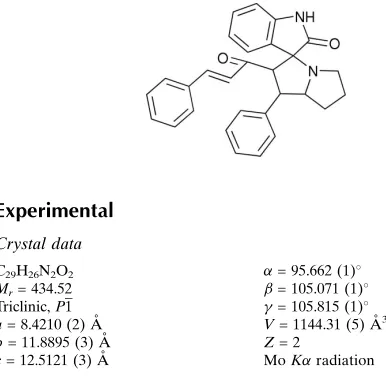
In the title compound, C29H26N2O2, one of the pyrrolidine
rings in the pyrrolizine system is disordered, with site occupancies of ca 0.55 and 0.45. Both components of the disordered pyrrolidine ring adopt envelope conformations, whereas the other pyrrolidine ring adopts a twist conforma-tion. The molecules are linked into centrosymmetric dimers by N—H O hydrogen bonds and the dimers are connectedvia
C—H interactions.
Related literature
For related literature, see: Araki et al.(2002); Caine (1993); Gore et al. (1991); Harris & Uhle (1960); Ho et al. (1986); James et al. (1991); Kobayashi et al. (1991); Ramesh et al.
(2007); Stevenson et al. (2000); Tietze et al.(1988). For ring puckering parameters, see: Cremer & Pople (1975).
Experimental
Crystal data
C29H26N2O2 Mr= 434.52 Triclinic,P1
a= 8.4210 (2) A˚
b= 11.8895 (3) A˚
= 95.662 (1) = 105.071 (1) = 105.815 (1) V= 1144.31 (5) A˚3 Z= 2
= 0.08 mm1 T= 293 (2) K
0.300.200.16 mm
Data collection
Bruker Kappa APEXII diffractometer
Absorption correction: multi-scan (Blessing, 1995)
Tmin= 0.977,Tmax= 0.987
30483 measured reflections 7422 independent reflections 4682 reflections withI> 2(I)
Rint= 0.024
Refinement
R[F2> 2(F2)] = 0.058 wR(F2) = 0.200 S= 1.04 7422 reflections
308 parameters
H-atom parameters constrained max= 0.43 e A˚
3
[image:1.610.48.241.565.753.2]min=0.23 e A˚3
Table 1
Hydrogen-bond geometry (A˚ ,).
D—H A D—H H A D A D—H A
N2—H2 O2i
0.86 2.02 2.854 (2) 162 C28—H28 Cg1ii 0.93 2.89 3.815 (3) 172
Symmetry codes: (i)xþ2;yþ2;zþ1; (ii)x1;y;z1.Cg1 is the centroid of the C8–C13 ring.
Data collection:APEX2(Bruker, 2004); cell refinement:APEX2
and SAINT (Bruker, 2004); data reduction: SAINT and XPREP
(Bruker, 2004); program(s) used to solve structure: SHELXS97
(Sheldrick, 2008); program(s) used to refine structure:SHELXL97
(Sheldrick, 2008); molecular graphics: ORTEP-3 (Farrugia, 1997); software used to prepare material for publication:PLATON(Spek, 2003).
SN thanks Professor M. N. Ponnuswamy, Department of Crystallography and Biophysics, University of Madras, India, for his guidance and valuable suggestions. SN also thanks the management of SRM, India, for their support.
Supplementary data and figures for this paper are available from the IUCr electronic archives (Reference: CI2643).
References
Araki, K., Suenaga, K., Sengoka, T. & Uemura, D. (2002).Tetrahedron,58, 1983–1996.
Blessing, R. H. (1995).Acta Cryst.A51, 33–38.
Bruker (2004).APEX2,SAINT andXPREP. Bruker AXS Inc., Madison, Wisconsin, USA.
Caine, B. (1993).Science,260, 1814.
Cremer, D. & Pople, J. A. (1975).J. Am. Chem. Soc.97, 1354–1358. Farrugia, L. J. (1997).J. Appl. Cryst.30, 565.
Gore, V. G., Chordia, M. D. & Narasimhan, N. S. (1991).Tetrahedron,46, 2483–2494.
Harris, L. S. & Uhle, F. C. (1960).J. Pharmacol. Exp. Ther.128, 353–363. Ho, C. Y., Haegman, W. E. & Perisco, F. (1986).J. Med. Chem.29, 118–121. James, D., Kunze, H. B. & Faulker, D. (1991).J.Nat. Prod.54, 1137–1140. Kobayashi, J., Tsuda, M., Agemi, K. & Vacelet, J. (1991).Tetrahedron,47,
6617–6622.
Ramesh, P., Murugavel, S., SubbiahPandi, A., Murugan, R. & Narayanan, S. S. (2007).Acta Cryst.E63, o4106–o4107.
Sheldrick, G. M. (2008).Acta Cryst.A64, 112–122. Spek, A. L. (2003).J. Appl. Cryst.36, 7–13.
Stevenson, G. I., Smith, A. L., Lewis, S., Michie, S. G., Neduvelil, J. G., Patel, S., Marwood, R., Patel, S. & Castro, J. L. (2000).Bioorg. Med. Chem. Lett.10, 2697–2704.
Tietze, L.-F., Schneider, G., Woelfling, J., Nobel, T. & Wulff, C. (1988).Angew. Chem. Int. Ed.37, 2469–2470.
Acta Crystallographica Section E Structure Reports Online
supporting information
Acta Cryst. (2008). E64, o1817 [doi:10.1107/S1600536808024781]
1-[2-Oxo-1
′
-phenyl-2
′
,3
′
,5
′
,6
′
,7
′
,7a
′
-hexahydroindoline-3-spiro-3
′
-1
′
H
-pyrrolizin-2
′
-yl]-3-phenylprop-2-en-1-one
S. Nirmala, R. Murugan, E. Theboral Sugi Kamala, L. Sudha and S. Sriman Narayanan
S1. Comment
Spiro-compounds are a particular class of naturally occurring substances characterized by highly pronounced biological
properties (Kobayashi et al., 1991; James et al., 1991). The spiro-pyrrolidine ring system is also found in phermones,
antibiotics (Gore et al., 1991) and antitumour agents (Tietze et al., 1988; Araki et al., 2002). Indole compounds can be
used as bioactive drugs (Stevenson et al., 2000). Indole derivatives exhibit anti-allergic, central nervous system
depressant and muscle relaxant properties (Harris & Uhle, 1960; Ho et al., 1986). In view of this biological importance,
the crystal structure of the title compound has been determined and the results are presented here.
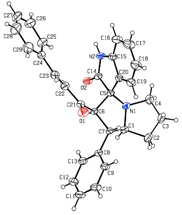
A displacement ellipsoid plot of the title compound is shown in Fig. 1. The pyrrolizine ring system is folded about the
bridging N1—C1 bond, as observed in related structures (Ramesh et al., 2007). The sum of angles at N1 (339.7°) is in
accordance with sp3 hybridization. The indole ring system (N2/C5/C14–C20) forms dihedral angles of 57.4 (6)° and
33.4 (5)°, respectively, with the C24—C29 and C8—C13 phenyl rings. The dihedral angle between the two phenyl rings
is 82.9 (7)°. In the pyrrolizine ring system, the pyrrolidine ring (N1/C1/C5/C6/C7) adopts a twist conformation with
Cremer & Pople (1975) puckering parameters q2 and φ of 0.419 (1) Å and 120.7 (2)°, respectively. Both major and minor
conformers of the disordered pyrrolidine ring adopt envelope conformations; the puckering parameters q2 and φ are
0.267 (4) Å and -68.4 (8)°, respectively, for the major conformer (N1/C1-C4), and 0.254 (8) Å and 108.3 (8)°,
respectively, for the minor conformer (N1/C1/C2/C3A/C4). Atom C3/C3A deviates by 0.411 (2)/0.389 Å from the
N1/C1/C2/C4 plane.
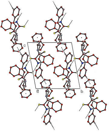
The crystal structure is stabilized by intermolecular N—H···O hydrogen bonds and C—H···π interactions involving the
C8-C13 phenyl ring (Table 1). The N—H···O hydrogen bonds link the molecules into centrosymmetric dimers (Fig. 2).
S2. Experimental
A solution of (1E,6E)-4-benzylidene-1,7-diphenylhepta-1,6-diene-3,5-dione (1 mmol), isatin (1 mmol) and L-proline (1
mmol) in aqueous methanol (20 ml) was refluxed until the disappearance of starting materials as evidenced by TLC. The
solvent was removed under reduced pressure and the crude product was purified by column chromatography using
petroleum ether-ethyl acetate (5:1) as eluent. The final product was recrystallized from ethanol-chloroform (2:8 v/v)
solution.
S3. Refinement
Atom C3 of one of the pyrrolidine rings is disordered over two positions (C3 and C3A) with site occupancies of
0.546 (12) and 0.454 (12). All H atoms were placed in idealized positions and allowed to ride on their parent atoms, with
Figure 1
The molecular structure of the title compound with 30% probability displacement ellipsoids. Only the major disorder
Figure 2
The packing of the molecules viewed along the a axis. Dashed lines indicate hydrogen bonds. H atoms not involed in
hydrogen bonds have been omitted. Only the major disorder component is shown.
1-[2-Oxo-1′-phenyl-2′,3′,5′,6′,7′,7a′-hexahydroindoline-3-spiro- 3′-1′H-pyrrolizin-2′ -yl]-3-phenylprop-2-en-1-one
Crystal data
C29H26N2O2 Mr = 434.52 Triclinic, P1 Hall symbol: -P 1 a = 8.4210 (2) Å b = 11.8895 (3) Å c = 12.5121 (3) Å α = 95.662 (1)° β = 105.071 (1)°
γ = 105.815 (1)° V = 1144.31 (5) Å3 Z = 2
F(000) = 460 Dx = 1.261 Mg m−3
Mo Kα radiation, λ = 0.71073 Å Cell parameters from 9449 reflections θ = 2.3–30.1°
T = 293 K Prism, yellow
0.30 × 0.20 × 0.16 mm
Data collection
Bruker Kappa APEXII diffractometer
Radiation source: fine-focus sealed tube Graphite monochromator
ω scans
Absorption correction: multi-scan (Blessing, 1995)
Tmin = 0.977, Tmax = 0.987
30483 measured reflections 7422 independent reflections 4682 reflections with I > 2σ(I) Rint = 0.024
θmax = 31.3°, θmin = 1.7° h = −12→11
k = −17→17 l = −18→18
Refinement
Refinement on F2 Least-squares matrix: full R[F2 > 2σ(F2)] = 0.058 wR(F2) = 0.200 S = 1.04 7422 reflections 308 parameters 0 restraints
Primary atom site location: structure-invariant direct methods
Secondary atom site location: difference Fourier map
Hydrogen site location: inferred from neighbouring sites
H-atom parameters constrained w = 1/[σ2(F
o2) + (0.1054P)2 + 0.1777P] where P = (Fo2 + 2Fc2)/3
(Δ/σ)max = 0.001 Δρmax = 0.43 e Å−3 Δρmin = −0.23 e Å−3
Special details
Geometry. All e.s.d.'s (except the e.s.d. in the dihedral angle between two l.s. planes) are estimated using the full covariance matrix. The cell e.s.d.'s are taken into account individually in the estimation of e.s.d.'s in distances, angles and torsion angles; correlations between e.s.d.'s in cell parameters are only used when they are defined by crystal symmetry. An approximate (isotropic) treatment of cell e.s.d.'s is used for estimating e.s.d.'s involving l.s. planes.
Refinement. Refinement of F2 against ALL reflections. The weighted R-factor wR and goodness of fit S are based on F2, conventional R-factors R are based on F, with F set to zero for negative F2. The threshold expression of F2 > σ(F2) is used only for calculating R-factors(gt) etc. and is not relevant to the choice of reflections for refinement. R-factors based on F2 are statistically about twice as large as those based on F, and R- factors based on ALL data will be even larger.
Fractional atomic coordinates and isotropic or equivalent isotropic displacement parameters (Å2)
x y z Uiso*/Ueq Occ. (<1)
O1 1.13350 (16) 0.65165 (11) 0.20138 (10) 0.0592 (3) O2 0.94660 (16) 0.99242 (9) 0.34629 (9) 0.0487 (3) N1 0.69132 (15) 0.79708 (12) 0.16496 (10) 0.0446 (3) N2 0.89750 (18) 0.84222 (11) 0.44618 (10) 0.0450 (3) H2 0.9240 0.8844 0.5119 0.054* C1 0.71391 (18) 0.77545 (15) 0.05236 (11) 0.0428 (3) H1 0.7437 0.8511 0.0259 0.051* C2 0.5380 (2) 0.6955 (2) −0.02380 (15) 0.0623 (5)
H3B 0.3205 0.6329 0.0175 0.065* 0.546 (12) C3A 0.4171 (6) 0.7056 (11) 0.0445 (4) 0.067 (2) 0.454 (12) H3C 0.3658 0.7674 0.0245 0.080* 0.454 (12) H3D 0.3248 0.6310 0.0294 0.080* 0.454 (12) C4 0.5126 (2) 0.7336 (2) 0.15888 (16) 0.0635 (5)
H4A 0.4454 0.7882 0.1586 0.076* 0.546 (12) H4B 0.5095 0.6932 0.2224 0.076* 0.546 (12) H4C 0.4673 0.7826 0.2006 0.076* 0.454 (12) H4D 0.5076 0.6622 0.1895 0.076* 0.454 (12) C5 0.83562 (17) 0.78291 (12) 0.25117 (11) 0.0367 (3)
C28 1.6530 (3) 0.8331 (3) 0.7128 (3) 0.1039 (10) H28 1.7444 0.8078 0.7497 0.125* C29 1.5644 (3) 0.7853 (3) 0.6008 (2) 0.0830 (7) H29 1.5949 0.7268 0.5632 0.100*
Atomic displacement parameters (Å2)
U11 U22 U33 U12 U13 U23
O1 0.0650 (8) 0.0620 (8) 0.0555 (7) 0.0293 (6) 0.0181 (6) 0.0059 (6) O2 0.0677 (7) 0.0387 (6) 0.0321 (5) 0.0100 (5) 0.0121 (5) −0.0012 (4) N1 0.0375 (6) 0.0593 (8) 0.0301 (6) 0.0104 (5) 0.0082 (5) −0.0051 (5) N2 0.0613 (8) 0.0429 (7) 0.0250 (5) 0.0086 (6) 0.0145 (5) −0.0029 (5) C1 0.0404 (7) 0.0532 (9) 0.0283 (7) 0.0103 (6) 0.0067 (5) −0.0011 (6) C2 0.0431 (8) 0.0851 (14) 0.0402 (9) 0.0100 (8) −0.0006 (7) −0.0092 (9) C3 0.0394 (18) 0.051 (2) 0.055 (2) 0.0010 (15) 0.0039 (14) −0.0054 (17) C3A 0.039 (2) 0.102 (6) 0.050 (2) 0.018 (2) 0.0014 (16) 0.011 (3) C4 0.0388 (8) 0.0880 (14) 0.0545 (10) 0.0097 (8) 0.0153 (7) −0.0037 (9) C5 0.0395 (6) 0.0390 (7) 0.0254 (6) 0.0050 (5) 0.0105 (5) −0.0041 (5) C6 0.0375 (6) 0.0365 (7) 0.0237 (6) 0.0056 (5) 0.0092 (5) −0.0005 (5) C7 0.0411 (6) 0.0379 (7) 0.0237 (6) 0.0049 (5) 0.0091 (5) −0.0015 (5) C8 0.0425 (7) 0.0419 (8) 0.0240 (6) 0.0110 (6) 0.0090 (5) 0.0009 (5) C9 0.0608 (9) 0.0423 (8) 0.0345 (7) 0.0121 (7) 0.0185 (7) 0.0004 (6) C10 0.0781 (12) 0.0610 (11) 0.0378 (8) 0.0248 (9) 0.0267 (8) −0.0002 (7) C11 0.0687 (11) 0.0753 (12) 0.0351 (8) 0.0228 (9) 0.0258 (8) 0.0106 (8) C12 0.0635 (10) 0.0562 (10) 0.0428 (9) 0.0092 (8) 0.0215 (8) 0.0129 (7) C13 0.0559 (8) 0.0451 (8) 0.0334 (7) 0.0109 (7) 0.0154 (6) 0.0009 (6) C14 0.0426 (7) 0.0405 (8) 0.0265 (6) 0.0086 (6) 0.0100 (5) −0.0028 (5) C15 0.0510 (8) 0.0436 (8) 0.0335 (7) 0.0086 (6) 0.0175 (6) 0.0022 (6) C16 0.0809 (12) 0.0603 (11) 0.0423 (9) 0.0185 (9) 0.0265 (9) 0.0127 (8) C17 0.1015 (16) 0.0559 (12) 0.0669 (13) 0.0207 (11) 0.0407 (12) 0.0247 (10) C18 0.0915 (14) 0.0417 (10) 0.0708 (13) 0.0098 (9) 0.0420 (11) 0.0086 (9) C19 0.0658 (10) 0.0420 (9) 0.0478 (9) 0.0030 (7) 0.0252 (8) −0.0028 (7) C20 0.0454 (7) 0.0407 (8) 0.0329 (7) 0.0050 (6) 0.0166 (6) −0.0009 (6) C21 0.0410 (7) 0.0495 (9) 0.0340 (7) 0.0113 (6) 0.0142 (6) 0.0075 (6) C22 0.0454 (8) 0.0581 (10) 0.0400 (8) 0.0157 (7) 0.0098 (6) 0.0084 (7) C23 0.0487 (8) 0.0655 (11) 0.0449 (9) 0.0204 (7) 0.0163 (7) 0.0158 (8) C24 0.0418 (8) 0.0762 (12) 0.0399 (8) 0.0111 (7) 0.0089 (6) 0.0195 (8) C25 0.0636 (11) 0.0808 (14) 0.0420 (9) 0.0191 (10) 0.0031 (8) 0.0104 (9) C26 0.0912 (16) 0.0830 (16) 0.0447 (11) 0.0016 (12) 0.0090 (10) 0.0056 (10) C27 0.0738 (14) 0.113 (2) 0.0494 (12) −0.0149 (14) −0.0117 (11) 0.0253 (13) C28 0.0616 (13) 0.159 (3) 0.0767 (17) 0.0308 (16) −0.0095 (12) 0.0473 (19) C29 0.0599 (11) 0.121 (2) 0.0753 (15) 0.0402 (12) 0.0142 (11) 0.0323 (14)
Geometric parameters (Å, º)
N1—C4 1.467 (2) C10—C11 1.362 (3) N1—C1 1.4772 (18) C10—H10 0.93 N2—C14 1.3477 (19) C11—C12 1.375 (3) N2—C15 1.398 (2) C11—H11 0.93 N2—H2 0.86 C12—C13 1.381 (2) C1—C2 1.525 (2) C12—H12 0.93 C1—C7 1.529 (2) C13—H13 0.93 C1—H1 0.98 C15—C16 1.375 (2) C2—C3 1.443 (5) C15—C20 1.387 (2) C2—C3A 1.511 (6) C16—C17 1.379 (3) C2—H2A 0.97 C16—H16 0.93 C2—H2B 0.97 C17—C18 1.377 (3) C2—H2C 0.96 C17—H17 0.93 C2—H2D 0.96 C18—C19 1.380 (3) C3—C4 1.503 (4) C18—H18 0.93 C3—H3A 0.97 C19—C20 1.379 (2) C3—H3B 0.97 C19—H19 0.93 C3A—C4 1.402 (5) C21—C22 1.481 (2) C3A—H3C 0.97 C22—C23 1.318 (2) C3A—H3D 0.97 C22—H22 0.93 C4—H4A 0.97 C23—C24 1.459 (2) C4—H4B 0.97 C23—H23 0.93 C4—H4C 0.96 C24—C29 1.383 (3) C4—H4D 0.96 C24—C25 1.394 (3) C5—C20 1.511 (2) C25—C26 1.380 (3) C5—C14 1.5502 (18) C25—H25 0.93 C5—C6 1.5623 (18) C26—C27 1.367 (4) C6—C21 1.502 (2) C26—H26 0.93 C6—C7 1.5268 (18) C27—C28 1.356 (4) C6—H6 0.98 C27—H27 0.93 C7—C8 1.5044 (18) C28—C29 1.382 (4) C7—H7 0.98 C28—H28 0.93 C8—C9 1.387 (2) C29—H29 0.93
C20—C5—C14 101.51 (11) C25—C26—H26 120.3 N1—C5—C6 102.60 (10) C28—C27—C26 120.9 (2) C20—C5—C6 113.67 (12) C28—C27—H27 119.6 C14—C5—C6 110.64 (11) C26—C27—H27 119.6 C21—C6—C7 116.10 (12) C27—C28—C29 120.4 (2) C21—C6—C5 113.56 (11) C27—C28—H28 119.8 C7—C6—C5 102.38 (10) C29—C28—H28 119.8 C21—C6—H6 108.1 C28—C29—C24 120.2 (3) C7—C6—H6 108.1 C28—C29—H29 119.9 C5—C6—H6 108.1 C24—C29—H29 119.9
C2—C1—C7—C8 85.07 (17) C21—C22—C23—C24 −171.52 (15) N1—C1—C7—C6 −31.99 (14) C22—C23—C24—C29 −167.19 (19) C2—C1—C7—C6 −149.10 (14) C22—C23—C24—C25 16.6 (3) C6—C7—C8—C9 132.52 (15) C29—C24—C25—C26 −0.2 (3) C1—C7—C8—C9 −109.94 (16) C23—C24—C25—C26 176.07 (19) C6—C7—C8—C13 −50.44 (19) C24—C25—C26—C27 1.4 (3) C1—C7—C8—C13 67.09 (18) C25—C26—C27—C28 −1.2 (4) C13—C8—C9—C10 0.3 (2) C26—C27—C28—C29 −0.2 (4) C7—C8—C9—C10 177.46 (15) C27—C28—C29—C24 1.4 (4) C8—C9—C10—C11 0.1 (3) C25—C24—C29—C28 −1.2 (3) C9—C10—C11—C12 −0.5 (3) C23—C24—C29—C28 −177.6 (2)
Hydrogen-bond geometry (Å, º)
D—H···A D—H H···A D···A D—H···A
N2—H2···O2i 0.86 2.02 2.854 (2) 162 C28—H28···Cg1ii 0.93 2.89 3.815 (3) 172