Original Article
Regulation and mechanisms of lncRNA H19 in COPD
Jiyou Zhang1, Jing Liu1, Ping Ju2, Chao Ren3, Zhao Ma4, Fangchao Jiang5
Departments of 1Pulmonary Disease, 2Nursing, 3Science and Education Section, 4Medical Section, Shandong
Qin-gdao Hospital of Integrated Traditional Chinese and Western Medicine, QinQin-gdao, Shandong, China; 5Hiser Medcal
Center of Qingdao, Qingdao, Shandong, China
Received March 28, 2019; Accepted July 11, 2019; Epub September 15, 2019; Published September 30, 2019
Abstract: Chronic obstructive pulmonary disease (COPD) is one of the most fatal diseases of the respiratory sys-tem. Moreover, lncRNA H19 is involved in the regulation of various diseases, including tumors. However, the roles of lncRNA H19 in COPD have not yet been elucidated. SD rats were randomly divided into the control group, model group, and H19 group. Real-time PCR was used to detect lncRNA H19 expression levels. H&E staining was used to analyze lung tissue pathological changes. Secretion of inflammatory factors IL-6, IL-8, and TNF-α was detected by ELISA. SOD and ROS content levels were also measured. The 16HBE cells were cultured in vitro and randomly divided into the control group, model group, and H19 group. This was followed by analysis of cell proliferation via MTT assays. Activity levels of caspase 3 and expression levels of Bax and Bcl-2 were analyzed by Western blotting. Compared with the control group, lncRNA H19 expression was significantly decreased in the COPD rat model group (P<0.05). Overexpression of lncRNA H19 improved lung tissue structure, downregulated secretion of IL-6, IL-8, and TNF-α, upregulated SOD expression, and inhibited ROS generation. Compared with the model group, differences were statistically significant (P<0.05). In addition, overexpression of lncRNA H19 in 16HBE cells promoted cell pro-liferation, decreased caspase 3 activity, decreased Bax expression, and increased Bcl-2 expression, with significant differences, compared to the control group (P<0.05). Expression of lncRNA H19 was decreased in COPD rats. In summary, upregulation of lncRNA H19 expression inhibits secretion of inflammatory cytokines, regulates oxidative stress, inhibits cell apoptosis, and promotes cell proliferation, alleviating COPD.
Keywords: Chronic obstructive pulmonary disease, lncRNA H19, inflammatory factor, apoptosis, oxidative stress
Introduction
Chronic obstructive pulmonary disease (COPD), or chronic obstructive pulmonary disease, is one of the most common respiratory diseases, worldwide. Incidence rates of this disease are second only to cardiovascular and cerebrovas-cular diseases [1]. According to statistics, deaths due to COPD are as high as 3 million people per year, ranking fourth in the world. Thus, it has been estimated to be third leading cause of deaths, worldwide, by 2020 [2, 3]. COPD can be found in all ages, but it is more common in middle-aged and elderly popula-tions. Incidence of COPD in Chinese patients over 40 years old is as high as 13.7% [4]. Pathological changes of COPD include chronic bronchitis and/or emphysema with chronic air-flow obstruction, chronic coughing, intolerance to exercise, shortness of breath when breath-ing, and difficulty breathing. These can further
develop into pulmonary heart disease. The common chronic disease of exhaustion is one of the most prone and deadliest diseases in the respiratory system, seriously affecting the qual-ity of life of patients and posing a serious thr- eat to social health [5, 6]. Despite current advances in the treatment of COPD, COPD has not been completely eradicated. Incidence and mortality rates of COPD remain high [7]. The pathogenesis of COPD has not yet been eluci-dated. Pathological manifestations are compli-cated. They can accumulate in lung tissue structures and pulmonary blood vessels [8]. Therefore, finding and clarifying the exact tar-gets of COPD is beneficial for treatment of COPD.
activities [9]. Moreover, lncRNAs have been thought to be the presence of transcriptional “noise” when initially recognized. Non-coding transcripts account for the vast majority of human genomes [10, 11]. A variety of biological activities and signaling pathways can be regu-lated by lncRNAs, including cell proliferation, differentiation, and other processes. Abnormal expression of lncRNAs has been closely related to tumors, neurological diseases, and autoim-mune diseases [12, 13]. Of note, lncRNA H19 may be involved in the regulation of various dis-eases, including tumors [14, 15]. It has been shown to be abnormally expressed in respira-tory diseases, including lung cancer [16]. How- ever, the roles of lncRNA H19 in COPD have not been elucidated.
Materials and methods
Animals
Healthy SD male rats, 6 weeks old, SPF grade, with body weights of 200 ± 20 g, were pur-chased from the Experimental Animal Center of Shandong University. They were fed in the SPF Animal Experiment Center with a temperature of 21 ± 1°C, relative humidity of 50-70%, and a 12/day cycle every 12 hours.
Experimental procedures were approved by the Animal Ethics Committee of Shandong Qingdao Hospital of Integrated Traditional Chinese and Western Medicine (Qingdao, Shandong, China).
Reagents and equipment
Primary normal human bronchial epithelial cells (16HBE) (product number: ATCC® CRL-2741TM) were purchased from ATCC, USA. TRIzol Reagent was purchased from Sigma (USA). Moreover, lncRNA H19 plasmids and lncRNA H19 adenoviruses were designed and synthesized by Shanghai Gene Co., Ltd. The RNA extraction kit, RT-PCR primer, reverse tran-scription (RT) kit, and Real-time PCR reagents were purchased from Axygen (USA). PVDF membranes were purchased from Pall Life Sciences. Western blotting related chemical reagent was purchased from Shanghai Bi- yuntian Biotechnology Co., Ltd. ECL reagent was purchased from Amersham Biosciences. Rabbit anti-human Bax monoclonal antibo- dies, rabbit anti-human Bcl-2 antibodies, and goat anti-rabbit Horseradish peroxidase (HRP)-labeled IgG secondary antibodies were pur-chased from Cell Signal, Inc. (USA). DMEM
medium, fetal bovine serum (FBS), and cyan chain double antibodies were purchased from Hyclone (USA). Dimethyl sulfoxide (DMSO) MTT powder was purchased from Gibco (USA). Trypsin digest was purchased from Sigma. IL-6, IL-8, and TNF-α ELISA kits were purchased from R&D (USA). Caspase 3 activity assay kit was purchased from Cell Signal, Inc. (USA). SOD activity detection kit and ROS activity kit were purchased from Wuhan Boster Biote- chnology Co., Ltd. ABI7900 HT Real-time PCR was purchased from ABI (USA). LabSystem Version 1.3.1 microplate reader was purchased from Bio-Rad Corporation of the United States. The CK2 fluorescence microscope was pur-chased from Olympus Corporation of Japan. Rat grouping and model preparation
Healthy male SD rats were randomly divided into 3 groups, with n=10 in each group. In the control group, SD rats were normally reared. Rats in the model group were used to prepare a COPD rat model using the cigarette smoke method. H19 group was transfected with H19 plasmid adenoviruses in COPD rats.
According to a literature report [17], the rats were placed in a self-made sealed fumigation box (80 × 60 × 58 cm). They were exposed to the smoke of 20 cigarettes (yellow fruit tree, each 15 mg tar, and 1.25 mg nicotine). They were ignited twice a day, burning up to 50 min-utes, twice a day for 16 weeks. Based on the preparation of rat COPD model, the H19 group received injections of lncRNA H19 plasmid adenovirus (108 pfu/500 μl) through the oral cavity.
Sample collection
A total of 5 mL of blood was collected through the tail veins. Blood was centrifuged at 3,000 rpm for 15 minutes to obtain serum, which was stored at -20°C for ELISA analysis. After the rats were sacrificed, the right middle lobes were frozen and fixed in liquid nitrogen or neu-tral paraformaldehyde.
H&E staining
seal-ing. The sealing film was observed under an optical microscope.
16HBE cell culturing, grouping, and processing
The 16HBE cells were taken out from liquid nitrogen, resuscitated, and cultured in a medi-um containing 10% FBS and 90% high glucose DMEM medium (containing 100 U/mL penicil-lin, 100 μg/mL streptomycin), at 37°C with 5% CO2. Cells in the logarithmic growth phase were used for experiments. Cells were randomly divided into 3 groups, including the control group, model group, and H19 group. Cigarette smoke extract was continuously added to model group and H19 group, while the H19 group was transfected with H19-pcDNA3.1 using Lipo2000 Reagent. The H19-pcDNA3.1 plasmid sequence was 5’-TAACACACAGGCA- CCAATTCATA-3’-3’.
Real-time PCR detection of lncRNA H19 ex-pression
Total RNA was extracted using TRIzol Reagent (American Axygen Corporation) for analysis of lncRNA H19 expression via real-time PCR. This method used primers designed by PrimerPremier6.0 (Table 1). GAPDH was used as a reference.
tion by MTT assays, according to manufacturer instructions. The experiment was repeated three more times.
ELISA analysis of levels of IL-6, IL-8 and TNF-α
Serum was collected, measuring levels of IL-6, IL-8, and TNF-α using ELISA, according to kit instructions.
Caspase 3 activity assays
Changes in caspase 3 activity were examined using 2 mM Ac-DEVD-pNA, according to kit instructions.
Western blot analysis of expression of Bax and Bcl-2
Total protein was extracted using RIPA lysis buf-fer and quantified using the BCA method. They were stored at -20°C for Western blot analysis, examining expression levels of Bax and Bcl-2. Data were analyzed using protein image pro-cessing system software and Quantity one soft-ware. The experiment was repeated four times.
Analysis of SOD activity and ROS activity
According to kit instructions, changes in SOD activity and ROS production in lung tissues of each group were analyzed.
Statistical analysis
Data are expressed as mean ± standard devia-tion (SD) and was processed using SPSS 11.5 statistical software. Mean values of the two groups were compared using Student’s t-tests and analyzed with analysis of variance (ANOVA). P<0.05 indicates statistical differences.
Results
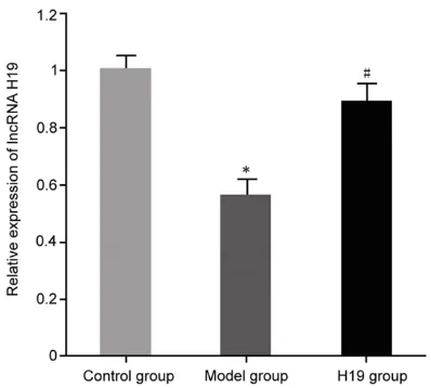
Expression of lncRNA H19 in a rat model of COPD
[image:3.612.89.358.83.125.2]Compared with the control group, expression of lncRNA H19 was significantly decreased in the COPD rat model (P<0.05). After transfection of H19 plasmid adenovirus into COPD rats,
Table 1. Primer sequences for real-time PCR Gene Forward 5’-3’ Reverse 5’-3’
GAPDH AGTACTCTGTCAGTGG TAATCGAATGTACGTGGT lncRNA H19 CATCTTGTCTTGGATGACTT CCTCACGCCAGGCTTTCACTG
MTT assays for detection of cell proliferation
The 16HBE cells (5 × 10³) were treated, as mentioned above, fol-lowed by analysis of cell
[image:3.612.89.288.145.324.2]expression of lncRNA H19 in COPD rats was sig-nificantly upregulated (P<0.05) (Figure 1). Upregulation of lnc RNA H19 expression in lung tissues of COPD rats
H&E staining was used to evaluate the effects of lncRNA H19 upregulation in COPD rats by transfecting H19 plasmid adenovirus on lung tissues. Results showed that lung tissues of the control group were intact, without inflam-matory cell infiltration, including a normal bron-chial structure and intact mucosa. However, lung tissues of COPD rats were narrowed or even broken. The blood vessel walls were thick-ened, with inflammatory cell infiltration and damaged bronchial mucosa, as well as a par-tially detached epithelium. Interestingly, over-expression of lncRNA H19 during COPD reduced inflammatory cell infiltration, improved lung tis-sue damage, reduced vessel wall thickness, and increased bronchial epithelial cell integrity (Figure 2).
Upregulation of lncRNA H19 expression on
IL-6, IL-8, and TNF-α
Secretion of inflammatory factors IL-6, IL-8, and TNF-α in the serum of COPD rats was increased significantly, compared with that in the control group (P<0.05). Overexpression of lncRNA H19 in COPD rats significantly inhibited the secre-tion of inflammatory cytokines IL-6, IL-8, and TNF-α, compared with the model group (P<0.05) (Figure 3).
Upregulation of lncRNA H19 expression on oxidative stress
Changes in SOD activity and ROS production in lung tissues of each group were analyzed. The activity of SOD in lung tissues of COPD rats was significantly decreased and the production of ROS was significantly increased, compared with levels in the control group (P<0.05). Overexpression of lncRNA H19 in COPD rats significantly upregulated SOD activity and inhib-ited ROS production, with statistical differenc-es, compared with the model group (P<0.05) (Figure 4).
Expression of lncRNA H19 in 16HBE cells
Expression of lncRNA H19 was significantly decreased in the COPD bronchial epithelial 16HBE cell injury model. It was prepared from cigarette smoke extract (P<0.05). Transfection of lncRNA H19 plasmids in the model group sig-nificantly promoted the increase of lncRNA H19 expression, compared with the model group (P<0.05) (Figure 5).
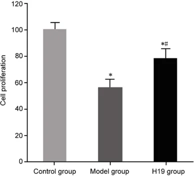
Upregulation of lncRNA H19 expression on proliferation of 16HBE cells
[image:4.612.94.522.72.189.2]In the model of COPD bronchial epithelial 16HBE cell injury, prepared by cigarette smoke Figure 2. Changes in lung tissues of COPD rats after upregulation of lncRNA H19 expression (× 100).
[image:4.612.89.289.229.395.2]expression on caspase3 activity
In the model of COPD bronchial epithelial 16HBE cell injury, prepared by cigarette smoke extract, the activity of caspase3 was significantly increased, compared with the con-trol group (P<0.05). Upregulation of lncRNA H19 expression in the model group of 16HBE cells significantly reduced caspase3 activity, compared with the model group (P<0.05) (Figure 7).
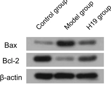
Upregulation of lnc RNA H19 expression on apoptosis of 16HBE cells
In the model of COPD bronchial epithelial 16HBE cell injury, prepared by cigarette smoke extract, expression of Bax-2 was increased and expression of anti-apoptotic protein Bcl-2 was decreased. Upregulation of lncRNA H19 expres-sion in the model group of 16HBE cells inhibit-ed Bax expression and increasinhibit-ed expression of Bcl-2 (Figure 8).
Discussion
[image:5.612.90.374.72.207.2]Prevalence and mortality rates of COPD are high. The high costs of treatment “three highs” poses a serious threat to public health and social economy [18]. The pathogenesis of COPD is very complicated, involving several factors, including chronic inflammation and oxidative stress. This can lead to chronic bronchial inflammation, bronchial epithelial cell damage, and destruction of lung parenchyma, ultimately leading to persistent airflow limitations and occurrence and progression of COPD [19]. Figure 4. Upregulation of expression of lncRNA H19 on oxidative stress in
COPD rats. A: SOD activity in COPD rats; B: ROS generation in COPD rats. Compared with control group, *P<0.05; compared with model group,
#P<0.05.
Figure 5. Expression of lncRNA H19 in 16HBE cells from COPD bronchial epithelium. Compared with control group, *P<0.05; compared with model group,
[image:5.612.89.288.277.459.2]#P<0.05.
Figure 6. Upregulation of expres-sion of lncRNA H19 on the prolif-eration of 16HBE cells; Compared with control group, *P<0.05; com-pared with model group, #P<0.05.
extract, cell proliferation was significantly reduced, com-pared with the control group (P<0.05). Upregulation of lnc- RNA H19 expression in the model group of 16HBE cells significantly promoted cell proliferation, compared with the model group (P<0.05) (Figure 6).
[image:5.612.92.286.531.707.2]Smoking can lead to the development of chronic lung diseases, such as COPD [20]. Therefore, the purpose of this study was to investigate the effects of cigarette smoke expo-sure on the lungs, aiming to evaluate possible therapeutic targets for treatment of chronic obstructive pulmonary disease. Moreover, lncRNAs do not encode proteins, have no bio-logical function, and are called dark substanc-es in gene transcription. However, they have been found to be involved in the regulation of protein coding, before and after transcriptional regulation, at epigenetic levels [21]. Additionally, lncRNA H19 has been confirmed to be abnor-mally expressed in respiratory diseases [16]. In this study, it was confirmed that expression of
lncRNA H19 was decreased in the COPD rat model. Expression in COPD rats was upregulat-ed after transfection with the H19 plasmid ade-novirus. Upregulation of lncRNA H19 expres-sion in COPD rats inhibited secretion of IL-6, IL-8, and TNF-α inflammatory factors and effec-tively improved the lung tissue structure of COPD. Results suggest that lncRNA H19 might be involved in the regulation of the develop-ment and progression of COPD and can be used as a target for prevention and treatment of COPD, as well as a biomarker.
[image:6.612.90.287.70.250.2]A previous study confirmed that all COPD patients have elevated oxidative stress and chronic inflammation [22]. In COPD patients, free radical production increases. Thus, the balance between reactive oxygen species pro-duction and antioxidant defense functions is impaired. This, in turn, aggravates the inflam-matory process [23]. Numerous studies have confirmed that increased oxidative damage in smokers leads to lung injuries through various biological effects. Oxidative stress index ROS is increased and SOD activity is decreased, which further aggravates inflammation [24]. The pres-ent study found that overexpression of lncRNA H19 in COPD rats improved lung tissue struc-ture, downregulated secretion of inflammatory factors IL-6, IL-8, and TNF-α, upregulated SOD activity, and inhibited ROS generation, suggest-ing that lncRNA H19 upregulation can inhibit COPD by regulating oxidative stress and inhibit-ing inflammation. Apoptotic protein Bax and anti-apoptotic protein Bcl-2 are important members of the apoptotic protein family. Abnormal expression of both can lead to abnormal expression of caspase3 activity and apoptosis, which leads to changes in cell bio-logical processes [20]. Present results con-firmed that Bax expression was decreased, Bcl-2 expression was increased, caspase3 activity was increased, bronchial epithelial cell apoptosis was promoted, and cell proliferation was inhibited. By upregulating lncRNA H19 expression in COPD bronchial epithelial cells, Bax expression was significantly inhibited, Bcl-2 expression was increased, and caspase3 activ-ity was decreased. These factors protected bronchial epithelial cells from apoptosis, pro-moting cell proliferation and preventing cell damage. Further investigation is required, ana-lyzing expression of lncRNA H19 in clinical sam-ples and examining possible mechanisms. This Figure 7. Upregulation of lncRNA H19 expression on
caspase3 activity in 16HBE cells; Compared with control group, *P<0.05; compared with model group,
#P<0.05.
[image:6.612.92.285.325.473.2]will provide reference for diagnosis and treat-ment of COPD.
In conclusion, expression of lncRNA H19 is decreased in COPD rats. Upregulation of lncRNA H19 expression in bronchial epithelial cells in COPD rats or COPD can inhibit oxidative stress, regulate oxidative stress, inhibit apopto-sis, and promote cell proliferation, alleviating COPD.
Disclosure of conflict of interest
None.
Address correspondence to: Dr. Fangchao Jiang, Hiser Medcal Center of Qingdao, No. 4 Renming Road, Shibei District, Qingdao, Shandong, China. Tel: +86-0532-83777196; Fax: +86-0532-8377- 7196; E-mail: jianglutao53@163.com
References
[1] Wielputz MO, Eichinger M, Wege S, Eberhardt R, Mall MA, Kauczor HU, Puderbach MU, Risse F, Heussel CP and Heussel G. Mid-term repro-ducibility of chest MRI in adults with clinically stable cystic fibrosis and chronic obstructive pulmonary disease. Am J Respir Crit Care Med 2019.
[2] Amegadzie JE, Badejo O, Gamble JM, Wright M, Farrell J, Jackson B, Sultana K, Hashmi M and Gao Z. Validated methods to identify pa-tients with asthma-COPD overlap in healthcare databases: a systematic review protocol. BMJ Open 2019; 9: e024306.
[3] Bunel V, Guyard A, Dauriat G, Danel C, Montani D, Gauvain C, Thabut G, Humbert M, Castier Y, Dorfmuller P and Mal H. Pulmonary arterial histologic lesions in patients with COPD with severe pulmonary hypertension. Chest 2019; 156: 33-44.
[4] Sun J and Zhao G. Clinical effects of lentinan combined with budesonide inhalation in treat-ing acute exacerbation of chronic obstructive pulmonary disease under mechanical ventila-tion. Exp Ther Med 2019; 17: 1503-1508. [5] Brozek GM, Nowak M, Zejda JE, Jankowski M,
Lawson J and Pierzchala W. Costs of pharma-cotherapy of chronic obstructive pulmonary disease in relation to changing global initiative for chronic obstructive lung disease 2007-2011-2017 guidelines. Pol Arch Intern Med 2019; 129: 308-315.
[6] Inomoto A, Yamato H, Michishita R, Jiang Y, Nishiyama S, Fukuda R and Deguchi J. Fre-quency of exposure to secondhand smoke out-side the home is associated with a lower FEV1/
FVC in male workers regardless of smoking status. J UOEH 2019; 41: 15-24.
[7] Sievi NA, Kohler M, Thurnheer R, Leuppi JD, Irani S, Frey M, Brutsche M, Brack T and Clar-enbach CF. No impact of exacerbation frequen-cy and severity on the physical activity decline in COPD: a long-term observation. Int J Chron Obstruct Pulmon Dis 2019; 14: 431-437. [8] Das N, Topalovic M, Aerts JM and Janssens W.
Area under the forced expiratory flow-volume loop in spirometry indicates severe hyperinfla-tion in COPD patients. Int J Chron Obstruct Pul-mon Dis 2019; 14: 409-418.
[9] Hu Y, He MY, Zhu LF, Yang CC, Zhou ML, Wang Q, Zhang W, Zheng YY, Wang DM, Xu ZQ, Wu YN and Liu LK. Tumor-associated macrophages correlate with the clinicopathological features and poor outcomes via inducing epithelial to mesenchymal transition in oral squamous cell carcinoma. J Exp Clin Cancer Res 2016; 35: 12.
[10] Zhang SR, Yang JK, Xie JK and Zhao LC. Long noncoding RNA HOTTIP contributes to the pro-gression of prostate cancer by regulating HOXA13. Cell Mol Biol (Noisy-le-grand) 2016; 62: 84-8.
[11] Chang L, Qi HL, Xiao YS, Li CS, Wang YT, Guo T, Liu ZS and Liu QY. Integrated analysis of non-coding RNAs and mRNAs reveals their poten-tial roles in the biological activities of the growth hormone receptor. Growth Horm IGF Res 2016; 29: 11-20.
[12] Cheng N and Guo Y. Long noncoding RNA NEAT1 promotes nasopharyngeal carcinoma progression through regulation of miR-124/ NF-kappa B pathway. Onco Targets Ther 2017; 10: 5843-5853.
[13] Wu J, Du MY, Zhang Q, Zhang WJ, Fan YX, Yin L, Fei Q, Jiang XS, Chen W, Zhu HF, Yan PW, He X and Bian XH. Long noncoding RNA UCA1 pro-motes the proliferation, invasion, and migra-tion of nasopharyngeal carcinoma cells via modulation of miR-145. Onco Targets Ther 2018; 11: 7483-7492.
[14] Basak P, Chatterjee S, Bhat V, Su A, Jin H, Lee-Wing V, Liu Q, Hu PZ, Murphy LC and Raouf A. Long non-coding RNA H19 acts as an estrogen receptor modulator that is required for endo-crine therapy resistance in ER+ breast cancer cells. Cell Physiol Biochem 2018; 51: 1518-1532.
[15] Zeng L, Sun SC, Han D, Liu Y, Liu HC, Feng HL and Wang YX. Long non-coding RNA H19/ SAHH axis epigenetically regulates odontogen-ic differentiation of human dental pulp stem cells. Cell Signal 2018; 52: 65-73.
development by regulating microRNA-107. Eur Rev Med Pharmacol Sci 2018; 22: 5946-5953.
[17] Kulvinskiene I, Raudoniute J, Bagdonas E, Ciu-zas D, Poliakovaite K, Stasiulaitiene I, Zabulyte D, Bironaite D, Rimantas Venskutonis P, Mar-tuzevicius D, Aldonyte R. Lung alveolar tissue destruction and protein citrullination in diesel exhaust-exposed mouse lungs. Basic Clin Pharmacol Toxicol 2019; 125: 166-177. [18] Stulce JM, Biddle C and Vacchiano C. Low-flow
domiciliary oxygen as a mechanism of ongo- ing oxidative stress. Respir Care 2019; [Epub ahead of print].
[19] Hsu PS, Lin CM, Chang JF, Wu CS, Sia KC, Lee IT, Huang KY and Lin WN. Participation of NADPH oxidase-related reactive oxygen spe-cies in leptin-promoted pulmonary inflamma-tion: regulation of cPLA2 and COX-2 expres-sion. Int J Mol Sci 2019; 20.
[20] Zhou TY, Hu Y, Wang YX, Sun C, Zhong YJ, Liao JP and Wang GF. Fine particulate matter (PM2.5) aggravates apoptosis of cigarette-in-flamed bronchial epithelium in vivo and vitro. Environ Pollut 2019; 248: 1-9.
[21] Bitarafan S, Yari M, Broumand MA, Ghaderian SMH, Rahimi M, Mirfakhraie R, Azizi F and Om-rani MD. Association of increased levels of ln-cRNA H19 in PBMCs with risk of coronary ar-tery disease. Cell J 2019; 20: 564-568. [22] Liu X, Deng K, Chen S, Zhang Y, Yao J, Weng X,
Gao T and Feng G. 8-Hydroxy-2’-deoxyguano-sine as a biomarker of oxidative stress in acute exacerbation of chronic obstructive pulmonary disease. Turk J Med Sci 2019; 49: 93-100. [23] Tesfaigzi Y, Petersen H, Celli B and Owen C.
Functional studies of single-nucleotide poly-morphisms suggest heterogeneity in chronic obstructive pulmonary disease due to suscep-tibility of different cell types. Ann Am Thorac Soc 2018; 15 Suppl 4: S285.