Original Article
Impact of amiodarone on cardiac
structural function and MMP-2 and TIMP-2
levels in atrial fibrillation radiofrequency ablation
Li’na Wang, Qiwei Liu, Xiaoyan Ma
Department of Cardiology, Baoji Hi-Tech People’s Hospital, Baoji, Shaanxi Province, China
Received November 14, 2018; Accepted December 7, 2018; Epub April 15, 2019; Published April 30, 2019
Abstract: Objective: The aim of this study was to determine the mechanisms of action of amiodarone on cardiac structure and function and on levels of matrix metalloproteinas-2 (MMP-2) and tissue inhibitor of metalloprotein-ase-2 (TIMP-2) after radiofrequency ablation of atrial fibrillation (AF). Methods: Eighty-six patients with AF, treated by radiofrequency ablation, were randomized into the experimental group (n=43) and control group (n=43). The experimental group received amiodarone hydrochloride tablets after the operation, while the control group did not receive any antiarrhythmic drugs. Electrocardiograms and echocardiography were used to determine the number of AF, duration of AF, left atrial end-diastolic diameter, left atrial end-systolic diameter, as well as left ventricular ejec-tion fracejec-tion (LVEF), before the operaejec-tion and 6 months after the operaejec-tion. Serum levels of MMP-2 and TIMP-2 were measured by ELISA and qRT-PCR, respectively, before and 6 months after the operation. Relevant data were ana-lyzed by statistics. Results: Six months after the operation, the number and duration of AF, left atrial end-diastolic diameter, and left atrial end-systolic diameter decreased significantly. LVEF increased significantly, compared to pre-operation values in both control and experimental groups (all P<0.05). Furthermore, the number and duration of AF, left atrial end-diastolic diameter, and left atrial end-systolic diameter were significantly lower. LVEF was significantly higher in the experimental group 6 months after the operation, compared to those in the control group, with statisti-cal significance (all P<0.05). Relevant data were analyzed by statistics. MMP-2 messenger ribonucleic acid and pro-tein levels in serum decreased significantly, while TIMP-2 mRNA and propro-tein levels increased significantly 6 months after the operation, in both groups, compared to pre-operation levels (all P<0.05). The experimental group had significantly lower MMP-2 mRNA and protein levels in serum and significantly higher TIMP-2 mRNA and protein lev-els, compared to the control group (all P<0.05), 6 months after the operation. Conclusion: After AF radiofrequency ablation, amiodarone can protect cardiac structure and function, reduce MMP-2 levels, and increase TIMP-2 levels.
Keywords: Atrial fibrillation radiofrequency ablation, amiodarone, the heart, matrix metalloproteinas-2, tissue in-hibitor of metalloproteinase-2
Introduction
Atrial fibrillation (AF) is a kind of arrhythmia characterized by fainting, fatigue, palpitations, and chest pain. Some patients, however, may have no specific symptoms [1, 2]. In recent years, epidemiological surveys have shown that the total prevalence rate of AF, world- wide, was 0.73%, while the clinical incidence rate in China was slightly higher (0.77%) [3]. Radiofrequency ablation of AF can improve symptoms and repair the structure of the left atrium [4]. However, it is still associated with a high recurrence rate after the operation. About one third of the AF patients that receive
of MMP-2 in patients with paroxysmal AF are significantly upregulated [10]. TIMP-2 is an endogenous specific inhibitor of MMPs. Inter- actions among MMPs, TIMPs, and their regula-tory factors determine the process of myocar-dial interstitial remodeling [11]. The effects of amiodarone on recurrence of AF after radiofre-quency ablation have been studied, but effects on cardiac structure and function, as well as levels of MMP-2 and TIMP-2, in patients with AF after radiofrequency ablation have not been studied.
In this study, 86 patients with AF, treated with radiofrequency ablation, were divided into the amiodarone group (experimental group) and non-amiodarone treated group (control group). Effects of amiodarone on cardiac function and MMP-2 and TIMP-2 levels were observed in both groups.
Methods and materials
Subjects and groupings
Eighty-six patients with AF, treated with radio-frequency ablation, in Baoji Hi-Tech People’s Hospital, from July 2014 to May 2016, were enrolled in the study. Ages of patients ranged from 41-75 years old, with 44 males and 42 females. The duration of atrial fibrillation was 0.5-3.1 years.
Inclusion criteria: Patients diagnosed with par-oxysmal atrial fibrillation, confirmed by clinical electrocardiogram (ECG); Patients without tu- mors; Patients without administration of antiar-rhythmic drugs; Patients that underwent first annular pulmonary vein ablation that reached the end point of ablation.
Exclusion criteria: Patients with coronary heart disease and hyperthyroidism; Patients whose AF was due to other diseases; Patients with cardiac diseases other than AF; Patients with contraindications of amiodarone.
Enrolled patients were randomized into the control group (n=43; 21 males and 22 fema- les) and experimental group (n=43; 23 males and 20 females). After the operation, experi-mental group patients were given amiodarone hydrochloride (0.2 g/tablet; 150267, Sanofi Pharmaceutical Co. Ltd., France) for 6 months. The dosage was 200 mg, three times a day, in
the first week. It was twice a day during the next two weeks and once a day thereafter. The control group did not receive any antiarrhyth-mic drugs after the operation. Informed con-sent was obtained from all patients and the experiment was approved by the Ethics Com- mittee of Baoji Hi-Tech People’s Hospital. ECG examinations
ECG examinations were conducted for all patients before the operation and 6 months after the operation. Before the ECG, patients were laid supine for three minutes and an electrocardiograph (Kenz108, Suzuken, Japan) was used to trace the lead V1 for 24 seconds. The calibration voltage was 20 mm/mV and the speed of recording paper was 50 mm/s. The amplitudes of wave in lead V1, which was not close to QRS synthesis wave, T wave, and U wave, were measured. A total of 20 waves we- re measured, consecutively, and the average value was calculated.
Thoracic echocardiography
Patients in the two groups were examined by echocardiography before the operation and 6 months after the operation. An ultrasound diag-nostic instrument (GE Vivid E9, USA) was used. Probe frequency was 2.5-10 MHz. Left atrial end-diastolic diameter and left atrial end-sys-tolic diameter were measured with M-mode ultrasound. Left ventricular ejection fraction (LVEF) was calculated using the Single-plane Simpson’s method.
ELISA detection of MMP-2 and TIMP-2 levels in the serum
37°C for 90 minutes. After washing the wells with detergent, 100 μL of the biotinylated anti-body was added and incubated at 37°C for 60 minutes. Following another wash, 100 μL of the enzyme-conjugated reactant solution was added and incubated at 37°C for 30 minutes, protected from light. The plates were washed 3 times and 100 µL of substrate was added and incubated at 37°C for 15 minutes. The stop solution was quickly added to terminate the reaction and the OD of each well was measur- ed at 450 nm with a universal enzyme stan-dard instrument (BioTek Synergy 2, USA), within 3 minutes. Based on OD values, a standard curve was drawn to calculate levels of MMP- 2 and TIMP-2. The experiment was repeated three times.
Quantitative RT-PCR of MMP-2, TIMP-2 mRNA levels
Fasting venous blood was collected from pa- tients and centrifuged at 4,000 rpm for 15 minutes at 4°C. Total RNA was extracted from the serum supernatant using TRIzol Reagent (Invitrogen, Cal, SUA). Reverse transcription was performed using the Primescript TM RTre- agent Kit reverse transcription kit (Product No. RRO37A, TaKaRa, Dalian, China). To amplify tar-get genes and the reference gene (GAPDH), PCR reactions were carried out as follows: 25
Observation index
ECG and thoracic echocardiography were per-formed before and 6 months after surgery in all patients. The number and duration of AF, left atrial end-diastolic diameter, left atrial end-sys-tolic diameter, and LVEF were recorded. Serum MMP-2 and TIMP-2 levels in the two groups of patients, before and 6 months after the opera-tion, were measured by ELISA. Their mRNA lev-els were measured by qRT-PCR before and 6 months after surgery. Changes in cardiac func-tion, before and after treatment with amioda-rone (left atrium end-diastolic diameter, left atrial end-systolic diameter, LVEF), and serum levels of MMP-2 and TIMP-2 were the main indicators.
Statistical analysis
Statistical analyses were performed using SPSS21.0 software (SPSS, Inc, Chicago, IL, USA). All experiments were repeated 3 times. Measurement data are expressed as mean ± standard deviation (_x ± sd) and were in accor-dance with normal distribution and homogene-ity of variance. An independent sample t-test was used for comparisons within groups, while a paired t-test was used for comparisons of multiple time points within a group. Quantitative data were compared using Chi-squared test.
μL 10× PCR Buffer, 2.5 μL 25 mm/L MgCl2, 1.5 μL 10 mmol/L dNTP, 0.5 μL 10 mmol/L Primer, 1 μL 1 nmol/L Probe, 0.25 μL Taq, 2.5 μL cDNA, and 15 μL of sterile distilled water. PCR was conducted in a fluores-cence quantitative PCR instrument (ABI 7500, Applied Biosystems, USA) with the following reaction conditions: denaturation at 94°C for 5 minutes, 94°C for 30 se- conds, 58°C for 45 seconds, and 72°C for 30 seconds, the denatur-ation cycled 40 times. After the denaturation cycles, a final elonga-tion was at 72°C for 10 minutes. Every reaction set three wells. The mRNA expression levels were cal- culated using the 2-ΔΔCt formula. The
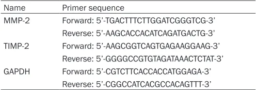
[image:3.612.89.353.84.177.2]experiment was repeated three times. Primer sequences are sh- own in Table 1.
Table 1. Primer sequences of PCR
Name Primer sequence
MMP-2 Forward: 5’-TGACTTTCTTGGATCGGGTCG-3’ Reverse: 5’-AAGCACCACATCAGATGACTG-3’ TIMP-2 Forward: 5’-AAGCGGTCAGTGAGAAGGAAG-3’
Reverse: 5’-GGGGCCGTGTAGATAAACTCTAT-3’ GAPDH Forward: 5’-CGTCTTCACCACCATGGAGA-3’
Reverse: 5’-CGGCCATCACGCCACAGTTT-3’
Note: MMP-2, matrix metalloproteinas-2; TIMP-2, tissue inhibitor of metal-loproteinase-2.
Table 2. Comparison of general data between the two groups (_x ± sd)
Item Experimental group (n=43) group (n=43) χControl 2/t P
Male (case) 23 21 0.186 0.666
Female (case) 20 22
Age (year) 65.3±9.6 67.5±8.4 1.131 0.261
[image:3.612.91.349.245.324.2]P<0.05 (bilateral) indicates statistical signifi- cance.
Results
Comparison of general data between the two groups
General clinical data of all patients are shown in Table 2. There were no significant differenc-es between the two groups in terms of age, gender, and medical history (all P>0.05). Effects of amiodarone on the reduction of AF after radiofrequency ablation
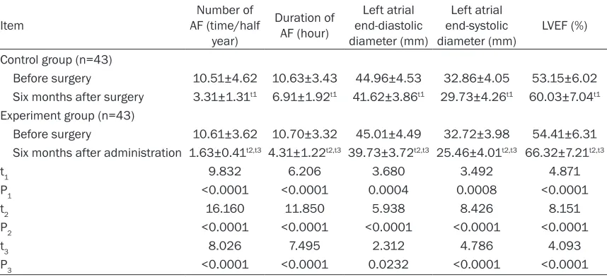
In control and experimental groups, the num-ber and duration of AF, left atrial end-diastolic diameter, and left atrial end-systolic diameter were significantly decreased at 6 months after surgery, compared to those before surgery. LVEF increased significantly, compared to pre-surgery values (all P<0.05). There were no sig-nificant differences in pre-surgery cardiac func-tion parameters between control and experi-mental groups (all P>0.05). At six months after surgery, however, left atrial end-systolic diame-ter, the number and duration of AF, and left atrial end-diastolic diameter values were sig-nificantly lower. LVEF was sigsig-nificantly higher in the experimental group, compared to that in the control group (all P<0.05). See Table 3. Taken together, amiodarone reduced AF and
improved cardiac structure and function after radiofrequency ablation of AF.
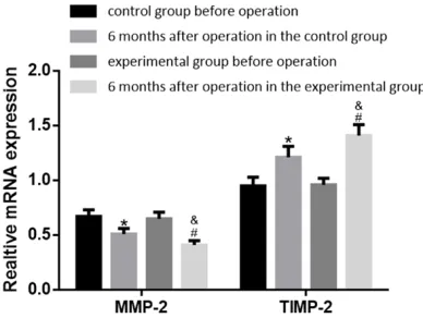
Effects of amiodarone on reductions of MMP-2 mRNA and protein levels and increases of TIMP-2 mRNA and protein levels in serum after radiofrequency ablation of AF
Results of ELISA and qRT-PCR are shown in Table 4 and Figure 1. In control and experi- mental groups, serum MMP-2 mRNA and pro-tein levels were significantly lower and TIMP-2 mRNA and protein levels were significantly higher at 6 months after surgery than those before surgery (all P<0.05). Although no signifi-cant differences were seen between control and experimental groups for pre-surgery ser- um MMP-2 and TIMP-2 mRNA and protein els (all P>0.05), MMP-2 mRNA and protein lev-els were significantly lower and TIMP-2 mRNA and protein levels were significantly higher in the experimental group than those in the con-trol group at 6 months after surgery (all P<0.05).
Discussion
[image:4.612.92.521.97.293.2]Studies have shown that MMP-2 plays a very important role in atrial structural reconstruc-tion in patients with mitral stenosis and AF [12]. MMP-2 in cardiomyocytes can degrade type IV collagen [13, 14]. Compared to patients with sinus rhythm, MMP-2 expression in the right Table 3. Comparison of the number and duration of AF, left atrial end-diastolic diameter, left atrial end-systolic diameter, and LVEF in two groups of patients before and 6 months after surgery (_x ± sd)
Item AF (time/half Number of
year)
Duration of AF (hour)
Left atrial end-diastolic diameter (mm)
Left atrial end-systolic
diameter (mm) LVEF (%) Control group (n=43)
Before surgery 10.51±4.62 10.63±3.43 44.96±4.53 32.86±4.05 53.15±6.02 Six months after surgery 3.31±1.31t1 6.91±1.92t1 41.62±3.86t1 29.73±4.26t1 60.03±7.04t1
Experiment group (n=43)
Before surgery 10.61±3.62 10.70±3.32 45.01±4.49 32.72±3.98 54.41±6.31 Six months after administration 1.63±0.41t2,t3 4.31±1.22t2,t3 39.73±3.72t2,t3 25.46±4.01t2,t3 66.32±7.21t2,t3
t1 9.832 6.206 3.680 3.492 4.871
P1 <0.0001 <0.0001 0.0004 0.0008 <0.0001
t2 16.160 11.850 5.938 8.426 8.151
P2 <0.0001 <0.0001 <0.0001 <0.0001 <0.0001
t3 8.026 7.495 2.312 4.786 4.093
P3 <0.0001 <0.0001 0.0232 <0.0001 <0.0001
Note: Compared with the control group before the operation, t1P<0.05; compared with the experimental group before the
operation, t2P<0.05; compared with the control group after the operation, t3P<0.05. AF, atrial fibrillation; LVEF, left ventricular
atrial appendage and free wall is significantly higher in patients with AF [15]. Related stu- dies have shown that expression of TIMP-2 in patients with AF decreases significantly [16, 17]. Related studies have shown that MMP-2 expression was elevated and that TIMP-2 ex- pression was decreased in patients with AF [18]. Other factors associated with AF include oxidative stress in the autonomic nervous sys-tem, ischemia, endocardial dysfunction, inflam-mation, and abnormal activity. Greenstein et al.
ration of AF, left atrial end-diastolic diameter, and left atrium end-systolic diameter were sig-nificantly lower. LVEF was sigsig-nificantly higher in the experimental group, which received amiod-arone, compared to those in the control group. Present findings clearly show that amiodarone can reduce AF and improve cardiac structure and function after radiofrequency ablation. Tsanaxidis et al. showed that ablation restored the AF rhythm to sinus rhythm, reversed atrial remodeling, and improved cardiac structure and function [21]. Amiodarone administration for 6 months after surgery in the experimental group significantly decreased serum MMP-2 levels and increased TIMP-2 levels. It affects cardiac electrophysiology and inhibits abnor-mal electrocardiographic trigger activity via the ion channels of myocardial cells, thus affecting MMP-2 and TIMP-2 levels in cardiomyocytes. Gai et al. reported MMP-2 upregulation and TIMP-2 downregulation in the atrial tissues of AF patients, consistent with present results. Taken together, amiodarone can improve the cardiac structure and function of patients after radiofrequency ablation of AF [22].
[image:5.612.93.390.97.269.2]However, the current study had some limita-tions. First, the cohort size was small, which narrowed the data source. Second, this study did not examine the underlying mechanisms of amelioration on cardiac structure and function after radiofrequency ablation, only analyzing Table 4. Comparison of serum MMP-2 and TIMP-2 protein levels in
the two groups of patients before and 6 months after surgery (_x ± sd)
Item MMP-2 (ng/mL) TIMP-2 (ng/mL)
Control group
Before surgery 386.74±36.84 64.53±6.06
Six months after surgery 320.62±32.63t1 90.86±8.62t1
Experimental group
Before surgery 382.96±37.46 63.96±6.38
Six months after administration 280.72±29.32t2,t3 108.62±9.68t2,t3
t1 8.810 16.390
P1 <0.0001 <0.0001
t2 14.090 25.260
P2 <0.0001 <0.0001
t3 5.964 8.985
P3 <0.0001 <0.0001
Note: Compared with the control group before the operation, t1P<0.05; compared with
the experimental group before the operation, t2P<0.05; compared with the control
group after the operation, t3P<0.05. MMP-2, matrix metalloproteinas-2; TIMP-2, tissue
inhibitor of metalloproteinase-2.
Figure 1. Comparison of serum MMP-2 mRNA and TIMP-2 mRNA levels in the experimental and control groups before and after surgery. Compared with the control group before surgery, *P<0.05; compared
with the experimental group before surgery, #P<0.05;
compared with the control group after the surgery,
&P<0.05. MMP-2, matrix metalloproteinas-2; TIMP-2,
tissue inhibitor of metalloproteinase-2.
[image:5.612.91.285.334.480.2]related indicators. Therefore, more studies are necessary to clarify this aspect.
In summary, the application of amiodarone in patients with AF after radiofrequency ablation can significantly reduce MMP-2 levels and in- crease TIMP-2 levels, showing protective effe- cts on the cardiac structure and function of patients.
Disclosure of conflict of interest
None.
Address correspondence to: Li’na Wang, Depart- ment of Cardiology, Baoji Hi-Tech People’s Hos- pital, No. 19 Gaoxin Forth Road, Weibin District, Baoji 721000, Shaanxi Province, China. Tel: +86-0917-3532131; E-mail: [email protected]
References
[1] Ejima K, Henmi R, Iwanami Y, Yagishita D, Sho-da M, Hagiwara N. Comparison of the efficacy of empiric thoracic vein isolation for the treat-ment of paroxysmal and persistent atrial fibril-lation in patients without structural heart dis-ease. J Cardiovasc Electrophysiol 2017; 28: 266-272.
[2] Fauchier L, Cinaud A, Brigadeau F, Lepillier A, Pierre B, Abbey S, Fatemi M, Franceschi F, Guedeney P, Jacon P, Paziaud O, Venier S, De-haro JC, Gras D, Klug D, Mansourati J, Mon-talescot G, Piot O, Defaye P. Device-related thrombosis after percutaneous left atrial ap-pendage occlusion for atrial fibrillation. J Am Coll Cardiol 2018; 71: 1528-1536.
[3] Stavrakis S, Garabelli P and Reynolds DW. Car-diac resynchronization therapy after atrioven-tricular junction ablation for symptomatic atrial fibrillation: a meta-analysis. Europace 2012; 14: 1490-1497.
[4] Montserrat S, Sitges M, Calvo N, Silva E, Tam-borero D, Vidal B, Berruezo A, Bernado C, Mont L and Brugada J. Effect of repeated radiofre-quency catheter ablation on left atrial function for the treatment of atrial fibrillation. Am J Car-diol 2011; 108: 1741-1746.
[5] Qian Y, Meng J, Tang H, Yang G, Deng Y, Wei D, Xiang B and Xiao X. Different structural remod-elling in atrial fibrillation with different types of mitral valvular diseases. Europace 2010; 12: 371-377.
[6] Rosso R, Chorin E, Levi Y, Rogowski O, Viskin S. Radiofrequency ablation of atrial fibrillation: nonrandomized comparison of circular versus point-by-point “smart” ablation for achieving circumferential pulmonary vein isolation and
curing arrhythmic symptoms. J Cardiovasc Electrophysiol 2016; 27: 1282-1287.
[7] Hughes BG and Schulz R. Targeting MMP-2 to treat ischemic heart injury. Basic Res Cardiol 2014; 109: 424.
[8] Thompson MM and Squire IB. Matrix metallo-proteinase-9 expression after myocardial in-farction: physiological or pathological? Cardio-vasc Res 2002; 54: 495-498.
[9] Liu WQ, Li WS and Ma QH. Salvianolic acid ef-fects of TNF alpha, MMP-2/TIMP-2 expression in the atrial fibrillation rat model randomized controlled study. J Practical Traditional Chi-nese Internal Medicine 2013; 27: 83-85. [10] An K, Zhu J, Ma N, Tang M and Mei J. Predictive
risk factors for recurrent atrial fibrillation after modified endoscopic ablation: a 2-year follow-up. Clin Cardiol 2018; 41: 372-377.
[11] Nagibin V, Egan Benova T, Viczenczova C, Szei-ffova Bacova B, Dovinova I, Barancik M and Tribulova N. Ageing related down-regulation of myocardial connexin-43 and up-regulation of MMP-2 may predict propensity to atrial fibrilla-tion in experimental animals. Physiol Res 2016; 65 Suppl 1: S91-S100.
[12] Marott SC, Benn M, Johansen JS, Jensen GB, Tybjaerg-Hansen A and Nordestgaard BG. YKL-40 levels and atrial fibrillation in the general population. Int J Cardiol 2013; 167: 1354-1359.
[13] Nielsen JC, Johannessen A, Raatikainen P, Hin-dricks G, Walfridsson H, Pehrson SM, Englund A, Hartikainen J, Mortensen LS and Hansen PS. Long-term efficacy of catheter ablation as first-line therapy for paroxysmal atrial fibrilla-tion: 5-year outcome in a randomised clinical trial. Heart 2017; 103: 368-376.
[14] Mesquita J, Ferreira A, Costa F, Cavaco D, Car-mo P, Morgado F, Mendes M and Adragao P. P881Atrial fibrillation relapse after a single catheter ablation procedure-an up to 10 years follow-up analysis. Europace 2017; 19: iii169-iii170.
[15] Pallisgaard JL, Gislason GH, Hansen J, Johan-nessen A, Torp-Pedersen C, Rasmussen PV and Hansen ML. Temporal trends in atrial fibril-lation recurrence rates after abfibril-lation between 2005 and 2014: a Nationwide Danish Cohort Study. Eur Heart J 2018; 39: 442-449. [16] Parkash R, Wells GA, Sapp JL, Healey JS, Tardif
[17] Khan I, Patel HC, Nanayakkara S, Raju H, Vos-koboinik A and Mariani JA. Trends in outpatient anti-arrhythmic prescriptions for atrial fibrilla-tion and left atrial ablafibrilla-tion in Australia: 1997-2016. Intern Med J 2018; 48: 427-432. [18] Roten L, Derval N and Jais P. Catheter ablation
for persistent atrial fibrillation: elimination of triggers is not sufficient. Circ Arrhythm Elec- trophysiol 2012; 5: 1224-1232.
[19] Greenstein D, Beau J, Gottlieb G, Teller D and Kulik A. Topical amiodarone during cardiac sur-gery: does epicardial application of amioda-rone prevent postoperative atrial fibrillation? J Thorac Cardiovasc Surg 2017; 154: 886-892. [20] Kamali A, Sanatkar A, Sharifi M and Moshir E.
Evaluation of amiodarone versus metoprolol in treating atrial fibrillation after coronary artery bypass grafting. Interv Med Appl Sci 2017; 9: 51-55.
[21] Tsanaxidis N, Aidonidis I, Hatziefthimiou A, Daskalopoulou SS, Giamouzis G, Triposkiadis F and Skoularigis I. Ranolazine added to amio-darone facilitates earlier conversion of atrial fibrillation compared to amiodarone-only ther-apy. Pacing Clin Electrophysiol 2017; 40: 372-378.