Original Article
Polyenylphosphatidylcholine alleviates palmitic
acid-induced apoptosis in HepG2 cells via
inhibiting endoplasmic reticulum stress
Gang Zhou1,3, Jiaheng Fang2, Yan Luo3, Junping Shi3
1Department of Gastroenterology, The Affiliated Hospital of Hangzhou Normal University, Hangzhou 310015, Zhejiang, P. R. China; 2Second Clinical Medical College, Zhejiang Chinese Medical University, Zhejiang, P. R. China;
3Center for Translational Medicine, The Affiliated Hospital of Hangzhou Normal University, Hangzhou 310015,
Zhejiang, P. R. China
Received November 16, 2016; Accepted November 5, 2017; Epub March 15, 2019; Published March 30, 2019
Abstract: Polyenylphosphatidylcholine (PPC) can reduce hyperlipidemia, relieve arteriosclerosis and decrease tri-glyceride. The aim of the present study was to investigate whether polyenylphosphatidylcholine (PPC) could alleviate apoptosis induced by palmitic acid (PA), and explore the possible molecular mechanisms in HepG2 cells. In this study, MTT assay was performed to identify the cell viability after PA (50, 100, 150 and 200 μmol/L) treatment for 24 h in HepG2 cells. The cell viability was significantly decreased by PA treatment in a dose dependent manner. Flow cytometry confirmed that co-incubation of PPC reduced PA-induced apoptosis. Moreover, the secretion level of tumor necrosis factor-α (TNF-α) was conspicuously reduced after PPC treatment using enzyme-linked immunosor-bent assay (ELISA). We observed that treatment of the cells with PA resulted in activation of Endoplasmic Reticulum Stress (ERS) associated proteins including glucose-regulated protein 78 (GRP78) and C/EBP homologous protein (CHOP). Western blot and real-time polymerase chain reaction (RT-PCR) confirmed that the expression levels of Bcl-2 associated X protein (Bax) and B cell lymphoma-Bcl-2 (Bcl-Bcl-2) were significantly regulated and the expression levels of GRP78 and CHOP were dramatically decreased by co-incubation of PPC. These results suggested that PPC could alleviate cell apoptosis and reduce the ERS-related proteins expressions after cells pre-treatment with PA, which showed that ERS might play an important role in cell apoptosis caused by PA.
Keywords: Polyenylphosphatidylcholine (PPC), palmitic acid (PA), endoplasmic reticulum stress (ERS), apoptosis, HepG2
Introduction
Non-alcoholic fatty liver disease (NAFLD) is, a metabolic stress-induced liver damage, closely related with insulin resistance (IR) and genetic susceptibility. NAFLD is considered to be benign lesions that could be treated. Otherwise, it could further develop into the end-stage liver disease, such as hepatic cirrhosis and liver cancer. Therefore, NAFLD is regarded as one of the major causes of liver failure [1-3]. With the improvement of living standards and the aging of population, obesity and metabolic syndrome have become an increasingly serious health problem among NAFLD patients [4]. Obesity can increase the content of free fat acid (FFA) in blood. The study found that free fat acid (FFA) could promote the formation of triglycerides,
which led to lipid accumulation in liver [5, 6]. PA is a central component of lipid in the body and a common high level of saturated fatty acid in food [7]. Some studies confirmed that PA could induce apoptosis in various cells [7-13]. Therefore, HepG2 cells were treated with PA to establish hyperlipidemia model in vitro in this study.
important step in onset and progression of NASH [12, 13].
PPC from soybean is a highly concentrated phospholipids and an essential component of cell membranes and sub cellular ones. The main active ingredient of PPC is poly phosphati-dylcholine diacylglycerol or polyketides lecithin [14]. Numerous of animal experiments have manifested that PPC could reduce cell apopto-sis, inhibit stress reaction, relieve and inflam-mation [15, 16]. Stimulation from various exter-nal environments can cause the imbalance of homeostasis in ER that results in ERS ultimate-ly [17, 18]. ERS can cause cell programmed death via up-regulating the expression levels of related genes including GRP78 and CHOP [18]. This study aimed at determining the anti-apoptosis effect of PPC on HepG2 cells treat- ed with PA and exploring underlying molecular mechanisms.
Materials and methods
Cell culture
HepG2 cells were obtained from cell bank of Shanghai Branch, Chinese Academy of Science (China) and incubated in Dulbecco’s modified Eagle’s medium (DMEM) (Hangzhou Sijiqing Co., Ltd, Zhejiang, China), 10% Fetal Bovine Serum (FBS) (Sijiqing), 100 U/ml penicillin and 100 μg/mL streptomycin (Solarbio, Beijing, China) at 37°C. The medium changed every other day. HepG2 cells were digested by 0.25% trypsin (Beyotime Biotechnology Co., Ltd, Sh- anghai, China) for sub-culturing when cells con-fluence reached 80%. HepG2 cells in expon- ential growth phase were chosen to do the experiment.
MTT assay
PA (Sigma, USA) was dissolved in 0.1 M NaOH in 70°C water bath to prepare 50 mM PA stock solution. HepG2 cells seeded in a 24-well plate at the density of 1×10⁶ cells/well with 100 mL
culture medium. After being cultured for 24 h, PA was added to the medium to the final con-centrations (50, 100, 150 and 200 μmol/L PA) and cultured for another 12, 24 and 48 h. Then 100 μL of MTT solution (Ameresco, USA) was added to each well and incubated in 5% CO2 at 37°C for 4 h. The optical density (OD) values were read at 492 nm by a microplate reader
(Thermo, USA). A Value of each experimental group was divided by that of control group, and multiplied by 100%, the resultant value was collected to access cell viability in each experi-mental group.
Flow cytometry assay
HepG2 cells was pre-treated with PPC (100 and 200 μmol/L) (Sanofi-Aventis Pharmaceutical Co., Ltd, Beijing, China) for 6 h, and 200 μmol/L of PA was added into the culture medium for 24 h. The apoptotic rate was analyzed by flow cytometer using an Annexin V-FITC apoptosis detection Kit (Beyotime Biotechnology Co., Ltd, Shanghai, China) according to the manufactur-er’s instructions. In brief, cells were harvested after being cultured for 24 h and washed with ice-cold PBS (Beyotime), resuspended in 500 μL of binding buffer, followed by addition of 5 μL of Annexin V stock solution and incubation for 10 min at 4°C. Propidium iodide (PI) (5 μL) was added to the cells, and they were immedi-ately analyzed using a flow cytometer (BD, San Jose, CA).
ELISA testing
HepG2 cells was pre-treated with PPC (100 and 200 μmol/L) (Sanofi-Aventis Pharmaceutical Co., Ltd, Beijing, China) for 6 h, and 200 μmol/L of PA was added into the culture medium for 24 h, cell supernatant was collected and centri-fuged at 3000 rpm with low temperature. The content of TNF-α was tested according to the manual of ELISA kit (Beyotime Biotechnology Co., Ltd, Shanghai, China).
Reverse transcription and RT-PCR
HepG2 cells were seeded at a density of 1×10⁵
(Applied Biosystems, Foster City, CA, USA), us- ing the SYBR Premix Ex Taq kit (Takara). The specific primer sequences for each gene were listed as follows: 5’ CTCAACATGGATCTGTTCCG 3’ and 5’ CCAGTTGCTGAATCTTTGGA 3’ for GR- P78 (product: 125 bp); 5’ CGCATGAAGGAGAAA- GAACA 3’ and 5’ CACCATTCGGTCAATCAGAG 3’ for CHOP (product: 125 bp) and 5’ TGGCACCCA- GCACAATGAA 3’ and 5’ CTAAGTCATAGTCCGCC- TAGAAGCA 3’ for actin (product: 125 bp). Data
analysis was done using the 2-ΔΔCT method for
relative quantification, and all samples were normalized to actin, which was used as an endogenous control.
Western blot
HepG2 cells were seeded at a density of 1×10⁵
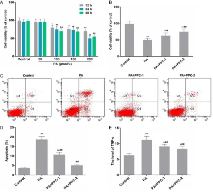
[image:3.612.92.523.73.462.2]cells/well in 6-well plates, cultured overnight and then pre-treated with PPC (100 and 200 Figure 1. PPC impacts on cell viability, cell apoptosis and the level of TNF-α in HepG2 cells with PA pre-treatment. A: Cells were treated with different doses of PA (0, 50, 100, 150 and 200 μmol/L) for 12, 24 and 48 h, and MTT were performed to identify the cell viability. Data were presented as mean ± SD, n=3, *P < 0.05 and **P < 0.01 vs.
control in 12 h; #P < 0.05 and ##P < 0.01 vs. control in 24 h; &P < 0.05 and &&P < 0.01 vs. control in 48 h. B: Cells
was pre-treated with PPC (100 and 200 μmol/L) for 6 h, and 200 μmol/L of PA was added into the culture medium for 24 h. MTT was then performed to identify the cell viability. C and D: Cells were pre-treated with 200 μmol/L of PA for 4 h, and PPC (100 and 200 μmol/L) was added into the culture medium, and incubated for 24 h. Flow cytometry was performed to detected cell apoptosis. E: Cells were pre-treated with 200 μmol/L of PA for 4 h, and PPC (100 and 200 μmol/L) was added into the culture medium, and incubated for 24 h. ELISA was performed to detected the level of TNF-α. Data were presented as mean ± SD, n=3, *P < 0.05 and **P < 0.01 vs. control; #P < 0.05 and ##P <
μmol/L) (Sanofi-Aventis Pharmaceutical Co., Ltd, Beijing, China) for 6 h, and 200 μmol/L of PA was added into the culture medium for 24 h. Cells were harvested and lysed in RIPA buffer containing 1 mM phenylmethylsulfonyl fluoride (PMSF). Equal amounts of protein were electrophoresed on 10% sodium dodecyl sul-fate polyacrylamide gel (SDS-PAGE), and the bands were transferred to polyvinylidene fluo-ride membrane (Millipore, USA). The membrane was blocked and then incubated with antibod-ies against Bax, Bcl-2 (Abcam), GRP78, CHOP (Beyotim) and actin (Santa). After washing, the membranes were incubated with peroxidase-conjugated secondary antibodies (Zhongshan, Beijing, China) for 1 h. Blots were visualized using enhanced chemiluminescence (ECL, Th- ermo Scientific, Shanghai, China). Relative pro-tein levels in each sample were normalized to actin to standardize the loading variations.
Statistical analysis
Data were expressed as the Mean ± SEM from at least three independent experiments. The differences between groups were analyzed us-
ing a Student t test when only 2 groups or 1-way analysis of variance in more than 2 groups were compared. All tests performed were 2-sided. P
< 0.05 was taken as statistical significance.
Results
PPC improve cell viability in HepG2 cells which suppressed by PA
In order to identify the effect of PA on the pro- liferation of HepG2 cells, MTT assay was em- ployed for cell viability analysis. After treatment of PA (50, 100, 150 and 200 μmol/L) for 12, 24 and 48 h, PA over the dose of 50 μmol/L obvi-ously suppressed the cell viability of HepG2 cells (Figure 1A). All the effects were in a time and dose dependent manner. In addition, PA inhibited the cell proliferation in a dose depen-dent manner (Figure 1A). The inhibitive effect of 200 μmol/L PA was the most significant at 24 h, therefore, 200 μmol/L of PA was deter-mined for the subsequent experiment. After PA/PPC co-treatment, the cell viability was in- creased by 100 and 200 μmol/L of PPC (Fig-
ure 1B). In addition, PPC increased the cell
[image:4.612.92.526.70.304.2]liferation in a dose dependent manner (Figure 1B).
PPC reduces PA-induced apoptosis and the level of TNF-α in HepG2 cells
In Figure 1, PA significantly promoted the
apoptosis of HepG2 cells compared with the control group, while PPC inhibited the apopto-sis caused by PA in HepG2 cells. The apoptoapopto-sis rate of HepG2 cells was 3.8 ± 0.28%, 18.7 ± 1.51%, 10.4 ± 0.97% and 5.1 ± 0.6%, respec-tively (Figure 1C and 1D). The level of TNF-α was significantly increased in PA treatment group compared with the control group and that was dramatically decreased in PA/PPC co-treatment group in a dose dependent manner
(Figure 1E). Compared with control group, the
expression level of Bax was increased and that of Bcl-2 was decreased after incubating with PA or/and PPC for 24 h (Figure 2). Bax expres-sion in PA/PPC co-treatment group was remark-ably less than PA treatment group, while Bcl-2 expression in PA/PPC co-treatment group was remarkably higher than PA treatment group. In addition, Bcl-2/Bax ratio in mRNA and protein
in Figure 3, GRP78 and CHOP genes were
evi-dently up-regulated with the induction of 200 μmol/L PA (P < 0.05). Compared with PA group, the expression levels of GRP78 and CHOP we- re evidently downregulated by PPC in a dose dependent manner.
Discussion
[image:5.612.91.379.71.284.2]NASH is a pivotal pathological stage during which NAFLD may progress to the late-stage liver disease [19, 20]. Intra-hepatic fatty acids and triglyceride accumulation are the patho-genesis basis of NASH progression. Never- theless, epidemiological research manifested that NAFLD arrested the phase of simple fatty liver in majority of patients, and only small parts of patients suffered from NASH. This pathogen-esis was still unclear [17, 18]. It was reported that elevated plasma FFAs concentration, fatty lesions and apoptosis of hepatocytes were sig-nificant features in NASH [21]. TNF-α plays an important role in cell apoptosis and can acti-vate Caspase. In addition, TNF-α can transmit death signal into cell. The highly expressed TNF-α promotes cell apoptosis. The increased Figure 3. PPC regulates the expression levels of ERS-related genes including
GRP78 and CHOP. A and B: Cells was pre-treated with PPC (100 and 200 μmol/L) for 6 h, and 200 μmol/L of PA was added into the culture medium for 24 h. The mRNA expression levels of GRP78 and CHOP were detected by RT-PCR. C and D: Cells was pre-treated with PPC (100 and 200 μmol/L) for 6 h, and 200 μmol/L of PA was added into the culture medium for 24 h. The protein expression levels of GRP78 and CHOP were detected by Western blot. GAPDH was also detected as the control of sample loading. Data were presented as mean ± SD, n=3, *P < 0.05 and **P < 0.01 vs. control; #P < 0.05
and ##P < 0.01 vs. PA.
levels of PA group was signifi-cantly decreased, compared to that of PA group, and Bcl-2/Bax ratio in mRNA and protein levels of PA/PPC co-treatment group was signifi-cantly increased, compared to that of PA group (Figure 2C
and 2F). The expression lev-els of Bax and Bcl-2 were sig-nificantly regulated by PA or/ and PPC treatment.
PPC reduces the expression levels of ERS-related genes in HepG2 cells
FFAs can cause ectopic accumulation of lipid in the liver to release excessive TNF-α by activat-ing ERS [22]. These inflammatory factors inter-fere with glucose and lipid metabolisms by binding cytokine receptors and pattern recogni-tion receptors in adipose tissue and other his-tocytes. Meanwhile, the sensitivity of hepato-cytes to inflammation responses and various damage elements is also enhanced. Moreover, lipotoxicity can lead to hepatocytes dysfunction or death, and lipoapoptosis can lead to cell apoptosis [23]. PA is an important fatty acid which constitutes liver TG for healthy people and NAFLD patients [24]. In our results, exces-sive PA inhibited the cell viability, caused cell apoptosis, increased the TNF-α level and ERS marker GRP78 and CHOP expressions (Figures 1 and 2). It showed that PA caused cell apopto-sis via up-regulating the ERS-related proteins expressions to activate ERS.
As one of physiological phospholipids, PPC has been reported its protective effects on liver [25, 26]. Firstly, PPC can inhibit lipid per-oxida-tion in hepatocytes induced by carbon tetra-chloride and arachidonic acid, as well as inhibit stress reaction and lipid per-oxidation caused by ethanol. A study showed that on one hand, PPC could suppress ethanol and LPS-induced TNF-α production and mitochondrial apoptosis in Kupffer cells [15]. On the other hand, PPC inhibited HSC activation and protects against liver fibrosis [27]. Therefore, PPC can alleviate PA-induced hepatocytes ERS and lipoapopto-sis. In this study, we detected the cell viability, cell apoptosis, TNF-α level and related genes expressions in PA/PPC co-treatment groups. Obviously, PPC increased the cell viability, re- duced the apoptosis and decreased the TNF-α level. It was a kind of clue for further resear- ches on anti-apoptotic molecular mechanism. Above results have confirmed that PA induced ERS in cells (Figure 2). Bax and Bcl-2 as impor-tant members of Bcl-2 protein family, can regu-late cell apoptosis [28].
The component proportion ratio of the member of Bcl-2 protein family is one of the key mecha-nisms for death receptor signaling pathway and mitochondria signaling pathway [29]. Especially, Bcl-2/Bax ratio directly determines cell surviv-al. In addition, the increased CHOP can change the expression levels of Bax and Bcl-2 to regu-late cell apoptosis. CHOP is a highly expression
in the process of ERS and can up-regulate the expression levels of Bax and down-regulate the expression levels of Bcl-2 [30-32]. In our results, the expression levels of GRP78 and CHOP were decreased by PPC treatment, and that of Bax was decreased and Bcl-2 was increased by PPC treatment. Bcl-2/Bax ratio was significantly decreased during PA-induced apoptosis in HepG2 cells. However, PPC pre-treatment could up-regulate Bcl-2/Bax ratio
(Figures 2 and 3). It showed that PPC inhibited
ERS and regulated Bax and Bcl-2 expressions.
In conclusion, PA could reduce significantly cell viability and promote cell apoptosis. ERS-related proteins were up-regulated significant- ly. It showed that ERS might be activated by PA. Therefore, PPC could alleviate PA-induced apoptosis in HepG2 cells via inhibiting ERS and regulating apoptosis related genes including Bax and Bcl-2, however, ER stress-related pa- thways still need further studies.
Disclosure of conflict of interest
None.
Address correspondence to: Junping Shi, Center for Translational Medicine, The Affiliated Hospital of Hangzhou Normal University, 126 Wenzhou Road, Hangzhou 310015, Zhejiang, P. R. China. Tel: 0086-571-88303699; Fax: 0086-0086-571-88303699; E-mail: wejklonnk@163.com
References
[1] Ratziu V, Bellentani S, Cortez-Pinto H, Day C and Marchesini G. A position statement on NAFLD/NASH based on the EASL 2009 special conference. J Hepatol 2010; 53: 372-384. [2] Waluga M, Kukla M, Zorniak M, Kajor M, Liszka
L, Dyaczynski M, Kowalski G, Zadlo D, Waluga E, Olczyk P, Buldak RJ, Berdowska A and Hart-leb M. Fibroblast growth factor-21 and omen-tin-1 hepatic mRNA expression and serum lev-els in morbidly obese women with nonalcoholic fatty liver disease. J Physiol Pharmacol 2017; 68: 363-374.
[3] Ying L, Yan F, Zhao Y, Gao H, Williams BR, Hu Y, Li X, Tian R, Xu P and Wang Y. (-)-Epigallocat-echin-3-gallate and atorvastatin treatment down-regulates liver fibrosis related genes in non-alcoholic fatty liver disease. Clin Exp Phar-macol Physiol 2017; 44: 1180-1191.
of nonalcoholic fatty liver disease (NAFLD): a population-based study in China. J Epidemiol 2013; 23: 115-121.
[5] Pessayre D, Berson A, Fromenty B and Man-souri A. Mitochondria in steatohepatitis. Semin Liver Dis 2001; 21: 57-69.
[6] Lee J, Narayan VP, Hong EY, Whang WK and Park T. Artemisia Iwayomogi extract attenuates high-fat diet-induced hypertriglyceridemia in mice: potential involvement of the adiponec-tin-AMPK pathway and very low density lipo-protein assembly in the liver. Int J Mol Sci 2017; 18.
[7] Abergel A, Sapin V, Dif N, Chassard C, Darcha C, Marcand-Sauvant J, Gaillard-Martinie B, Rock E, Dechelotte P and Sauvant P. Growth arrest and decrease of alpha-SMA and type I collagen expression by palmitic acid in the rat hepatic stellate cell line PAV-1. Dig Dis Sci 2006; 51: 986-995.
[8] Liu C, Fu Y, Li CE, Chen T and Li X. Phycocyanin-functionalized selenium nanoparticles reverse palmitic acid-induced pancreatic beta cell apoptosis by enhancing cellular uptake and blocking reactive oxygen species (ROS)-medi-ated mitochondria dysfunction. J Agric Food Chem 2017; 65: 4405-4413.
[9] Jiang XS, Chen XM, Wan JM, Gui HB, Ruan XZ and Du XG. Autophagy protects against palmit-ic acid-induced apoptosis in podocytes in vitro. Sci Rep 2017; 7: 42764.
[10] Yan PS, Tang S, Zhang HF, Guo YY, Zeng ZW and Wen Q. Nerve growth factor protects against palmitic acid-induced injury in retinal ganglion cells. Neural Regen Res 2016; 11: 1851-1856.
[11] Yan P, Tang S, Zhang H, Guo Y, Zeng Z and Wen Q. Palmitic acid triggers cell apoptosis in RGC-5 retinal ganglion cells through the Akt/FoxO1 signaling pathway. Metab Brain Dis 2017; 32: 453-460.
[12] Correction: palmitate activates autophagy in INS-1E beta-cells and in isolated rat and hu-man pancreatic islets. PLoS One 2015; 10: e0122235.
[13] Kim JE, Ahn MW, Baek SH, Lee IK, Kim YW, Kim JY, Dan JM and Park SY. AMPK activator, AICAR, inhibits palmitate-induced apoptosis in osteoblast. Bone 2008; 43: 394-404.
[14] Aleynik SI, Leo MA, Takeshige U, Aleynik MK and Lieber CS. Dilinoleoylphosphatidylcholine is the active antioxidant of polyenylphosphati-dylcholine. J Investig Med 1999; 47: 507-512. [15] Xu Y, Leo MA and Lieber CS. DLPC attenuates
alcohol-induced cytotoxicity in HepG2 cells ex-pressing CYP2E1. Alcohol Alcohol 2005; 40: 172-175.
[16] Aleynik SI and Lieber CS. Polyenylphosphatidyl-choline corrects the alcohol-induced hepatic
oxidative stress by restoring s-adenosylmethio-nine. Alcohol Alcohol 2003; 38: 208-212. [17] Pai H, Kang CI, Byeon JH, Lee KD, Park WB,
Kim HB, Kim EC, Oh MD and Choe KW. Epide-miology and clinical features of bloodstream infections caused by AmpC-type-beta-lacta-mase-producing Klebsiella pneumoniae. Anti-microb Agents Chemother 2004; 48: 3720-3728.
[18] Mengesdorf T, Althausen S and Paschen W. Genes associated with apoptotic and pro-tective mechanisms are affected differently on exposure of neuronal cell cultures to arsenite. No indication for endoplasmic reticulum stress despite activation of grp78 and gadd153 ex-pression. Brain Res Mol Brain Res 2002; 104: 227-239.
[19] Guo Y, Dong C, Lin H, Zhang X, Wen H, Shen Y, Wang T, Chen S, Liu Y and Chen X. Evaluation of non-alcoholic fatty liver disease using acous-tic radiation force impulse imaging elastogra-phy in rat models. Ultrasound Med Biol 2017; 43: 2619-2628.
[20] Hagstrom H, Nasr P, Ekstedt M, Hammar U, Stal P, Hultcrantz R and Kechagias S. Fibrosis stage but not NASH predicts mortality and time to development of severe liver disease in biopsy-proven NAFLD. J Hepatol 2017; 67: 1265-1273.
[21] Caldwell S. NASH (nonalcoholic steatohepati-tis): a case of multiorganelle failure. Free Rad-ic Biol Med 2014; 75 Suppl 1: S6.
[22] Jiao P, Ma J, Feng B, Zhang H, Diehl JA, Chin YE, Yan W and Xu H. FFA-induced adipocyte in-flammation and insulin resistance: involve-ment of ER stress and IKKbeta pathways. Obe-sity (Silver Spring) 2011; 19: 483-491. [23] Marra F and Lotersztajn S. Pathophysiology of
NASH: perspectives for a targeted treatment. Curr Pharm Des 2013; 19: 5250-5269. [24] Araya J, Rodrigo R, Videla LA, Thielemann L,
Orellana M, Pettinelli P and Poniachik J. In-crease in long-chain polyunsaturated fatty acid n-6/n-3 ratio in relation to hepatic steatosis in patients with non-alcoholic fatty liver disease. Clin Sci (Lond) 2004; 106: 635-643.
[25] Baird AC, Lloyd F and Lawrance IC. Prostaglan-din E(2) and polyenylphosphatidylcholine pro-tect against intestinal fibrosis and regulate myofibroblast function. Dig Dis Sci 2015; 60: 1603-1616.
[26] Shimizu Y and Kusano M. The efficacy of poly-enylphosphatidylcholine for liver disease. Hep-atol Res 2006; 34: 74-75.
[28] SIRT1 against breast cancer through downreg-ulating Bcl-2 protein. J Clin Oncol 2012; 30: 34.
[29] Teymournejad O, Mobarez AM, Hassan ZM and Talebi Bezmin Abadi A. Binding of the helico-bacter pylori OipA causes apoptosis of host cells via modulation of Bax/Bcl-2 levels. Sci Rep 2017; 7: 8036.
[30] Boyce M and Yuan J. Cellular response to en-doplasmic reticulum stress: a matter of life or death. Cell Death Differ 2006; 13: 363-373.
[31] Gardner BM, Pincus D, Gotthardt K, Gallagher CM and Walter P. Endoplasmic reticulum stress sensing in the unfolded protein re-sponse. Cold Spring Harb Perspect Biol 2013; 5: a013169.