warwick.ac.uk/lib-publications
Manuscript version: Author’s Accepted Manuscript
The version presented in WRAP is the author’s accepted manuscript and may differ from the published version or Version of Record.
Persistent WRAP URL:
http://wrap.warwick.ac.uk/126172
How to cite:
Please refer to published version for the most recent bibliographic citation information. If a published version is known of, the repository item page linked to above, will contain details on accessing it.
Copyright and reuse:
The Warwick Research Archive Portal (WRAP) makes this work by researchers of the University of Warwick available open access under the following conditions.
Copyright © and all moral rights to the version of the paper presented here belong to the individual author(s) and/or other copyright owners. To the extent reasonable and
practicable the material made available in WRAP has been checked for eligibility before being made available.
Copies of full items can be used for personal research or study, educational, or not-for-profit purposes without prior permission or charge. Provided that the authors, title and full
bibliographic details are credited, a hyperlink and/or URL is given for the original metadata page and the content is not changed in any way.
Publisher’s statement:
Please refer to the repository item page, publisher’s statement section, for further information.
Unravelling Photoprotection in Microbial Natural Products
Jack M. Woolleya* and Vasilios G. Stavrosa*
a University of Warwick, Department of Chemistry, Library Road, Coventry, CV4 7AL
* Corresponding author: v.stavros@warwick.ac.uk
* Corresponding author: j.woolley.1@warwick.ac.uk
Abstract: Mycosporine-like amino acids have long been known as a natural form of photoprotection for fungi and cyanobacteria. This review will highlight the key time-resolved experimental and theoretical techniques unravelling their photochemistry and photophysics, and directly link this to their use in commercial skin-care products, namely as sunscreen filters. Three case studies have been selected, each having aided advancement in this burgeoning field of research. We discuss these studies in the context of photoprotection and conclude by evaluating the necessary future steps towards translating the photochemistry and photophysics insight of these nature derived sunscreen filters to commercial application.
1.
Mycosporine and Mycosporine-like amino acids
Ultraviolet radiation (UVR) can be both beneficial and deleterious to life.1–4 To combat the
detrimental effects, nature has evolved to include several collections of UVR absorbing compounds, each biologically engineered to protect their host, while allowing sufficient absorption in different regions of the electromagnetic spectrum for important biological processes.5–8 The recent controversy surrounding artificial sunscreen filters focuses largely on their potential to act as endocrine disruptors and skin irritants to humans.9–12 Alongside these, there are growing concerns surrounding the environmental impact sunscreens filters are having; for example the common UV sunscreen filter oxybenzone has been linked to coral bleaching and reproductive toxicity aquatic life.9,13–15 These findings have therefore fuelled research into alternative filters, such as those derived from nature.16,17 One such class of natural UVR absorbers, is that of mycosporine/mycosporine-like amino acids and their derivatives (both termed MAAs hereon).* MAAs are a common form of photoprotectant species, found in fungi, cyanobacteria and phytoplankton. This review focuses on their photochemical and photophysical (collectively termed photochemical hereon) properties and how this knowledge can be utilised to develop next generation sunscreen filters. Importantly, the reader is directed to the following excellent reviews for further details regarding MAA biosynthesis and extraction, areas that have already received considerable attention. 7,8,18–23
MAAs present a promising form of future sunscreen filter given their strong UVR absorption, with typical extinction coefficients in the region of 30,000 M-1 cm-1.8 Currently, approximately 30 MAAs have been isolated which, combined, cover the ultraviolet region (UVA = 400-315 nm, UVB = 315-280 nm and UVC =280-100 nm) of the electromagnetic spectrum.7,8,24 Derived
from a basic cyclohexenone or cyclohexenimine core, Figure 1(a), each having a differing absorption maximum, these properties lend themselves towards use in future cosmeceutical applications, largely due to the ability to choose the range over which particular MAAs absorb. This variability in absorption is achieved through selective biosynthesis of MAAs with differing functional groups onto either position one and or three of the core structure (Figure 1(a)).25 Substitutions far removed from these two positions have little effect unless a large change in conjugation is encountered, resulting in a change to the overall chromophore (highlighted in red in Figure 1a).23 The exploration of differing functionalisation on positions
* Mycosporine refers to systems based on the cyclohexenone core, and mycosporine-like amino acid refers to
one and three not only allows for variability in the electronic ground state absorption spectrum of the MAA, but is necessary for widespread commercialisation as natural source extraction and synthetic preparation yields only small quantities of natural MAAs (typically 15 steps, 1% of overall yield).8,26–28
Sunscreen filters work by facilitating the safe dissipation of absorbed, potentially harmful, UVR as heat to the surrounding environment.29 This review will highlight the main
experimental, and to a lesser extent, complementary theoretical techniques used to track molecule-photon interactions (photochemistry), and hence unravel the photoprotection mechanisms underpinning MAAs. This, in turn yields important insight into why nature has chosen these particular sunscreen filters as photoprotective species. Furthermore, we will briefly discuss how we can adapt these techniques to accommodate a truer cosmeceutical environment. This review will then discuss three key articles – termed Case Studies – which put into practise these experimental and theoretical techniques. Using findings from natural MAAs and other sources,16,17 ‘rules’ for efficient molecular design can be established in relation to increasing the efficacy of next generation sunscreen filters. This review closes by discussing future steps towards translating photochemical insight into application.
Figure 1: a) Core structures of mycosporine (top) and mycosporine-like amino acids (bottom), with the
chromophore highlighted in red. b) Common mycosporine and mycosporine-like amino acid structures along
with their reported absorption maxima adapted from reference 8.
a) b)
Mycosporine-Glycine (310 nm)
Shinorine (334 nm)
Palythine (320 nm) Usujirene (357 nm)
1
2
3
4 6
[image:4.595.155.450.166.389.2]2.
Tracking the energy flow within molecules.
There are many articles and reviews in the literature that discuss methods to track population flow within a molecule’s excited states, along with their advantages and disadvantages. The reader is therefore referred to the following excellent sources for such detailed discussion.30–35 What follows is a brief overview of the commonly used experimental and theoretical techniques used to track the evolution of photoexcited states pertinent to the Case Studies to be discussed in Section 3.
2.1Transient electronic absorption spectroscopy
Time-resolved pump-probe spectroscopic techniques such as transient electronic absorption spectroscopy provide powerful insight into the fate of photoexcited molecules.34,36 An initial femtosecond (fs; where 1 fs = 1x10-15 s) laser pulse within the UVA/UVB region photoexcites, or pumps, the sample of interest to an electronically excited state. This photoprepared excited state is then interrogated with a second laser pulse termed the ‘probe’, typically a white light supercontinuum spanning wavelengths between 300-700 nm.37 The relative time-delay (t) between the pump and probe is varied, usually, from femtoseconds to nanoseconds (ns; where 1 ns = 1x10⁻⁹ s). Changes in optical density (OD) are calculated between the probe, I0(λ,t)
traversing a photoexcited (via interaction by the pump) and unperturbed sample I0(λ).
[image:5.595.78.519.451.554.2]A schematic of a typical transient electronic absorption spectroscopy system is displayed in Figure 2, presented as three key sections. Firstly, in Figure 2(a), ultrafast pulse generation is commonly produced through the use of a Ti:sapphire oscillator combined with regenerative amplification to generate an 800 nm fundamental fs pulse train with a pulse energy of a few mJ,38,39 and a pulse duration of 10s of fs; these levels of output energy allow for the generation of both the pump and probe pulses through nonlinear optical effects.37,40,41 Secondly, in Figure 2(b) the output of the laser system is split to generate the pump and probe pulses. Probe pulses
Figure 2: Key stages of a transient electronic absorption spectroscopy system. a) Ultrafast pulse generation,
b) pump-probe setup, and c) sample interaction and signal recovery.
Diode Laser
Ti:sapphire Oscillator
Chirped Pulse Amplification System
OPA Delay Stage
Chopper
Spectrometer Δt
Circulating Sample
follow an optical delay line (in this example) that includes mirrors and a retroreflector mounted on a delay stage, which enables t to be varied (by virtue of increasing/decreasing the optical path length of the probe) before focussing the 800 nm beam onto a calcium fluoride (CaF2)
window to generate the aforementioned broadband supercontinuum.40 The probe pulses are
then focussed into the solution (within a liquid flow cell, see below) containing the solute under investigation. Lastly, in Figure 2(c), the transmitted probe is collimated (not explicitly shown) and sent into a spectrometer.
Pump pulses are typically generated through optical parametric amplification of the 800 nm beam, which allows for the variability of pump wavelengths across the UV spectrum (in the present Case Studies UVA/UVB) and beyond. The pump beam is spatially overlapped with the
probe beam within a liquid flow cell containing the sample solution. The solution is recirculated which enables fresh sample to be interrogated for each measurement. Importantly, a chopper wheel blocks every second pump pulse to allow for calculations of the change in optical density (OD; see Figure 2(c) for details) between pulse pairs (i.e.pump ‘on’ and pump
‘off’). The collected data, termed transient absorption spectra (TAS), are then commonly displayed as false colour heat maps (vide infra). These 2-dimensional plots show the full probe
continuum as a function of pump-probe time-delay, with the change in optical density shown on the z-scale as changes in colour.
2.2 Photoacoustic Calorimetry
[image:6.595.76.521.548.672.2]Photothermal techniques such as photoacoustic calorimetry provide quantitative information on the energy flow between solute and solvent environment.31–33 Photoacoustic calorimetry
Figure 3: Sample interaction for photoacoustic calorimetry with a) right angled and b) front facing geometry.
Incoming laser pulse is shown in green, with induced pressure changes shown in orange. These changes are
detected by a transducer connected to an oscilloscope. c) Expected signal from a photoacoustic calorimetry
measurement showing the initial amplitude H.
Transducer Transducer
Dichroic Mirror b)
a) c)
H
Tr
ans
duc
er
S
ig
nal
allows for precise thermodynamic properties and lifetimes of a photochemical reaction to be derived through monitoring of the pressure changes induced through photoexcitation.42 These pressure changes, depicted by orange bars in Figure 3, are initiated usually by ns pulsed UV lasers, depicted by the green bar in Figure 3, which traverse the solvated sample held in a cuvette at a given temperature. The transducer monitors the pressure change commonly in a right angled orientation or alternatively a forward facing orientation, in which case a dichroic mirror is placed between the sample and transducer; these are shown, respectively, by Figures 3(a) and 3(b).31,33 A typical photoacoustic signal is displayed in Figure 3(c), this signal formed
from two contributions: 1) the thermal expansion of the solute upon the heat being delivered through radiationless relaxation; and 2) the volume change induced in the solute and solvent.
28,29 Through performing the experiment at multiple temperatures and comparing with a known
calorimetric standard, the initial peak to peak amplitude can be assigned as the energy lost from the solute, H in Figure 3(c).28,29 A calorimetric standard is usually a compound that presents a similar absorption and undergoes no radiative transitions, i.e., fluorescence or phosphorescence.43
2.3Theoretical methods
Computational exploration of the ground and excited electronic states of molecules can allow for complementary insight into the molecular processes undergone in molecules following photoexcitation, some of which may be undetectable utilising the experimental techniques discussed supra. Electronic state calculations for the electronic ground states can be considered routine for numerous systems, given a single electronic configuration.30 Challenges for electronically excited states often arise from the need to consider numerous electronic configurations, as well as different ranged interactions, such as those discussed below. Intermolecular interactions (such as hydrogen bonding, clustering, and aggregation) can also alter the excited electronic states of systems further to the intramolecular considerations which one encounters in vacuo. Polarizable continuum and conductor like screening models can be applied to account for the electrostatic nature of the solvent interaction.44,45 However, to fully
capture the explicit nature of solvent interaction and systems such as those described above, particularly hydrogen bonding for example, multiple solvation shells are regularly required along with one of the aforementioned models to achieve suitable levels of accuracy.46–48
prescribed number of electrons. A particular approach, known as complete active space self-consistent field (CASSCF), has advantages in finding degeneracies between excited states, so called conical intersections (CI), which often drive non-radiative relaxation of the excited state population (crucial for an effective sunscreen filter, as will be discussed later).49,50 The disadvantage of such a method is the computational cost associated with such calculations.† To remedy this, such calculations often only involve key orbitals and electrons (often those located within the light absorbing ‘chromophoric’ component of the molecule); reducing the number
of orbitals and electrons considered reduces the quality of the result.30,51,52 Furthermore
additional techniques such as Time Dependent Density Functional Theory (TDDFT) which works analogues to density functional theory, by applying functionals to model the spatially dependent electronic density of the molecule under study are regularly used to reduce these computational costs for vertical excitations and excited state geometries,53 however TDDFT is unable to locate CIs.
3.
Case Studies
Above (in Section 2) we have discussed three key techniques which allow for insight into the excited state dynamics of systems of interest herein. We will now highlight three key papers (along with associated works), pertaining to the excited state dynamics of MAAs, which have each represented an important step forward in the photochemistry and photophysics of natural sunscreen filter research. We note that these Case Studies display a small, but we hope representative, selection of this burgeoning area of research. Case studies 1 and 2 detail both theoretical and experimental excited state lifetimes of MAA-inspired systems. Case study 3 discusses how energy flows within a solution from the photoexcited solute to the surrounding solvent environment.
† CASSCF is also commonly employed with a Møller-Plesset perturbation (CASPT2) to aid the accuracy of
3.1Case Study 1:
Losantos R, Funes-Ardoiz I, Aguilera J, Herrera-Ceballos E, Garcia-Iriepa C, Campos P.J, Sampedro D, Rational Design and Synthesis of Efficient Sunscreens to Boost the Solar Protection Factor. Angew. Chemie. Int. Ed. 2017; 56: 2632–2635.
One of the major challenges towards commercialisation of MAAs is the lack of their simple organic synthesis. Whilst protocols have been reported for their synthesis or extraction, most
require multiple steps and have low percentage yields.26,28 Work by Losantos et al. has explored the functionalisation of the core moieties of MAAs (shown in Figure 4) and their consequent excited state dynamics properties theoretically, through the use of CASSCF methodology (see Section 2.3), in an effort to mimic the natural (MAA) systems. Importantly, this reduces the complexity of organic synthesis, as these MAA-inspired systems require fewer synthetic steps along with a greater percentage yield.27
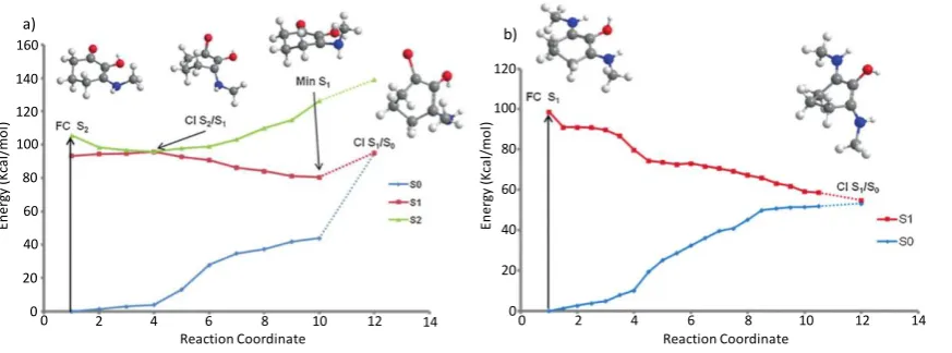
Employing a computational approach, Losantos et al. extracted key points along the minimum energy pathway (CIs and potential energy surface minima) for the core structures with differing functional groups, based upon the three cyclohexenone and five imine core structures given in Figure 4. Their findings show that MAA-inspired moieties that possess a cyclohexenone core prevent repopulation of the electronic ground state; following photoexcitation to the second excited electronic state (S2), excited state population traverses through an S2/S1 CI before being
trapped in the excited state minimum of the first excited electronic state (S1). While Losantos
et al. did locate a CI between the S1 and ground electronic state (S0), this is energetically
inaccessible as it is located above the S1 minimum. This is summarised by Figure 5(a). In
contrast, MAA-inspired moieties that are formed of a cyclohexenimine core repopulate S0
Figure 4: Chemical structures of systems studied by Losantos et al. a) cyclohexenone core structures, b)
cyclohexenimine core structures.Adapted from reference 27.
1. R1= OH, R2=
2. R1= H, R2=
3. R1= H, R2= H
4. R1= OH, R3= CH3
5. R1= H, R3= CH3
6. R1= OH
7. R1= H
8. R1= CH3
a) b)
1 2
[image:9.595.74.522.215.388.2]following photoexcitation, this time directly to S1: excited state population passes through an
energetically accessible S1/S0 CI, as shown by Figure 5(b). All the systems studied show that
the geometry of the CI involves an out of plane movement for both substituents at positions 1 and 3 on the ring (see Figure 4(b)). Furthermore, non-adiabatic molecular dynamics simulations conducted on molecules 6 and 8 (Figure 4(b)) predict an excited state (S1) lifetime
of approximately 200 fs. This lifetime is shorter than that of current commercial filters such as menthyl anthranilate and avobenzone.54–56 The shorter excited state lifetime is beneficial to potential sunscreen filters; the probability for competing (reactive) pathways within the sunscreen filter itself, or side reactions between the electronically excited sunscreen filter and additional compounds (within a commercial formulation) is minimised. These competing side reactions can also induce unfavourable reactions some of which have been known to cause skin irritation.11,12
Given their findings regarding the differing core structures, Losantos et al. proceed to suggest 16 additional structures based upon the imine core moiety, each allowing for variation in the electronic ground state absorption to fully cover the UVA and UVB region of the electromagnetic spectrum. They also further explore the photostability and perform solar protection factor (SPF) measurements on mixtures of these systems.27 These findings are in
[image:10.595.85.522.76.238.2]notable contrast to commercial formulations, demonstrating significant increases in SPF. Of these 16 suggested structures, Losantos et al. have subsequently published a follow-on paper, on three of these proposed structures, employing both transient electronic absorption spectroscopy (see Section 2.1) and fluorescence up conversion to monitor the excited state lifetimes. These experimental studies are complemented by CASPT2 calculations (see Section 2.3).57 Their recent experiments agree with their computational findings (vida supra), and Case
Figure 5: Calculated minimum energy pathways for systems studied by Losantos et al. a) structure 1, and b)
structure 6, from Figure 4. Adapted from reference 27.
En er gy ( K ca l/ m o l) 20 40 60 100 80 120 160 140 20 40 60 100 80 120 0 0
0 2 4 6 8 10 12 14 0 2 4 6 8 10 12 14
En er gy ( K ca l/ m o l)
Reaction Coordinate Reaction Coordinate
Study 2 of an easily accessible S1/S0 CI promoting rapid repopulation of the electronic ground
state.57
3.2Case Study 2:
Woolley J.M, Staniforth M, Horbury M.D, Richings G.W, Wills M, Stavros V.G,
Unravelling the Photoprotection Properties of Mycosporine Amino Acid Motifs. J. Phys. Chem. Lett. 2018; 9: 3043-3048.
Building on the work by Losantos et al., an experimental investigation into two of the systems, a cyclohexenone and cyclohexenimine core shown as in Figure 6, was performed by Woolley
et al.16 They performed transient electronic absorption spectroscopy (see Section 2.1) on both
systems in a variety of solvents (acetonitrile and methanol), to unravel the effect of solvent environment on the photoexcited-state dynamics.
Woolley et al.’s experimental TAS, shown in the form of false colour heat maps, in Figure 7, revealed that, following photoexcitation at the absorption maximum of the cyclohexenone core (see Figure 6), the vibrationally hot S1 state undergoes vibrational cooling, leading to
population of the S1 minimum. Excited state population localised within the S1 minimum is
subsequently trapped for at least 2.5 ns (a consequence of the maximum time-window of the experimental setup used by Woolley et al.). On close inspection of the false colour map of Figure 7(a), the initial broad absorption (340-550 nm) is assigned to a initially populated, vibrationally hot, excited state which then vibrationally cools (spectrally blue shifts) on the S1
state. The strong absorption (shown in red), which persists for the maximum time-window of the experiment, describes the trapped excited state population at the S1 minimum. While the
[image:11.595.110.516.317.413.2]change of solvent environment did affect the observed spectra, mainly in terms of signal amplitude (cf. Figure 7(a), and 7(b)), Woolley et al. concluded that the excited state dynamics
Figure 6: Chemical structures of systems studied by Woolley et al., a) cyclohexenone core structure and b)
imine core structure. c) The absorption maximum for each system in the two solvents used in the study.
Adapted from reference 16.
a)
b)
Structure/Solvent acetonitrile methanol
a 272 nm 285 nm
b 328 nm 328 nm
followed the same relaxation mechanism regardless of solvent environment; that of excited state population trapped in the S1 state.
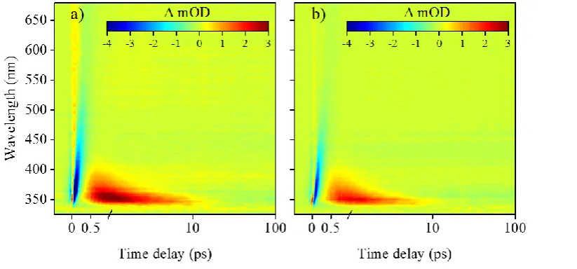
[image:12.595.76.522.113.313.2]For the cyclohexenimine core photoexcited at the absorption maximum (see Figure 6), the TAS is once again presented as false colour heat maps in Figure 8, display short lived stimulated emission and ground state bleach features (shown in blue) that persist for approximately 500 fs. The data then displays characteristic properties of a vibrationally hot electronic ground state (shown in red):58 a sharp absorption that rapidly spectrally blue shifts and decays after a few ps. Woolley et al. assign these features to excited state population traversing from the initial
Figure 8: False colour heat maps of transient absorption spectroscopy data collected by Woolley et al. for the
imine core structure in two solvents; (a) acetonitrile and (b) methanol. Adapted from reference 16. Further
[image:12.595.109.521.512.704.2]permissions related to this material should be directed to the ACS.
Figure 7: False colour heat maps of transient absorption spectroscopy data collected by Woolley et al. for the
cyclohexenone core structure in two solvents; (a) acetonitrile and (b) methanol.Adapted from reference 16.
Franck-Condon window towards the S1/S0 CI, giving rise to the blue stimulated emission. This
is subsequently followed by vibrational energy transfer to the solvent (shown in red), possibly enhanced through the charged nature of the species (see Figure 6(b)).
Overall the work performed by Woolley et al. corroborates the theoretical and experimental investigations performed by Losantos et al. discussed in Case Study 1. In particular, the results obtained for the cyclohexenone core unit from both experiment and theory clearly demonstrates that repopulation of the ground electronic state is prevented, shown by the long lived absorption in the transient absorption spectra attributed to the S1 state absorption (Figure 7(a)). In the
context of a photoprotectant species, the long-lived excited state population is potentially detrimental; the extended lifetime (>2.5 ns) in the S1 state increases the likelihood for
competing photochemical processes, for example triplet-state formation (which may subsequently be quenched by triplet oxygen to generate cytotoxic singlet oxygen). Interestingly gadusol and mycosporine-glycine (Figure 1(b)), which have a cyclohexenone core, have been shown to demonstrate high photostability.59–61 This may imply that further modification of the core unit makes the S1/S0 CI energy accessible to excited state population, possibly through
additional substitution to differing ring positions (Figure 6(a)). Further work is certainly warranted in this regard, to assess how molecular complexity of the cyclohexenone core influences the photoexcited state dynamics. Conversely studies based on the cyclohexenimine core should also be carried out to fully understand the excited state landscape associated with this rapid relaxation.
3.3Case Study 3:
Conde, F.R, Churio, M.S, Previtali C.M, The Deactivation Pathways of the Excited-States of the Mycosporine-Like Amino Acids Shinorine and Porphyra-334 in Aqueous Solution. Photochem. Photobiol. Sci. 2004; 3: 960-967.
Case Studies 1 and 2, vide supra, focussed on tracking the intramolecular energy transfer within MAA-inspired structures using either theory and computation or time-resolved pump-probe spectroscopy. Work by Conde et al.62 uses the advantages of photoacoustic calorimetry
flash photolysis to study the triplet state absorption. The values for photodecomposition and fluorescence quantum yield are then compared to the already reported data for porphyra-334.63
Photoacoustic calorimetry performed on both shinorine and porphyra-334 (both excited at 355 nm) showed equivalent values of the initial amplitude, H, (see section 2.2 for further explanation) between the solute and the reference sample, indigo carmine.43 This equivalence (between solute and standard) is an indication that the energy absorbed by the MAAs is rapidly released as heat to the surrounding solvent. Conde et al. note that the percent conversion of this energy transfer (solute to solvent) is 96-98%,62 leaving some residual energy trapped within the MAA which undergoes intersystem crossing to populate a triplet state. The authors confirm the presence of the triplet state for both shinorine and porphyra-334 with the use of laser flash photolysis;63,64 this triplet state accounts for the remaining energy trapped within the MAAs. To corroborate their photoacoustic calorimetry measurements, Conde et al. preformed steady-state experiments to fully describe the photodecomposition and relaxation dynamics of shinorine and porphyra-334. These results validate the photoacoustic calorimetry measurements with negligible fluorescence quantum yields determined, with values on the order of 10-4. The rate of photodecomposition was found to be of the same magnitude, leading
the authors to attribute a similar relaxation pathway as described for the MAA-inspired structures above (Case Study 1, Figure 5(b)).
The authors close by placing their research in the wider field of natural sunscreens highlighting that their results strongly favour the use of both shinorine and porphyra-334 in an in-vivo
[image:14.595.120.522.123.278.2]environment as natural sunscreen filters, even though a small proportion of the excess energy remains trapped as a triplet population with a significantly longer lifetime.62,63,65 Relating this work to Case Studies 1 and 2, the proposed overall dynamics of the system agree with those of
Figure 9: Chemical structures of systems studied by Conde et al. a) Shinorine and b) Porphyra-334.Adapted
from reference 62.
b)
a)
the MAA-inspired systems by Losantos et al. and Woolley et al.:16,27 rapid relaxation through an accessible CI resulting in a vibrationally excited electronic ground state.
4.
Conclusions and Outlook
Given the continuing increase in skin cancer cases and recent concerns raised over current sunscreen filters such as phototoxicity and photdegradation,9,10,13–15,66–68 unravelling photoprotection mechanisms through an intrinsic understanding of the photochemistry of both nature based (and inspired) and artificial sunscreen filters continues to grow as a field of research.16,27,69 Studies on exploring the synthesis and isolation of MAAs in nature provides complementary aspects of this research.7,70,71 That being said, more work is certainly warranted. Notably, identification of additional MAA-inspired sunscreens is essential: although 30 MAAs have been isolated, regions of the UVR spectrum remain poorly protected from using pure MAA products alone, which is undoubtedly an area where functionalisation of core MAA structures can play a key role.8,27 Additionally, a greater photochemical exploration of MAAs should be undertaken to consolidate the relaxation pathways of MAAs, such that we are then best positioned to propose ‘rules’ for efficient molecular design, leading
to MAA-inspired sunscreen filters. Undeniably, this will necessitate the use of both experiment and theory.72,73
5.
Acknowledgements
J. M. W. is grateful to EPSRC and Newport Spectra-Physics Ltd for a joint studentship. V. G. S. thanks the HO2020 FET-OPEN Grant BoostCrop for financial support.
6.
References
1. Crespo-Hernández CE, Cohen B, Hare PM, et al. Ultrafast excited-state dynamics in nucleic acids. Chem Rev. 2004; 104: 1977–2019.
2. Pfeifer GP, You YH, Besaratinia A. Mutations induced by ultraviolet light. Mutat Res - Fundam Mol Mech Mutagen. 2005; 571: 19–31.
3. Sinha RP, Häder D-P. UV-induced DNA damage and repair: a review. Photochem Photobiol Sci. 2002; 1: 225–236.
4. de Gruijl FR. Skin cancer and solar UV radiation. Eur J Cancer. 1999; 35: 2003–2009. 5. Trione EJ, Leach CM, Mutch JT. Sporogenic substances isolated from fungi. Nature.
1966; 212: 163–164.
6. Fraser CM, Chapple C. The Phenylpropanoid Pathway in Arabidopsis. Epub ahead of print 2011. DOI: 10.1199/tab.0152.
7. Cardozo KHM, Guaratini T, Barros MP, et al. Metabolites from algae with economical impact. Comp Biochem Physiol Part C Toxicol Pharmacol. 2007; 146: 60–78.
8. Bandaranayake WM. Mycosporines : are they Nature’s Sunscreens ? Nat Prod Rep. 1998; 15: 159–172.
9. Ghazipura M, McGowan R, Arslan A, et al. Exposure to benzophenone-3 and reproductive toxicity: A systematic review of human and animal studies. Reprod Toxicol. 2017; 73: 175–183.
10. Krause M, Klit A, Blomberg Jensen M, et al. Sunscreens: Are they beneficial for health? An overview of endocrine disrupting properties of UV-filters. Int J Androl. 2012; 35: 424–436.
Arch Dermatol. 2010; 146: 753–757.
12. Karlsson I, Vanden Broecke K, Mårtensson J, et al. Clinical and experimental studies of octocrylene’s allergenic potency. Contact Dermatitis. 2011; 64: 343–352.
13. Coronado M, De Haro H, Deng X, et al. Estrogenic activity and reproductive effects of the UV-filter oxybenzone (2-hydroxy-4-methoxyphenyl-methanone) in fish. Aquat Toxicol. 2008; 90: 182–187.
14. Downs CA, Kramarsky-Winter E, Segal R, et al. Toxicopathological Effects of the Sunscreen UV Filter, Oxybenzone (Benzophenone-3), on Coral Planulae and Cultured Primary Cells and Its Environmental Contamination in Hawaii and the U.S. Virgin Islands. Arch Environ Contam Toxicol. 2016; 70: 265–288.
15. Blüthgen N, Zucchi S, Fent K. Effects of the UV filter benzophenone-3 (oxybenzone) at low concentrations in zebrafish (Danio rerio). Toxicol Appl Pharmacol. 2012; 263: 184–194.
16. Woolley JM, Staniforth M, Horbury MD, et al. Unravelling the Photoprotection Properties of Mycosporine Amino Acid Motifs. J Phys Chem Lett. 2018; 9: 3043–3048. 17. Baker LA, Horbury MD, Greenough SE, et al. Ultrafast Photoprotecting Sunscreens in
Natural Plants. J Phys Chem Lett. 2016; 7: 56–61.
18. Fernanda Pessoa M. Algae and aquatic macrophytes responses to cope to ultraviolet radiation – a Review. Emirates J Food Agric. 2012; 24: 527–545.
19. Colabella F, Moline M, Libkind D. UV Sunscreens of Microbial Origin: Mycosporines and Mycosporine- like Aminoacids. Recent Pat Biotechnol. 2015; 8: 179–193.
20. Osborn AR, Almabruk KH, Holzwarth G, et al. De novo synthesis of a sunscreen compound in vertebrates. Elife. 2015; 1–15.
21. Sinha RP, Klisch M, H D. Ultraviolet-Absorbing / Screening Substances in Cyanobacteria , Phytoplankton and Macroalgae. J Photochem Photobiol B Biol. 1998; 1344: 83–94.
22. Schmidt EW. An Enzymatic Route to Sunscreens. ChemBioChem. 2011; 12: 363–365. 23. Gao Q, Garcia-Pichel F. Microbial Ultraviolet Sunscreens. Nat Rev Microbiol. 2011; 9:
24. Whitehead K, Hedges JI. Photodegradation and photosensitization of mycosporine-like amino acids. J Photochem Photobiol B Biol. 2005; 80: 115–121.
25. Balskus EP, Walsh CT. The Genetic and Molecular Basis for Sunscreen Biosynthesis in Cyanobacteria. Science (80- ). 2010; 329: 1653–1656.
26. White JD, Cammack JH, Sakuma K, et al. Transformations of Quinic Acid. Asymmetric Synthesis and Absolute Configuration of Mycosporin I and Mycosporin-gly. J Org Chem. 1995; 60: 3600–3611.
27. Losantos R, Funes-Ardoiz I, Aguilera J, et al. Rational Design and Synthesis of Efficient Sunscreens To Boost the Solar Protection Factor. Angew Chemie Int Ed. 2017; 56: 2632– 2635.
28. White JD, Cammack JH, Sakuma K. The Synthesis and Absolute Configuration of Mycosporins. A Novel Application of the Staudinger Reaction. J Am Chem SOC. 1989; 111: 8970–8972.
29. Rodrigues NDN, Stavros VG. From fundamental science to product : a bottom-up approach to sunscreen development. Sci Prog. 2018; 101: 8–31.
30. González L, Escudero D, Serrano-Andrés L. Progress and challenges in the calculation of electronic excited states. ChemPhysChem. 2012; 13: 28–51.
31. Gensch T, Viappiani C. Time-resolved photothermal methods: Accessing time-resolved thermodynamics of photoinduced processes in chemistry and biology. Photochem Photobiol Sci. 2003; 2: 699–721.
32. Braslavsky SE, Helbel GE. Time-Resolved Photothermal and Photoacoustic Methods Applied to Photoinduced Processes in Solution. Chem Rev. 1992; 92: 1381–1410. 33. Arnaut LG, Caldwell RA, Elbert JE, et al. Recent advances in photoacoustic calorimetry:
Theoretical basis and improvements in experimental design. Rev Sci Instrum. 1992; 63: 5381–5389.
34. Baker LA, Stavros VG. Observing and Understanding the Ultrafast Photochemistry in Small Molecules : Applications to Sunscreens. Sci Prog. 2014; 99: 282–311.
nucleotides, amino acids and beyond. Proc R Soc London A Math Phys Eng Sci.; 469. 36. Zewail AH. Laser Femtochemistry. Science (80- ). 1988; 242: 1645.
37. Megerle U, Pugliesi I, Schriever C, et al. Sub-50 fs broadband absorption spectroscopy with tunable excitation: putting the analysis of ultrafast molecular dynamics on solid ground. Appl Phys B Lasers Opt. 2009; 96: 215–231.
38. Strickland D, Mourou G. Conpression of Amplified Chirped Optical Pulses. Opt Commun. 1985; 55: 447–449.
39. Treacy E. Optical pulse compression with diffraction gratings. IEEE J Quantum Electron. 1969; 5: 454–458.
40. Bradler M, Baum P, Riedle E. fsec continuum generation in bulk laser host materials with sub uj pump pulses.pdf. Appl Phys B Lasers Opt. 2009; 97: 561–574.
41. Berera R, van Grondelle R, Kennis JTM. Ultrafast transient absorption spectroscopy: Principles and application to photosynthetic systems. Photosynth Res. 2009; 101: 105– 118.
42. Callis JB, Parson WW, Gouterman M. Fast changes of enthalpy and volume on flash excitation of Chromatium chromatophores. Biochim E,T Biophys - Bioenerg. 1972; 267: 348–362.
43. Abbruzzetti S, Viappiani C, Murgida DH, et al. Non-toxic, water-soluble photocalorimetric reference compounds for UV and visible excitation. Chem Phys Lett. 1999; 304: 167–172.
44. Cossi M, Barone V, Mennucci B, et al. Ab initio study of ionic solutions by a polarizable continuum dielectric model. Chem Phys Lett. 1998; 286: 253–260.
45. Klamt A, Schüürmann G. COSMO: A new approach to dielectric screening in solvents with explicit expressions for the screening energy and its gradient. J Chem Soc Perkin Trans 2. 1993; 0: 799–805.
46. Turner MAP, Horbury MD, Stavros VG, et al. Determination of Secondary Species in Solution through Pump-Selective Transient Absorption Spectroscopy and Explicit-Solvent TDDFT. J Phys Chem A. 2019; 123: 873–880.
colours of a solvated organic dye: The example of nile red. J Chem Phys. 2017; 146: 146–159.
48. Santoro F, Barone V, Gustavsson T, et al. Solvent Effect on the Singlet Excited-State Lifetimes of Nucleic Acid Bases: A Computational Study of 5-Fluorouracil and Uracil in Acetonitrile and Water. J Am Chem Soc. 2006; 128: 16312–16322.
49. Kühl A, Domcke W. Effect of a dissipative environment on the dynamics at a conical intersection. Chem Phys. 2000; 259: 227–236.
50. Worth GA, Cederbaum LS. BEYOND BORN-OPPENHEIMER: Molecular Dynamics Through a Conical Intersection. Annu Rev Phys Chem. 2004; 55: 127–158.
51. Roos B, Taylor PR. A Complete Active Space SCF Method (CASSCF) Using a Density Matrix: Formulated Super-CI Approach. Chem Phys. 1980; 48: 157–173.
52. Roos BO. The complete active space SCF method in a fock‐matrix‐based super‐CI formulation. Int J Quantum Chem. 1980; 18: 175–189.
53. Stratmann RE, Scuseria GE, Frisch MJ. An efficient implementation of time-dependent density-functional theory for the calculation of excitation energies of large molecules. J Chem Phys. 2002; 109: 8218–8224.
54. Baker LA, Horbury MD, Greenough SE, et al. Probing the ultrafast energy dissipation mechanism of the sunscreen oxybenzone after UVA irradiation. J Phys Chem Lett. 2015; 6: 1363–1368.
55. Dunkelberger AD, Kieda RD, Marsh BM, et al. Picosecond Dynamics of Avobenzone in Solution. J Phys Chem A. 2015; 119: 6155–6161.
56. Rodrigues NDN, Cole-Filipiak NC, Horbury MD, et al. Photophysics of the sunscreen ingredient menthyl anthranilate and its precursor methyl anthranilate: A bottom-up approach to photoprotection. J Photochem Photobiol A Chem. 2018; 353: 376–384. 57. Losantos R, Lamas Frejo I, Montero R, et al. Photophysical Characterization of New
and Efficient Synthetic Sunscreens. Phys Chem Chem Phys. 2019; 21: 11376–11384. 58. Pecourt JML, Peon J, Kohler B. DNA excited-state dynamics: Ultrafast Internal
59. Moliné M, Arbeloa EM, Flores MR, et al. UVB Photoprotective Role of Mycosporines in Yeast: Photostability and Antioxidant Activity of Mycosporine-Glutaminol-Glucoside. Radiat Res. 2011; 175: 44–50.
60. Arbeloa EM, Bertolotti SG, Churio MS. Photophysics and reductive quenching reactivity of gadusol in solution. Photochem Photobiol Sci. 2011; 10: 133–42.
61. Rastogi RP, Incharoensakdi A. Analysis of UV-absorbing Photoprotectant Mycosporine-like Amino Acid (MAA) in the Cyanobacterium Arthrospira sp. CU2556.
Photochem Photobiol Sci. 2014; 13: 1016–1024.
62. Conde FR, Churio MS, Previtali CM. The deactivation pathways of the excited-states of the mycosporine-like amino acids shinorine and porphyra-334 in aqueous solution.
Photochem Photobiol Sci. 2004; 3: 960–967.
63. Conde FR, Churio MS, Previtali CM. The photoprotector mechanism of mycosporine-like amino acids. Excited-state properties and photostability of porphyra-334 in aqueous solution. J Photochem Photobiol B Biol. 2000; 56: 139–144.
64. Porter G, Topp M. Nanosecond Flash Photolysis. Proc R Soc A Math Phys Eng Sci. 1970; 315: 163–184.
65. Conde FR, Churio MS, Previtali CM. Experimental Study of the Excited-State Properties and Photostability of the Mycosporine-Like Amino Acid Palythine in Aqueous Solution. Photochem Photobiol Sci. 2007; 6: 669–674.
66. WHO world Health Organisation Ultraviolet Radiation: Skin cancers. How common is Skin Cancer, https://www.who.int/uv/faq/skincancer/en/index1.html (accessed 29 April 2019).
67. Sharma A, Bányiová K, Babica P, et al. Different DNA damage response of cis and trans isomers of commonly used UV filter after the exposure on adult human liver stem cells and human lymphoblastoid cells. Sci Total Environ. 2017; 593–594: 18–26.
68. Kockler J, Oelgemöller M, Robertson S, et al. Photostability of sunscreens. J Photochem Photobiol C Photochem Rev. 2012; 13: 91–110.
70. Garcia-Pichel F, Castenholz RW. Occurence of UV-absorbing, Mycosporin-like compounds among cyanobacterial isolates and estimates of their screening capacity.
Appl Environ Microbiol. 1993; 59: 163–169.
71. Carreto JI, Carignan MO. Mycosporine-like amino acids: Relevant secondary metabolites. chemical and ecological aspects. Mar Drugs. 2011; 9: 387–446.
72. Losantos R, Churio MS, Sampedro D. Computational exploration of the photoprotective potential of gadusol. ChemistryOpen. 2015; 4: 155–160.