Partial maintenance of organ-specific epigenetic marks
during plant asexual reproduction leads to heritable
phenotypic variation
Anjar Wibowoa,b,1, Claude Beckerb,c,1,2, Julius Durra, Jonathan Pricea, Stijn Spaepend,e, Sally Hiltona, Hadi Putraa, Ranjith Papareddya,2, Quentin Saintaina, Sarah Harveya,f, Gary D. Bendinga, Paul Schulze-Lefertd,e, Detlef Weigelb,3, and Jose Gutierrez-Marcosa,3
aSchool of Life Sciences, University of Warwick, CV4 7AL Coventry, United Kingdom;bDepartment of Molecular Biology, Max Planck Institute for
Developmental Biology, 72076 Tübingen, Germany;cGregor Mendel Institute of Molecular Plant Biology, Austrian Academy of Sciences, Vienna Biocenter,
1030 Vienna, Austria;dDepartment of Plant Microbe Interactions, Max Planck Institute for Plant Breeding Research, 50829 Cologne, Germany;eCluster of
Excellence on Plant Sciences, Max Planck Institute for Plant Breeding Research, 50829 Cologne, Germany; andfDepartment of Biology, University of York,
YO10 5DD York, United Kingdom
Contributed by Detlef Weigel, July 31, 2018 (sent for review March 28, 2018; reviewed by Xiaofeng Cao and Mary Gehring)
Plants differ from animals in their capability to easily regenerate fertile adult individuals from terminally differentiated cells. This unique developmental plasticity is commonly observed in nature, where many species can reproduce asexually through the ectopic initiation of organogenic or embryogenic developmental programs. While organ-specific epigenetic marks are not passed on during sexual reproduc-tion, the fate of epigenetic marks during asexual reproduction and the implications for clonal progeny remain unclear. Here we report that organ-specific epigenetic imprints inArabidopsis thalianacan be par-tially maintained during asexual propagation from somatic cells in which a zygotic program is artificially induced. The altered marks are inherited even over multiple rounds of sexual reproduction, be-coming fixed in hybrids and resulting in heritable molecular and phys-iological phenotypes that depend on the identity of the founder tissue. Consequently, clonal plants display distinct interactions with beneficial and pathogenic microorganisms. Our results demonstrate how novel phenotypic variation in plants can be unlocked through altered inheritance of epigenetic marks upon asexual propagation.
epigenetics
|
Arabidopsis thaliana|
DNA methylation|
transgenerational inheritance|
asexual reproductionC
ompared with animals, in plants somatic cells can be muchmore easily coaxed into regenerating entire individuals (1). A potential reason why differentiated plant cells can rapidly acquire
“stemness”is that the epigenome of plants is much more flexible than
that of animals. Thus, asexual reproduction is much more common in plants than in animals (2, 3), and this has been traditionally exploited by humans for the clonal propagation and genetic manipulation of many economically important plant species, including grapevines, nearly all tuber and root crops, and fruit and forest trees (4).
Although clonal propagation provides ecological and evolu-tionary benefits, the resulting restricted genetic variation could be detrimental to fitness. Notably, clonally propagated plants are not always phenotypically identical to their parents, a phenom-enon termed somaclonal variation. While somaclonal variation is often attributed to the accumulation of random genetic mutations in form of single-base changes or transposon activation (5, 6), in-creasing evidence suggests that genetic changes are not solely re-sponsible. For example, genome-wide DNA methylation patterns can be perturbed during clonal propagation through hormone-dependent tissue culture, and these epimutations can be
associ-ated with altered expression of protein-coding genes (7–10). In
some cases, such tissue-culture induced epimutations can be stably inherited across multiple sexual generations (7, 8, 10) and can be responsible for phenotypes that distinguish clonal descendants from their parents (11). Increased genetic and epigenetic diversity may be deleterious, but also potentially result in advantageous traits. For all these reasons, we would like to better understand the
precise origin and mechanistic basis of the molecular and pheno-typic changes created during plant regeneration.
Depending on the species, different types of organs, such as roots, stems, and leaves, as well as entire embryos, can be used for clonal propagation. The starting material thus includes tissue with different patterns of gene expression and epigenetic
pro-files. For example, 1.6% of methylated regions in theArabidopsis
thalianagenome differ in methylation status between shoots and roots, and more than 2,000 genes differ in expression levels (12).
Significance
While clonally propagated individuals should share identical genomes, there is often substantial phenotypic variation among them. Both genetic and epigenetic modifications in-duced during regeneration have been associated with this phenomenon. Here we investigated the fate of the epigenome after asexual propagation by generating clonal individuals from differentiated somatic cells through the manipulation of a zygotic transcription factor. We found that phenotypic novelty in clonal progeny was linked to epigenetic imprints that reflect the organ used for regeneration. Some of these organ-specific imprints can be maintained during the cloning process and subsequent rounds of meiosis. Our findings are fundamental for understanding the significance of epigenetic variability arising from asexual reproduction and have significant impli-cations for future biotechnological appliimpli-cations.
Author contributions: A.W., C.B., G.D.B., P.S.-L., D.W., and J.G.-M. designed research; A.W., C.B., J.D., H.P., R.P., Q.S., and S. Harvey performed research; A.W., C.B., J.P., S.S., S. Hilton, D.W., and J.G.-M. analyzed data; and A.W., C.B., D.W., and J.G.-M. wrote the paper.
Reviewers: X.C., Institute of Genetics and Developmental Biology, Chinese Academy of Sciences; and M.G., Massachusetts Institute of Technology.
A.W., C.B., D.W., and J.G.-M. are inventors on patent application“Stable epigenetic plant variants”(PCT/EP2016/055377 and WO2016146552A1) filed by University of Warwick and Max Planck Society.
This open access article is distributed underCreative Commons Attribution-NonCommercial-NoDerivatives License 4.0 (CC BY-NC-ND).
Data deposition: Sequencing reads have been deposited at the European Nucleotide Archive (accession nos.PRJEB26932andPRJEB14117). DNA methylation data have been uploaded to the EPIC-CoGe epigenome browser and can be accessed at
https://genomevolution.org/CoGe/NotebookView.pl?lid=835. 1A.W. and C.B. contributed equally to this work.
2Present address: Gregor Mendel Institute of Molecular Plant Biology, Austrian Academy
of Sciences, Vienna Biocenter, 1030 Vienna, Austria.
3To whom correspondence may be addressed. Email: weigel@weigelworld.org or j.f.
gutierrez-marcos@warwick.ac.uk.
This article contains supporting information online atwww.pnas.org/lookup/suppl/doi:10. 1073/pnas.1805371115/-/DCSupplemental.
GENET
An attractive hypothesis is that complete or partial mainte-nance of variant epigenetic landscapes present in the starting material will contribute to phenotypic variation among
regen-erants. We have tested this hypothesis by regeneratingA.
thali-ana plants from different postembryonic organs. Frequent
limitations of plant regeneration are the lengthy periods of tissue culture and the hormone mixtures that need to be optimized for each tissue, which confounds the interpretation of phenotypic differences in regenerated plants. To circumvent this problem, we have exploited the fact that somatic embryogenesis can be
induced in different tissues of A. thalianaby ectopic
expres-sion of certain zygotic factors (2). Here we used transgenic
lines carrying an inducible version of the RWP-RK
DOMAIN-CONTAINING 4 (RKD4) transcription factor gene (13). We provide evidence that certain organ-specific epigenetic
differ-ences are partially maintained during regeneration ofA. thaliana.
Whole plants derived from roots inherit many aspects of root-specific methylation and gene expression patterns not just in roots, but also in leaves. These epigenetic profiles and the resulting macroscopic and molecular phenotypes are stably transmitted during meiosis for at least four self-crossing gener-ations. Our findings demonstrate that plants with novel methyl-ation and gene expression patterns, as well as physiological phenotypes, can be created using specific regeneration strategies.
Results
Tissue Origin of Regenerants Affects Activity of Defense-Related Genes. Plants can reproduce asexually from both belowground and aboveground organs, which are known to be epigenetically distinct (12, 14). We took advantage of this situation to determine the extent to which the organ-specific origin of the epigenome could influence phenotypes of clonal progeny. To mimic naturally occurring events associated with asexual propagation (2, 15), we did not resort to hormone-induced regeneration in tissue culture, but instead produced somatic embryos from distinct root (root
origin; RO) and leaf (leaf origin; LO) tissues ofA. thaliana
(Col-0 strain) by controlled expression of the RKD4 zygotic factor (13)
(SI Appendix, Fig. S1). We collected seeds from independently
regenerated G0 individuals after self-pollination and further
propagated each line by selfing for over three consecutive
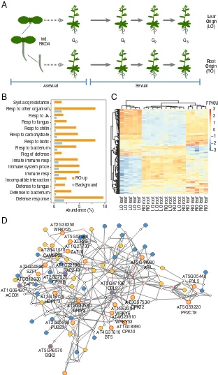
gen-erations (G1–G3) (Fig. 1A). Visual examination revealed no
ob-vious morphological differences between RO and LO plants. To determine any potential differences at the molecular level, we performed whole-genome transcriptome analyses in five
ran-domly selected G2lines. Using stringent thresholds [false
discov-ery rate (FDR) <0.01; absolute log-twofold change >1.5], only
13 differentially expressed genes (DEGs) differentiated roots of RO plants from roots of LO plants, but almost 20-fold more
DEGs (n =239) were identified between leaves of RO and LO
plants (SI Appendix, Fig. S2andDataset S1). Gene ontology (GO)
analysis revealed that these DEGs were enriched for stress- and
defense-related genes (FDR <0.05) (Fig. 1B). The 51 genes in
these two categories were mostly up-regulated in leaves of RO plants and primarily involved in cellular signaling (27%) and transcriptional regulation (22%). Clustering of all samples based on DEG expression levels suggested that leaves of RO plants had partial root characteristics, sharing apparent similarity with roots from both RO and LO plants (Fig. 1C); in contrast, leaves of LO plants formed a single distinct group (Fig. 1C). To gain further insight into this transcriptional variation, we performed a network analysis of DEGs that distinguished leaves of RO and LO plants,
based on a less stringent threshold (FDR<0.05) (16). This
net-work contained the 239 stringent DEGs (SI Appendix, Fig. S2and
Dataset S1), of which 213 had known interactions with other DEGs and 69 were part of a single functional network (Fig. 1D).
This network included five genes (WRKY6,SZF1,PYL5,PUB23,
andDRIP2) that had been previously implicated in the negative
regulation of abiotic and biotic stress responses (17–21).
Tissue of Origin of Regenerants Affects Interaction with Microbes.
We next sought to define whether the gene expression differ-ences between RO and LO plants were functionally meaningful and affected whole-plant or tissue-specific functions. Plant microbiota are known to be affected by developmental factors as well as immune system activity (22, 23), and both the altered expression of tissue-specific genes in RO plants as well as the enriched GO categories suggested that it would be relevant to test for a change in the interaction with the biotic environment. We grew plants in natural soils and after 4 weeks assessed the bacterial communities that became associated with their roots. Bacterial communities from roots of RO plants differed from
those of both nonregenerated plants (ANOSIM,r=0.485,P=
0.007) and LO plants (ANOSIM,r=0.216,P=0.056) (Fig. 2A
andSI Appendix, Fig. S3). In addition, in vitro root colonization
byBacillus amyloliquefaciensFZB42, a soil-borne plant growth-promoting bacterium (24), differed significantly between RO and
nonregenerated plants (Wilcoxon rank-sum test,W=3862.5,P=
5.9e-05) (SI Appendix, Fig. S4). Finally, we inoculated roots of
regenerated and nonregenerated plants with synthetic communi-ties (SynComs) consisting of abundant soil- and root-derived bacterial isolates (25). Again, the root bacterial communities dif-fered between regenerated and nonregenerated individuals (7.3%
variance explained by genotype; permutation-based ANOVA,P=
0.07) (SI Appendix, Fig. S5); this was especially obvious for
the Alcaligenaceae family (Betaproteobacteria) (Fig. 2B and
SI Appendix, Fig. S5). Similarly, when analyzing leaves of
SynCom-inoculated plants (9.6% variance explained by genotype;
P = 0.023), the most notable differences were observed for
Xanthomonadaceae (Gammaproteobacteria) (ANOVA, P <
0.05) (SI Appendix, Figs. S5 and S6), which include several
phy-topathogenic strains.
These differences in microbiota led us to further assess the response to known pathogens. For this, we inoculated leaves with
the bacteriumPseudomonas syringaepv. tomato strain DC3000
and the oomyceteHyaloperonospora arabidopsidisisolate Noks1,
as theA. thalianaCol-0 strain lacks gene-for-gene resistance to
both of these pathogens (26, 27). We found that RO plants were more sensitive than LO plants to infection by either pathogen, and that these differences were stably inherited for at least three
generations (Fig. 2C and D). This result was in line with our
previous observation of the up-regulation of negative regulators of biotic stress in RO plants (Fig. 1D).
Heritable Differences in Genome-Wide DNA Methylation in Regenerants. Given the unexpected widespread and heritable differences between transcriptional profiles and microbe re-sponses in RO and LO plants, which should have very few, if any, group-wise genetic differences, we investigated the potential epigenomic basis for phenotypic differentiation between RO and LO plants. DNA methylation is an important epigenetic mark, for which excellent statistical methods for genome-wide com-parisons are available (28). Moreover, dynamic changes in DNA methylation during sexual reproduction have been well docu-mented (29). We used whole-genome bisulfite sequencing of leaves and roots from RO and LO individuals to monitor methylome changes over three consecutive generations (Dataset S2). Principal component analysis (PCA) of 736,413 differen-tially methylated positions (DMPs) discovered in pairwise con-trasts revealed clear differences between root and leaf samples from regenerated and nonregenerated plants (PC1; Fig. 3A). Regenerated samples clustered according to their tissue of origin before regeneration and not, as might be expected, by tissue identity at the time of DNA extraction (PC2; Fig. 3A). When repeating PCA with the methylation level of positions not clas-sified as DMPs, we found little residual variance, indicating a low
Because the functional relevance of individual DMPs in plants is unclear, we also analyzed differentially methylated regions (DMRs). Cluster analysis based on 765 DMRs identified in
pairwise comparisons between all G2leaf samples validated the
use of the independent RO and LO lines as replicate groups
(Fig. 3B). We found 255 consistent RO vs. LO DMRs in G2
leaves (Dataset S3). Compared with methylated regions in gen-eral, RO vs. LO DMRs were overrepresented in exons of gene
coding sequences and in 2-kb regions flanking genes (SI
Ap-pendix, Fig. S8). Similar distributions of DMRs across genomic
Asexual Sexual
Leaf
Origin (LO)
Root
Origin (RO)
G0 G1 G2 G3
Abundance (%)
C
B
Defense response Defense to bacterium Defense to fungus Incompatible interaction Immune resp Immune system proce Innate immune resp Reg of defense Resp to bacterium Resp to biotic Resp to carbohydrate Resp to chitin Resp to fungus Resp to JA Resp to other organism Syst acq resistance
0 5 10
RO up Background
−3 −2 −1 0 1 2 3 FPKM
LO leaf LO leaf LO leaf LO leaf LO leaf RO root RO root LO root LO root LO root RO root LO root RO root LO root RO root RO leaf RO leaf RO leaf RO leaf RO leaf
AT1G18890 CPK10 ind.
RKD4
A
D
AT3G16720 HCF173
AT2G30580 DRIP2
AT5G46570 BSK2
AT4G37610 BT5
AT4G23810 WRKY53 AT2G35930
PUB23
AT2G44080 ARL AT1G22190
RAP2-13 AT2G37220
CP29B AT3G55980
SZF1
AT3G12630 SAP5 AT1G06460
ACD31
AT5G47100 CBL9
AT1G62300 WRKY6
AT3G57530 CPK32
AT5G05440 PYL5
AT5G59220 PP2C78 AT2G30250
WRKY25
AT2G41010 CAMBP25
AT1G27730 ZAT10 AT5G57560
[image:3.585.134.446.52.589.2]XTH22
Fig. 1. Differential expression of defense-related genes in RO and LO plants. (A) Experimental design. RO and LO regenerated plants (n=5 independent lines each) were propagated through self-fertilization over three generations. (B) GO analysis of DEGs (FDR<0.01; absolute log-twofold change>1.5) be-tween leaves of RO and LO G2plants revealing enrichment for defense-related functions. (C) Heatmap of scaled, normalized log-transformed read counts for
genes underlying the significant enrichment of GO terms inB. FPKM, fragments per kilobase per million. (D) Interaction gene network of DEGs. To facilitate network analysis, we lowered the FDR threshold to<0.05. Of the resulting 4,585 DEGs, 2,752 were represented in the ANAP protein interaction database. Nodes represent genes; triangles highlight defense-related genes fromB; edges indicate evidence for gene interaction. Blue filled circles, high expression in LO plants; orange filled circles, high expression in RO plants; red outlined circles, genes with stress-related GO terms.
GENET
features have been reported in the context of biotic and abiotic
stresses (30–32), suggesting that gene-proximal DNA methylation
changes might be a common feature of induced epimutations. Methylation patterns are known to differ between shoots and
roots in A. thalianaand closely related species (12, 14).
Meth-ylation in LO leaves was similar to that in nonregenerated leaves (PC 1 in Fig. 3C); likewise, roots from RO samples showed patterns similar to those in nonregenerated roots. However, leaves from RO populations had methylation patterns closer to those of roots, especially in a symmetric cytosine context (Fig. 3C). In contrast, methylation levels at these DMRs in roots of LO plants were similar to those seen in nonregenerated root samples (Fig. 3C). As expected, methylation levels at non-DMRs grouped samples primarily by their tissue identity, regardless of regenerant origin (Fig. 3C).
Our DMR analysis also revealed that leaves from RO plants had less overall CG and CHG (but not CHH) methylation in the identified DMRs compared with leaves of nonregenerated plants or LO plants (Fig. 3D). To account for sampling bias and sto-chastic effects, we repeated the analyses using randomly selected non-DMRs, which did not produce any evidence of reduced methylation in roots of RO plants (Fig. 3D). The reduced DNA methylation in RO leaves was stably inherited over at least three
generations (Fig. 3CandD), indicating that root-specific DNA
methylation patterns are not lost during subsequent sexual reproduction.
To understand the importance of the methylome status at the time of regeneration, we compared the leaf methylome of RO plants with the methylation profiles of roots from nonregenerated
seedlings and mature plants (SI Appendix, Fig. S9). The RO leaf
methylome was closer to that of roots from nonregenerated seed-lings, implying that regeneration had been induced at the seedling stage and that the cell-specific methylation pattern was maintained throughout the regeneration process and in subsequent sexually reproduced progeny.
Because there is evidence of methylation information being transferable between chromosomes (33, 34), we tested whether
such information transfer could occur intransin our system. To
this end, we performed reciprocal crosses between RO and
nonregenerated plants. DNA methylation in both F1hybrids was
at midparent values, indicating that the DMRs on chromosomes inherited from the RO parent retained their hypomethylated
status (Fig. 3E and SI Appendix, Fig. S10). We assessed the
heritability of the observed differential methylation at these loci
by sequencing individual F2 plant progenies. More than
two-thirds (80%) of DMRs with midparental methylation in the F1
hybrid retained their hypomethylated state in at least one F2
descendant, indicating that allele-specific methylation was stably inherited through both mitotic and meiotic divisions (Fig. 3F).
The establishment and maintenance of DNA methylation in plants rely on a series of partially interconnected pathways, depending on the genomic features that are methylated (35). Consequently, when genome-wide demethylation is induced by various mutations, some regions can be remethylated upon res-toration of the methylation machinery, while others cannot, and
this is a function of the underlying methylation pathways (36–
38). To gain insight into the mechanisms that supported the partial maintenance of root-specific methylation patterns in RO plants, we investigated whether methylated regions that became hypomethylated in RO plants were under the control of a specific epigenetic pathway, by comparing the methylation changes in different mutant contexts (39). We found that CG methylation in such regions was affected in the chromatin
re-modeling mutantddm1and in the DNA methylation maintenance
mutantsmet1andvim1 vim2 vim3, while CHG methylation was
altered in the de novo methyltransferase cmt3 mutant and in
mutants with a compromised H3K9 methylation machinery (SI
Appendix, Fig. S11).
Contrary to our expectations, the regions that were hypo-methylated in RO plants were not affected in the triple ros1 dml2 dml3 (rdd) mutant, which lacks three DNA deme-thylases, suggesting that DNA hypomethylation in RO plants is due to differences in the establishment and/or maintenance of DNA methylation in root initials during embryogenesis, rather than to subsequent active demethylation. Induced methylation changes are often associated with changes in activity of trans-posable elements (TEs), which in turn is reflected in altered expression patterns of siRNAs, which are a central component of TE silencing pathways that involve RNA-directed DNA
meth-ylation (RdDM) (40–42). LO vs. RO DMRs tend to not directly
overlap with TEs and 24-nt siRNA loci (43), but are often closer
to them than would be expected by chance (SI Appendix,
Fig. S12).
Differential Methylation Affects Expression ofRSM1.To establish a connection between the molecular and phenotypic variation generated by plant regeneration, we searched for correlations between changes in DNA methylation (765 DMRs) and gene expression in leaves of regenerated plants, using a combined set
of 1,537 DEGs from G1and G2generations (FDR<0.01). We
found 29, mostly hypomethylated, DMRs that were within 2 kb
upstream or downstream of DEGs (SI Appendix, Fig. S13 and
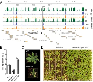
Dataset S4). To confirm that such DMRs can indeed affect gene expression, we selected a DMR approximately 1 kb downstream
of RADIALIS-LIKE SANT/MYB1 (RSM1)/MATERNAL
EF-FECT EMBRYO ARREST 3(MEE3), a regulator of growth and flowering time (44, 45). This DMR was hypomethylated in RO
leaves compared with LO leaves, in both G1and G2generations
NR LO RO 1 2 3 4 5 6 7 8 9 10
NR LO fls2-1
G2 LO G3 RO G2 RO G3 log 10 (cfu/cm 2) *** NS ** *** 0 2 4 6 8 10 12 14 16 Sporangiophores/plant NS LO G2 LO G3 RO G2 RO G3 eds1-1 NR *** −0.6 −0.3 0.0 0.3 0.6
−1.0 −0.5 0.0 0.5 CPCoA 1 (80.16%)
Root
CPCoA 2 (19.84%)
NR LO RO
A
B
[image:4.585.45.286.52.252.2]C
D
Fig. 2. Plants regenerated from roots or leaves interact differently with beneficial and pathogenic microbes. (A) PCA of Bray–Curtis distances of bacterial communities present in roots of nonregenerated and regenerated plants grown in natural soils (n =10). (B) Canonical analysis of principal coordinates (based on Bray–Curtis distances) showing different root-associated communities of SynComs colonized on nonregenerated and regenerated plants (n = 12). (C) Susceptibility of nonregenerated and regenerated plants toP. syringaepv. tomato strain DC3000 infection. Bac-terial growth was determined at 3 d after inoculation (100 cfu mL−1). Data
(SI Appendix, Fig. S14). To manipulate DNA methylation ex-perimentally, we introduced an inverted repeat (IR) hairpin to force DNA hypermethylation at the RSM1-DMR (RSM1-IR) by RdDM. Bisulfite sequencing confirmed that the targeted ge-nome region became specifically hypermethylated in IR trans-genic lines (Fig. 4A), and this was accompanied by reduced
RSM1expression (Fig. 4B). RSM1_IR lines suffered from
pleio-tropic developmental defects, including accelerated senescence and earlier flowering (Fig. 4C). When we transformed RSM1_IR plants with a synthetic RSM1 construct (synRSM1) resistant to RSM1_IR targeting, the RSM1-IR developmental phenotypes
were suppressed (Fig. 4D), indicating thatRSM1is the causative
gene responsible for the underlying developmental defects ob-served in IR lines. In aggregate, these observations are consistent with the DMR containing a regulatory region, the activity of which is influenced by DNA methylation.
Discussion
We have set out to determine whether epigenetic phenomena contribute to phenotypic variation arising from asexual propaga-tion in plants. Our data show that the epigenetic profile of certain somatic cells can be at least partially maintained when clonal regenerants are produced by inducing an embryogenic de-velopmental program in these somatic cells. Inappropriate maintenance of somatic epigenetic profiles has also been ob-served when mammalian clones are produced by nuclear trans-fer, where it can result in embryonic lethality and postnatal growth defects (46, 47).
Because somatic embryogenesis closely mimics many aspects of normal embryogenesis (48), the partial retention of tissue-specific epigenetic signatures in the primary regenerants is not so surprising, as these marks have not passed through gametogenesis
C
D
[image:5.585.41.539.51.453.2]A
B
E
F
Fig. 3. DNA methylation variation in regenerated plants is stably inherited after selfing and back-crossing. (A) PCA of DNA methylation levels at DMPs identified from pairwise sample comparisons. Numbers in brackets indicate the fraction of overall variance explained by the respective PC. (B) Clustering of LO and RO leaf samples in generation G2, based on 765 DMRs identified in all-against-all pairwise comparisons. (C) PCA of methylation at 255 DMRs identified in
G2RO vs. LO leaf comparison, divided by cytosine sequence context. (Right) PCA on methylation in all contexts within randomly chosen non-DMRs. Numbers
in brackets indicate the amount of variance explained by the respective PC. (D) Gains and losses of DNA methylation in DMRs identified between RO and LO leaves in the G2generation. Color keys indicate methylation rate differences in relation to leaves of nonregenerated plants. (Right) Differences in a random
subset of non-DMRs. (E) Methylation frequencies at DMRs in leaves of nonregenerated Col-0, RO, and reciprocal crosses (F1) between nonregenerated
Col-0 and RO plants. (F) Methylation analysis of progeny from F1reciprocal hybrids. The heatmap shows DMR methylation levels in individual F1hybrid plants
(#7 and #16) and each of four independent descendants. The bar plot shows the frequency of hypomethylation in F2plants of DMRs that were
hypo-methylated in the F1hybrid.
GENET
and early stages of embryogenesis. A less expected finding is that progeny of regenerated plants, which have undergone multiple cycles of sexual reproduction, can continue to retain some organ-specific epigenetic marks, as well as transcriptional and pheno-typic signatures pheno-typical of the founder tissue used for the initial propagation. The stability of these epigenetic marks and the re-duced methylation levels of the affected loci resemble epialleles induced by the inactivation of positive regulators of DNA meth-ylation. Such epialleles have been shown to be heritable over multiple generations in epigenetic recombinant inbred lines (36, 37, 49), where they are also associated with stable phenotypic variation (36, 50).
Plant regeneration induced by phytohormones can lead to heritable genome-wide DNA hypomethylation that is correlated with altered
gene expression. In bothA. thalianaand rice, such epimutations seem
to occur in a stochastic manner (7, 9), but they appear to be more consistent with and to be targeted to specific genomic regions in maize (10). While we also observe some random epigenetic changes after somatic embryogenesis induced by zygotic factors, most meth-ylation changes following tissue-specific regeneration are reproducible and reflect the epigenetic state of the tissue of origin.
InA. thaliana, asymmetric CHH methylation is actively reprog-rammed during male gametogenesis and proper methylation is
rapidly restored in the embryo after fertilization (51–53). On the
other hand, symmetric CHG and CG methylation is thought to remain stable throughout both male and female gametogenesis and
in embryos (51–55). Unfortunately, detailed methylation maps of
embryonic lineages and stem cells are not available, but the leaf methylation pattern can be envisioned to resemble a ground state more closely than the root methylation pattern, which is distin-guished from the leaf pattern primarily by hypomethylation. With
roots being further away from the ground state than leaves, one might then expect RO plants to be epigenetically more distinct from nonregenerated plants compared with LO plants.
Targeted hypomethylation likely takes place already during embryogenesis, when root cell initials are first specified, because hypomethylation is a characteristic of many cell types in the postembryonic root (51). The mechanisms underlying epigenetic reprogramming in roots are unknown, but methylation differ-ences between roots and leaves have been found in other plant species as well (12, 14, 56, 57). One possibility is that such marks are involved in regulating distinct tissue-specific transcriptional responses to environmental factors, such as exposure and
inter-actions with microorganisms. In support of this argument, A.
thaliana leaves challenged with bacterial pathogens rapidly re-model their DNA methylation profiles (31), and root resistance to fungal pathogens requires active DNA demethylation (58). However, whether DNA methylation is the primary epigenetic mark that changes upon regeneration and whether it is the most important cause for the expression and phenotypic differences observed remains to be clarified.
In summary, we propose that organ-specific epigenetic marks captured during somatic embryogenesis are also likely to contribute to the phenotypic somaclonal variation observed in plants propa-gated in vitro (5, 11, 59, 60), as well as in natural asexually repro-ducing plant populations (61). The partial maintenance of organ-specific epigenetic marks during cloning might not be a unique feature of plants, and may also occur in clonal animals. Our findings thus not only suggest new and exciting possibilities for enhancing or, perhaps more importantly, limiting phenotypic variation in clonally propagated elite lines (11), but also raise pertinent questions
B
A
WT
RSM1-IR
0 2 4 6 8 10
RSM1-IR WT
RSM1-IR;synRSM1
Rel. expression (log
2
)
9.22 9.23
9.24 9.25
9.26 9.27
9.28 9.29
9.30
AT2G21550.1
AT2G21600.1
AT2G21650.1
AT2G21700.1
AT2G21750.1
AT2G21800.1
AT2G21840.1 CG
CHG
CHH
CG
CHG
CHH
Wild type
RSM1_IR-DMR
[image:6.585.135.457.50.333.2]C
D
RSM1-IR RSM1-IR; synRSM1Fig. 4. Activity of anRSM1regulatory element is affected by DNA methylation. (A) Snapshot of DNA methylation in different sequence contexts at theRSM1 locus in wild-type (WT) Col-0 plants and plants carrying an inverted repeat transgene (RSM1-IR). The black horizontal bar indicates the region targeted by RSM1-IR–induced RdDM for DNA hypermethylation. Green ticks, CG methylation; blue, CHG methylation; orange, CHH methylation. (B)RSM1expression in leaves and roots of WT plants, RSM1-IR plants, and RSM1-IR plants complemented with synRSM1. White bar, leaf; black bar, root. (C) Induced DNA hyper-methylation of RSM1-DMR affects morphology. (D) Genetic complementation of RSM1-IR with a synthetic RSM1 transgene (synRSM1) resistant to RSM1-IR–
regarding the adaptive significance of developmental epigenomic changes captured during asexual reproduction in plants.
Materials and Methods
A. thalianaCol-0 was grown in a controlled environment (16-h light/8-h dark, 22 °C). For the direct plant regeneration of plants from differenti-ated organs, we used transgenic lines carrying a dexamethasone-inducible transgene to overexpress the GRANDE (GRD)/RWPRK motif-containing (RKD4) transcription factor (13). Seeds from a transgenic indRKD4 line were germinated on Murashige and Skoog (MS) plates. After 4–6 d, plants were transferred to MS plates with 20μM dexamethasone, on which they were incubated for 7 d. Plants were transferred to dexamethasone-free MS plates to allow the formation of somatic embryos, which were dissected manually by micromanipulation using tungsten needles. Isolated somatic embryos were transferred to MS plates to aid whole plant regeneration from leaves (LO) or roots (RO). We generated 10 independent regenerants from each organ (G0
generation), which were grown in soil to produce seeds. We grew 24 plants
from each line (G1generation) and selected 10 individuals at random for each
regenerated line to produce seed. We continued the sexual propagation of each regenerant population for two additional generations (G2and G3)
fol-lowing the same scheme.
Additional information is provided inSI Appendix, Materials and Methods.
ACKNOWLEDGMENTS. We thank C. Lanz and J. Hildebrandt for help with Illumina sequencing, J. Engelhorn for assistance with RNA sampling, Rainer Borriss for providing theB. amyloliquefaciensFZB42 strain, and Liliana M. Costa for discussions and comments on the manuscript. This work was supported by a European Research Council (ERC) Marie Sklodowska-Curie Fellowship (751204-H2020-MSCA-IF-2016, to A.W.); the ERC AdG IMMUNEMESIS Project, the Deut-sche Forschungsgemeinschaft SPP1529 Program, and the Max Planck Society (D.W.); the ERC AdG ROOTMICROBIOTA Project, the Cluster of Excellence on Plant Sciences, and the Max Planck Society (P.S.-L.); and the Biotechnology and Biological Sciences Research Council (Grants BB/L025892/1, to G.D.B., and BB/ L003023/1, BB/N005279/1, BB/N00194X/1, and BB/P02601X/1, to J.G.-M.).
1. Sugimoto K, Gordon SP, Meyerowitz EM (2011) Regeneration in plants and animals: Dedifferentiation, transdifferentiation, or just differentiation?Trends Cell Biol21: 212–218.
2. Ikeuchi M, Ogawa Y, Iwase A, Sugimoto K (2016) Plant regeneration: Cellular origins and molecular mechanisms.Development143:1442–1451.
3. Sugimoto K, Jiao Y, Meyerowitz EM (2010)Arabidopsisregeneration from multiple tissues occurs via a root development pathway.Dev Cell18:463–471.
4. McKey D, Elias M, Pujol B, Duputié A (2010) The evolutionary ecology of clonally propagated domesticated plants.New Phytol186:318–332.
5. Jiang C, et al. (2011) RegenerantArabidopsislineages display a distinct genome-wide spectrum of mutations conferring variant phenotypes.Curr Biol21:1385–1390. 6. Miyao A, et al. (2012) Molecular spectrum of somaclonal variation in regenerated rice
revealed by whole-genome sequencing.Plant Cell Physiol53:256–264.
7. Stroud H, et al. (2013) Plants regenerated from tissue culture contain stable epige-nome changes in rice.eLife2:e00354.
8. Stelpflug SC, Eichten SR, Hermanson PJ, Springer NM, Kaeppler SM (2014) Consistent and heritable alterations of DNA methylation are induced by tissue culture in maize. Genetics198:209–218.
9. Tanurdzic M, et al. (2008) Epigenomic consequences of immortalized plant cell sus-pension culture.PLoS Biol6:2880–2895.
10. Han Z, et al. (2018) Heritable epigenomic changes to the maize methylome resulting from tissue culture.Genetics209:983–995.
11. Ong-Abdullah M, et al. (2015) Loss of Karma transposon methylation underlies the mantled somaclonal variant of oil palm.Nature525:533–537.
12. Widman N, Feng S, Jacobsen SE, Pellegrini M (2014) Epigenetic differences between shoots and roots in Arabidopsisreveals tissue-specific regulation.Epigenetics9: 236–242.
13. Waki T, Hiki T, Watanabe R, Hashimoto T, Nakajima K (2011) TheArabidopsisRWP-RK protein RKD4 triggers gene expression and pattern formation in early embryogenesis. Curr Biol21:1277–1281.
14. Seymour DK, Koenig D, Hagmann J, Becker C, Weigel D (2014) Evolution of DNA methylation patterns in the Brassicaceae is driven by differences in genome organi-zation.PLoS Genet10:e1004785.
15. Garcês HM, Koenig D, Townsley BT, Kim M, Sinha NR (2014) Truncation of LEAFY COTYLEDON1 protein is required for asexual reproduction inKalanchoë daigremontiana. Plant Physiol165:196–206.
16. Warde-Farley D, et al. (2010) The GeneMANIA prediction server: Biological network integration for gene prioritization and predicting gene function.Nucleic Acids Res38: W214–W220.
17. Lackman P, et al. (2011) Jasmonate signaling involves the abscisic acid receptor PYL4 to regulate metabolic reprogramming inArabidopsisand tobacco.Proc Natl Acad Sci USA108:5891–5896.
18. Qin F, et al. (2008) ArabidopsisDREB2A-interacting proteins function as RING E3 ligases and negatively regulate plant drought stress-responsive gene expression. Plant Cell20:1693–1707.
19. Robatzek S, Somssich IE (2002) Targets of AtWRKY6 regulation during plant senes-cence and pathogen defense.Genes Dev16:1139–1149.
20. Sun J, et al. (2007) The CCCH-type zinc finger proteins AtSZF1 and AtSZF2 regulate salt stress responses inArabidopsis.Plant Cell Physiol48:1148–1158.
21. Trujillo M, Ichimura K, Casais C, Shirasu K (2008) Negative regulation of PAMP-triggered immunity by an E3 ubiquitin ligase triplet inArabidopsis.Curr Biol18: 1396–1401.
22. Lebeis SL, et al. (2015) Salicylic acid modulates colonization of the root microbiome by specific bacterial taxa.Science349:860–864.
23. Chaparro JM, Badri DV, Vivanco JM (2014) Rhizosphere microbiome assemblage is affected by plant development.ISME J8:790–803.
24. Chowdhury SP, et al. (2013) Effects ofBacillus amyloliquefaciensFZB42 on lettuce growth and health under pathogen pressure and its impact on the rhizosphere bacterial community.PLoS One8:e68818.
25. Bai Y, et al. (2015) Functional overlap of the Arabidopsis leaf and root microbiota. Nature528:364–369.
26. Tomé DF, Steinbrenner J, Beynon JL (2014) A growth quantification assay for Hyaloperonospora arabidopsidisisolates inArabidopsis thaliana.Methods Mol Biol 1127:145–158.
27. Whalen MC, Innes RW, Bent AF, Staskawicz BJ (1991) Identification ofPseudomonas syringaepathogens ofArabidopsisand a bacterial locus determining avirulence on bothArabidopsisand soybean.Plant Cell3:49–59.
28. Hagmann J, et al. (2015) Century-scale methylome stability in a recently diverged Arabidopsis thalianalineage.PLoS Genet11:e1004920.
29. Quadrana L, Colot V (2016) Plant transgenerational epigenetics.Annu Rev Genet50: 467–491.
30. Wibowo A, et al. (2016) Hyperosmotic stress memory inArabidopsisis mediated by distinct epigenetically labile sites in the genome and is restricted in the male germline by DNA glycosylase activity.eLife5:e13546.
31. Dowen RH, et al. (2012) Widespread dynamic DNA methylation in response to biotic stress.Proc Natl Acad Sci USA109:E2183–E2191.
32. Popova OV, Dinh HQ, Aufsatz W, Jonak C (2013) The RdDM pathway is required for basal heat tolerance inArabidopsis.Mol Plant6:396–410.
33. Greaves IK, Groszmann M, Wang A, Peacock WJ, Dennis ES (2014) Inheritance of trans chromosomal methylation patterns fromArabidopsisF1hybrids.Proc Natl Acad Sci USA111:2017–2022.
34. Greaves IK, et al. (2012) Trans chromosomal methylation inArabidopsishybrids.Proc Natl Acad Sci USA109:3570–3575.
35. Matzke MA, Mosher RA (2014) RNA-directed DNA methylation: An epigenetic path-way of increasing complexity.Nat Rev Genet15:394–408.
36. Cortijo S, et al. (2014) Mapping the epigenetic basis of complex traits.Science343: 1145–1148.
37. Reinders J, Paszkowski J (2009) Unlocking theArabidopsisepigenome.Epigenetics4: 557–563.
38. Teixeira FK, et al. (2009) A role for RNAi in the selective correction of DNA methyl-ation defects.Science323:1600–1604.
39. Stroud H, Greenberg MV, Feng S, Bernatavichute YV, Jacobsen SE (2013) Compre-hensive analysis of silencing mutants reveals complex regulation of theArabidopsis methylome.Cell152:352–364.
40. Wei L, Cao X (2016) The effect of transposable elements on phenotypic variation: Insights from plants to humans.Sci China Life Sci59:24–37.
41. Zhai J, et al. (2008) Small RNA-directed epigenetic natural variation inArabidopsis thaliana.PLoS Genet4:e1000056.
42. Borges F, Martienssen RA (2015) The expanding world of small RNAs in plants.Nat Rev Mol Cell Biol16:727–741.
43. Rajagopalan R, Vaucheret H, Trejo J, Bartel DP (2006) A diverse and evolutionarily fluid set of microRNAs inArabidopsis thaliana.Genes Dev20:3407–3425. 44. Hamaguchi A, et al. (2008) A small subfamily ofArabidopsis RADIALIS-LIKE SANT/MYB
genes: A link toHOOKLESS1-mediated signal transduction during early morpho-genesis.Biosci Biotechnol Biochem72:2687–2696.
45. Li C, Zhou Y, Fan L-M (2015) A novel repressor of floral transition, MEE3, an abiotic stress regulated protein, functions as an activator of FLC by binding to its promoter in Arabidopsis.Environ Exp Bot113:1–10.
46. Wong AHC, Gottesman II, Petronis A (2005) Phenotypic differences in genetically identical organisms: The epigenetic perspective.Hum Mol Genet14:R11–R18. 47. Rideout WM, 3rd, Eggan K, Jaenisch R (2001) Nuclear cloning and epigenetic
re-programming of the genome.Science293:1093–1098.
48. Zimmerman JL (1993) Somatic embryogenesis: A model for early development in higher plants.Plant Cell5:1411–1423.
49. Johannes F, et al. (2009) Assessing the impact of transgenerational epigenetic vari-ation on complex traits.PLoS Genet5:e1000530.
50. Kooke R, et al. (2015) Epigenetic basis of morphological variation and phenotypic plasticity inArabidopsis thaliana.Plant Cell27:337–348.
51. Kawakatsu T, Nery JR, Castanon R, Ecker JR (2017) Dynamic DNA methylation re-configuration during seed development and germination.Genome Biol18:171. 52. Bouyer D, et al. (2017) DNA methylation dynamics during early plant life.Genome
Biol18:179.
53. Walker J, et al. (2018) Sexual-lineage-specific DNA methylation regulates meiosis in Arabidopsis.Nat Genet50:130–137.
GENET
54. Calarco JP, et al. (2012) Reprogramming of DNA methylation in pollen guides epi-genetic inheritance via small RNA.Cell151:194–205.
55. Hsieh P-H, et al. (2016)Arabidopsismale sexual lineage exhibits more robust mainte-nance of CG methylation than somatic tissues.Proc Natl Acad Sci USA113:15132–15137. 56. Chwialkowska K, Nowakowska U, Mroziewicz A, Szarejko I, Kwasniewski M (2016) Water-deficiency conditions differently modulate the methylome of roots and leaves in barley (Hordeum vulgareL.).J Exp Bot67:1109–1121.
57. Ferreira LJ, Azevedo V, Maroco J, Oliveira MM, Santos AP (2015) Salt-tolerant and -sensitive rice varieties display differential methylome flexibility under salt stress.PLoS One10:e0124060.
58. Le TN, et al. (2014) DNA demethylases target promoter transposable elements to positively regulate stress responsive genes inArabidopsis.Genome Biol15:458. 59. Miguel C, Marum L (2011) An epigenetic view of plant cells cultured in vitro:
Soma-clonal variation and beyond.J Exp Bot62:3713–3725.
60. Zhang D, et al. (2014) Tissue culture-induced heritable genomic variation in rice, and their phenotypic implications.PLoS One9:e96879.