M I N I R E V I E W
Open Access
Translational models for vascular cognitive
impairment: a review including larger
species
Atticus H. Hainsworth
1,2*, Stuart M. Allan
3, Johannes Boltze
4,5, Catriona Cunningham
3, Chad Farris
6,7,
Elizabeth Head
8, Masafumi Ihara
9, Jeremy D. Isaacs
1,2, Raj N. Kalaria
10, Saskia A. M. J. Lesnik Oberstein
11,
Mark B. Moss
6,7, Björn Nitzsche
12,13,14, Gary A. Rosenberg
15, Julie W. Rutten
11,18, Melita Salkovic-Petrisic
16and Aron M. Troen
17Abstract
Background:
Disease models are useful for prospective studies of pathology, identification of molecular and
cellular mechanisms, pre-clinical testing of interventions, and validation of clinical biomarkers. Here, we review
animal models relevant to vascular cognitive impairment (VCI). A synopsis of each model was initially presented by
expert practitioners. Synopses were refined by the authors, and subsequently by the scientific committee of a
recent conference (International Conference on Vascular Dementia 2015). Only peer-reviewed sources were cited.
Methods:
We included models that mimic VCI-related brain lesions (white matter hypoperfusion injury, focal
ischaemia, cerebral amyloid angiopathy) or reproduce VCI risk factors (old age, hypertension, hyperhomocysteinemia,
high-salt/high-fat diet) or reproduce genetic causes of VCI (CADASIL-causing Notch3 mutations).
Conclusions:
We concluded that (1) translational models may reflect a VCI-relevant pathological process, while not
fully replicating a human disease spectrum; (2) rodent models of VCI are limited by paucity of white matter; and (3)
further translational models, and improved cognitive testing instruments, are required.
Keywords:
Vascular dementia, Vascular cognitive impairment, VCID, Experimental models, In vivo models, Translational
models
Introduction
Vascular cognitive impairment (VCI) is a spectrum of
clinical disease states [1
–
4] that range from
post-stroke mild cognitive impairment or dementia following a
large artery stroke, through
‘
sporadic
’
small vessel
dis-ease (SVD), to pure genetic small vessel arteriopathy
(CADASIL, CARASIL,
COL4A1/4A2
mutations) [1, 5, 6].
The most common pathology underlying VCI is cerebral
SVD, which leads to focal lacunar ischaemic infarcts,
diffuse white matter lesions, and small haemorrhages in
deep brain areas [3, 4]. These disease states manifest in a
spectrum of cognitive impairments. Further complexity
arises as most clinical dementia in older persons is likely
to be
‘
mixed
’
as a result of Alzheimer
’
s disease (AD)
com-bined with vascular pathology [7, 8]. While
characterisa-tion of the neuropathological and radiological features of
human VCI has improved over the last two decades (see
adjoining articles) the molecular changes that underpin
these characteristics remain elusive [6]. VCI currently
lacks symptomatic treatment (comparable to donepezil for
AD) and molecular targets (comparable to tau, amyloid
precursor protein (APP) and
β
-amyloid (A
β
)).
Because VCI arises from a spectrum of diseases, no single
model will reproduce all pathological and cognitive features
of SVD or VCI [6, 9
–
12] (Table 1). Furthermore, as with
any animal model for dementia, the behavioural-cognitive
phenotype of any given model can never fully represent
hu-man cognitive deficits. We define a
‘
translational
’
model as
* Correspondence:ahainsworth@sgul.ac.uk
1Clinical Neurosciences (J-0B) Molecular and Clinical Sciences Research Institute,
St George’s University of London, Cranmer Terrace, London SW17 0RE, UK
2Department of Neurology, St George’s University Hospitals NHS Foundation
Trust, London, UK
Full list of author information is available at the end of the article
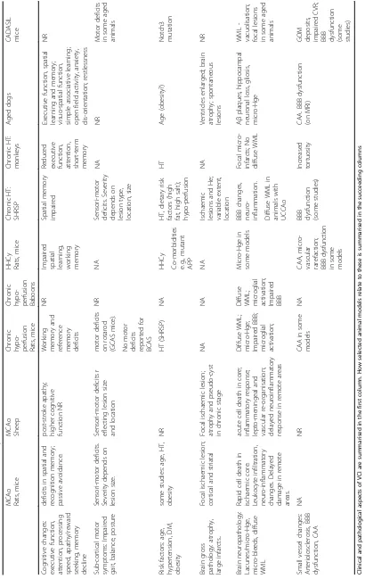
Table
1
Features
of
VCI,
as
related
to
experimental
models
considered
MCAo Rats, mice MCAo Sheep Chron ic hypo -perfusi on Rats, mice Chron ic hypo -perfusi on Babo ons HHCy Rats, mice Chron ic HT: SHRS P Chron ic HT: monk eys Aged dogs CADA SIL mi ce Cogni tive chang es: execut ive function, atte ntion, proces sing spee d, apathy/reward seeking , me mory decline deficits in spati al and recogni tion me mory; passive avoid ance. post-stroke apath y; higher cogn itive function NR Working memor y an d referenc e memor y defici ts NRImpaired spatial learning, working memor
y Spatial memor y impaired Redu ced exec utive function , atte ntion, short -term memor y Execut ive function, spa tial learn ing and memor y; visuo-spatial fu nction, simpl e asso ciative learn ing; ope n field activ ity, anxiet y, dis-orie ntation; res tlessne ss NR Sub-co rtical motor symp toms: Impaired gait, bal ance, posture Sensori -moto r de ficits. Seve rity depe nds on lesion size. Sensori -motor defi cits r eflecti ng le sion size and loc ation moto r de ficits on rotar od (GCA S mice) . NR NA Sensori -moto r deficits . Seve rity depe nds on lesion type, locatio n, size NA NR Mot or de ficits in som e aged anim als No motor defici ts repo rted for BCAS Risk facto rs: age, hypert ension, DM, obe sity som e studies: age, HT, obesity NR HT (SHR SP) NA HHCy HT, dietary ris k factors (high fat, high salt); hypo -perfusion HT Age (obes ity?) Not ch3 mu tation Co-m orbiditie s e.g., mu tant APP Brain gross patho logy: atrop hy, large infarcts .. Focal isch aemic lesion; cortical and striat al Focal ischae mic lesion; atrophy and pseud o-cyst in chron ic st age NA NA NA Ischae mic lesions an d He; variable exte nt, locatio n NA Ven tricles enlarge d; brain atrop hy; spon taneous lesions NR Brain neu ropath ology: Lacune s/micro-Hge , micro-blee ds, diff use WML Rapid ce ll death in ischae mic core. Leuk ocyte inf iltration, neuro -inflammatory chang es. Del ayed damage in remo te areas. acute cell death in core ; inflammatory respon se; lepto -mening eal and vascular re-organisat ion; delayed neu roinflam matory response in remot e area s Diff use WM L; micro-Hge ; Impaired BBB; microg lial activ ation; Diff use WML; microg lial activ ation; Impaired BBB Micro-Hge in some model s BBB changes , neuro -inflam mation. Focal micro-infarc ts; No diffus e WML A β pla ques, hipp ocampal neu ronal loss , glio sis, micro-Hge WML -vac uolisation; focal lesions in som e aged anim als Diffuse WML in anim als with UCCA o Small ves sel chang es: Arteri olosclero sis, BBB dysfunc tion, CAA NA NR CAA in som e mod els NA CAA,
micro-vascular rarefaction; BBB
[image:2.595.64.474.87.736.2]one that impacts on clinical practice [13]. Therefore, in
order to be translational an animal model should
repro-duce at least one of the pathological processes in human
VCI [6, 12, 14]. A fully translational model would permit
(1) prospective studies of the timescale and the sequence
of events during development of the pathological process,
(2) identification of novel molecular, cellular and
physio-logical mechanisms, (3) pre-clinical testing of drugs and
other interventions, for proof-of-concept studies, (4)
pre-clinical testing of safety profile of drugs, optimal dosing
and time-scale, and (5) validation of clinical biomarkers
and endpoints such as radiological or biochemical
signa-tures. Models representing the initiating factors would
allow translation of preventive strategies, whereas models
of advanced disease states allow testing of therapeutic
interventions. It is appropriate and timely to seek
inter-national accord on such models [15]. Following the recent
NIH-sponsored Alzheimer
’
s Disease-Related Dementias
2016 Summit
(https://aspe.hhs.gov/alzheimers-disease-re-lated-dementias-adrd-summit-2016-prioritized-research-mi
lestones), the number one recommendation for VCI was to
“
Establish new animal models that: (i) reproduce small
ves-sel disease and other key pathogenic processes thought to
re-sult in cognitive impairment; (ii) are easily applicable to
both VCID and AD research for advances in mixed etiology
dementias; (iii) address vascular contributions to dementia
via both white matter and grey matter or (iv) include genetic
and acquired conditions that are associated with VCID
”
.
Here, we review published models relevant to VCI,
in-cluding rodents and emphasising larger species. This
re-view is the result of discussions between experts from 12
laboratories across seven countries. Relevant systematic
reviews are available [10, 12].
Overview of experimental species
Rodents
We have included models of focal ischaemia (middle
cerebral artery occlusion; MCAo) [16
–
19] as this is a
validated, translational model of cerebrovascular injury.
Global hypoperfusion models include bilateral carotid
artery occlusion (BCAo) in rats [20] and bilateral carotid
artery stenosis (BCAS) using wire coils in mice [21, 22].
A refinement of the BCAo protocol employs constrictor
cuffs to give a gradual arterial occlusion over
approxi-mately 1
–
2 days [20]. These global models produce
is-chaemic white matter lesions, likely reflecting the low
baseline perfusion of white matter. Other pathologies
can also occur, including hippocampal cell death, small
haemorrhages and vascular amyloid deposition. Genetic
alterations include inbred strains (e.g., SHR, stroke-prone
spontaneously hypertensive rats (SHRSP)) [23
–
26] or
transgenic manipulations (e.g.,
Notch3
mutant strains)
[27
–
29]. VCI-relevant animals can also result from
ma-nipulation of risk factors, such as age, hypertension,
diabetes mellitus, hyperhomocysteinemia or a high-salt/
high-fat (
‘
fast food
’
) diet [14, 25, 26, 30, 31].
Larger species
Larger animals have a longer natural life span than
ro-dents. Experimental ruminants (sheep, goats) are
predom-inantly used to simulate acute cerebrovascular pathologies
such as ischaemic stroke [32
–
34] and cerebral
haemor-rhage [35]. In domestic dogs, hypercaloric or unbalanced
diet, lack of physical exercise and dyslipidemia are
preva-lent [36]. As in humans, hypertension [37] and cerebral
arteriosclerosis [38] are often observed in older subjects.
Consequently, a canine cognitive dysfunction syndrome,
featuring some clinical aspects of VCI, has been described,
particularly in breeds living long enough (>9 years) to fully
develop a neurological phenotype [39
–
42]. In cats, less is
known about the relation between aging, vascular
patholo-gies and cognitive decline. A
β
and tau pathologies have
been described in cats showing clinical signs of cognitive
decline [43
–
45]. Hypertension associated with
arterio-sclerosis, as well as small, multifocal cerebral
haemor-rhages, have also been reported for felines [46].
Behavioural paradigms for cognitive assessment in
lar-ger species have been reported from specialist centres for
sheep, pigs and cattle [41, 47
–
51]. The most advanced
cognitive abilities are seen in primates, for which
so-phisticated cognitive tools have been developed [52, 53].
Hypercaloric diet can decelerate aging and prevent
microvascular pathologies and cognitive decline in
primates [54, 55], without changing the lifespan [56].
Nevertheless, physiological aging can take decades in
primates, and studies relevant to VCI may be
re-stricted to specialised colonies [57, 58].
Large animal models allow clinical neuroimaging
with-out significant limitations in resolution, acquisition time
or data analysis. MRI protocols are now available for
dogs [59], cats [60], non-human primates [61
–
63], pigs
[64, 65] and sheep [66]. MRI (T1, T2, FLAIR) is
advanta-geous for analysis of tissue volume and lesions [66], as
well as for anatomical evaluation of particular brain
areas [67]. Perfusion and diffusion-weighted sequences
reveal cerebral blood flow (CBF) dynamics and vascular
permeability [68]. Templates, automatic segmentation
and labelling routines for larger species are essential for
studies aiming at quantitative morphometric analysis of
MRI and/or PET images. Automatic labelling and
process-ing routines have been developed for rhesus and
cynomol-gus monkeys [61, 69, 70], sheep [67], pigs [71, 72], and
dogs [73]; this enables efficient, observer-independent
analysis of grey and white matter regions.
Review methods
the review. All synopses were circulated for editing by
all authors, and subsequently by the scientific committee
of an international conference (International Conference
on Vascular Dementia, ICVD2015, Ljubjiana, Slovenia).
Only peer-reviewed sources in English were included.
Ethical statements on animal data
Sheep experiments from which data were derived were
approved by the responsible authorities for University of
Lübeck and University of Leipzig, Germany (animal
protocol numbers TVV33/09, TVV09/11, TVV33/12).
Experiments using monkeys were approved by the
Insti-tutional Animal Care and Use Committee of Boston
University Medical Center. All procedures with dogs were
conducted in accordance with University of Kentucky
ap-proved animal protocols (2009-0483) and the NIH Policy
on Humane Care and Use of Laboratory Animals.
Expert reviews of specific models
Large Vessel Ischaemia
–
Middle Cerebral Artery Occlusion
(MCAo) in Rodents
MCAo induces acute focal ischaemia bordered by a
par-tially ischaemic penumbra [74, 75]. While recovery of
sensorimotor function is well-characterised using
behav-ioural tests, there is less literature on cognitive
impair-ment [76]. Spatial learning, assessed by Y- and T-maze
tests, is hippocampus-dependent, but as other regions
are also required, including prefrontal cortex and basal
forebrain, these tests are relevant to the MCAo model
[77]. Following MCAo, male rats showed decreased rates
of spontaneous alternation compared with sham-operated
animals at day 21 post-stroke [78]. At 4 days post-MCAo,
male mice spend less time exploring a novel object than
sham animals [79]. Fear-motivated tasks such as passive
avoidance have also been used to assess cognitive
impair-ment after stroke [80]. While passive avoidance is a simple
task, it is stressful so could confound results of other
be-havioural tests [76].
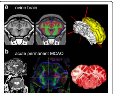
Larger species: sheep with vascular ischaemic lesions
Permanent [32] and transient [34] MCAo have been
per-formed in sheep, resulting in well controlled and
reprodu-cible lesion sizes (Fig. 1). Histopathological investigations
revealed both grey and white matter changes, including
glial scar formation, microglial activation and replacement
of the tissue by new formation of blood vessels and foamy
fat cells [33]. Moreover, ovine models have been
success-fully employed to test experimental therapeutic paradigms
in short- [81] and longer-term (up to 7 weeks) approaches
[33], during which benefits of single- and multi-mode
im-aging protocols became evident.
A caveat in this species (and other domestic mammals)
is the
rete mirabile epidurale rostrale
, a local arborisation
within the carotid artery [82]. This often necessitates a
transcranial approach for MCAo. Leaving the trepanation
covered only by soft tissue reduces intracranial pressure,
which greatly increases long-term survival. In mild and
se-vere global cerebral ischaemia models in sheep, it became
evident that the basilar artery can contribute a higher
pro-portion of CBF than in humans [83]. After prior bilateral
clamping of both common carotid arteries for 4
–
30 min,
no lesions were found in brains of sheep subjected to the
method for less than 10 min. Longer duration produced
neuronal changes of several brain regions, similar to those
described in other species.
Primates and rodents: chronic brain hypoperfusion
With the assumption that reducing CBF is a common
feature of VCI [3, 84, 85], the original mouse BCAS
model was developed by placing microcoils on the
ca-rotid arteries to induce cerebral hypoperfusion [86].
While complete ligation of the carotid arteries (i.e.,
BCAo) substantially increased mortality, mice can
with-stand up to 50% BCAS [22, 87]. Monitoring cognitive
function using the Y, radial arm, Barnes maze and
Morris water maze has provided robust evidence that
the BCAS model replicates some features of VCI, in
Fig. 1Focal ischaemic lesions in ovine brain.aAdult sheep brain in coronal section. T1-weighted population-averaged brain template (left), depiction of grey and white matter, as well as cerebrospinal fluid (middle panel, overlay on template) and surface reconstruction of white (white) and grey matter (yellow) in stereotactic space (right).Grey
[image:4.595.306.539.86.280.2]particular the deficit of working memory [10, 86, 87]. In
BCAS, global CBF drops rather abruptly. With the same
principle as BCAS, ameroid micro-constrictors made of
casein (which swells on absorbing water) were placed
around the carotid arteries to provide a more gradual
stenosis [20]. Ameroid constrictors have also been
ap-plied to spontaneously hypertensive rats [20]. Further
re-finements have allowed the development of mice models
that exhibit subcortical infarcts and white matter
dam-age by surgical implantation of an ameroid constrictor
to the right common carotid artery and placement of a
microcoil to the left common carotid artery to induce
approximately 50% arterial stenosis; this is referred to as
gradual carotid artery stenosis [88]. There was gradual
reduction of CBF over 28 days, and multiple infarct
damage in right subcortical regions, including the
cor-pus callosum, internal capsule, hippocampal fimbria, and
caudoputamen in 81% of mice [88, 89]. These
hypoper-fusion models are discussed further elsewhere [12].
A baboon (
Papio anubis
) model evaluated whether
partial cerebral ischaemia or oligaemia resulting from
re-duced blood flow to the brain induces white matter
pathology consistent with SVD or AD-like changes. The
baboon model is ideal to relate to AD because it exhibits
both a
β
and tau pathology with ageing and carries
APOE4
associated with AD pathology. Adult, male baboons were
subjected to three-vessel occlusion by complete ligation of
the internal carotid arteries bilaterally, and occlusion of
the left vertebral artery. We have recently reported
subcortical and white matter changes in animals to
28 days after three-vessel occlusion [90]. This model
is useful to evaluate interventions at various stages
and specifically examine the effects of ageing, high-fat
diet, hypertension and neuroinflammation. Ameroid
constrictors to replicate a gradual reduction in CBF
may be a future refinement [84, 85].
SHRSP with modified diet or hypoperfusion
Hypertensive rat strains can undergo white matter
changes [23
–
26, 91]. SHRSP typically live for 9
–
12
months before developing ischaemic and haemorrhagic
stroke lesions [12, 92]. When a low-protein, high-salt diet
is given to the SHRSP, lesions and death are accelerated
[93]. Starting the diet after 6 weeks of life leads to
haemor-rhagic strokes, but delaying the onset of the diet until the
12th month slows the onset of strokes and allows the
damage to the white matter to occur earlier [25]. The
white matter damage results from hypoxic hypoperfusion
[94]. In a recent study, minocycline, a tetracycline
deriva-tive with the ability to inhibit matrix metalloproteinases,
reduced white matter damage and reversed the
behav-ioural changes in SHRSP [26]. For a more extensive
dis-cussion of SHRSP, see [12, 92].
Dietary induction of hyperhomocysteinemia
Elevated circulating homocysteine
(hyperhomocysteine-mia) is caused by a variety of genetic, physiologic and
diet-ary conditions extensively studied in rodents [95
–
98].
These cause cognitive impairment in
ApoE
null mice,
transgenic mouse models of Alzheimer
’
s disease, and
wild-type mice and rats [31, 99, 100], with surprisingly little
neurodegeneration or inflammation. Feeding wildtype
C57BL6J mice a diet deficient in three B-vitamins (folate,
B12 and B6) for 10 weeks resulted in
hyperhomocysteine-mia, microvascular rarefaction and impaired performance
in the Morris water maze [31, 100]. The same dietary
regime in APP transgenic mice worsened cognitive
impairment [101], and in combination with excess
methionine in dual mutant APP/PS1 mice, the diet
induced the redistribution of amyloid from brain
par-enchyma to the microvasculature along with
micro-haemorrhages, as determined by histology and MRI
[30, 102]. In Sprague
–
Dawley rats, folate-deficiency alone
was sufficient to induce homocysteinemia and cognitive
impairment, and to reduce cerebral blood volume and
re-activity measured by absolute, non-invasive, near-infrared
spectroscopy [103
–
105]. For further discussion of
hyper-homocysteinemia models, see [12].
Dietary modification can be applied to most species,
models and co-morbidities. Caveats are that dietary
models typically have higher variability and more subtle
effects than genetic or pharmacological models.
Out-comes are sensitive to dietary formulation and feeding.
This underscores the need for biochemical and
meta-bolic verification of the diet in brain and the periphery.
While chronic folate and B12 deficiency in humans
causes macrocytic anaemia and myeloneuropathy, these
outcomes are almost never observed in rodent models.
Associations between microvascular rarefaction and
cog-nitive impairment, in the absence of neurodegenerative
changes have been observed in other models, including
mice fed a high-fat diet [106], aged rats [107], and
irradi-ated rats [108].
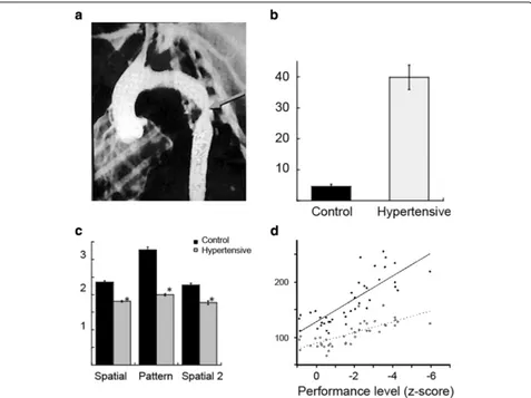
Primates with chronic hypertension
The basis of this model is the induction of hypertension
by surgical coarctation of thoracic aorta in the rhesus
monkey [52, 109
–
111]. A segment of the thoracic aorta
is mobilised and dissected without injuring the
medias-tinal and intercostal branches. The external diameter of
the same segment is measured and then narrowed to a
luminal diameter of 2.0
–
2.5 mm (Fig. 2). A pressure
transducer inserted into the femoral artery is advanced
through the surgical site. Typically, systolic/diastolic
pressure is 170/100 mmHg above the coarctation and
80/50 mmHg (normal for rhesus monkeys) below.
these domains were assessed in adult primates (5
–
11
years of age). The tasks consisted of an automated task of
simple attention, two tasks of memory function, the
de-layed non-matching to sample task (DNMS) [112, 113]
and the delayed recognition span task [114, 115], and a
primate analogue to the Wisconsin Card Sort task, the
Conceptual Set-Shifting Task (CSST) [116]. Performance
was compared with sham-operated controls that
under-went every stage of the surgical procedures up to, but not
including, narrowing of the aorta. Animals with
coarcta-tion were grouped into borderline (135
–
150 mmHg) or
hypertensive (> 150 mmHg).
On the task of simple attention in which monkeys are
required to select the same target stimulus on the
touch-screen, there was a positive correlation between response
time and systolic and mean blood pressure; hypertensive
(but not borderline) animals were significantly impaired
relative to the sham-operated group. Hypertensive
mon-keys were impaired on a task that required orienting to,
and then responding by touching, a randomly-presented
visual stimulus. Unlike normotensive animals,
hyperten-sive monkeys did not benefit from the presentation of a
cue that preceded the target stimulus. The effect did not
appear to be related to motivational state as there was no
difference in the number of missed trials. These findings
suggest a reduction in the speed of processing in the
stimulus
–
response chain.
The findings on memory assessment revealed a
signifi-cant difference among the groups on the DNMS up to
12 months post-surgery. Hypertensive monkeys re-learned
the DNMS task less efficiently than sham-operated
con-trols (Fig. 2). On both the spatial and pattern conditions
of the delayed recognition span task, the performance of
the hypertensive monkeys was significantly impaired with
Fig. 2VCI in adult monkeys with surgically-induced chronic hypertension.aArteriogram showing surgical coarctation of the thoracic aorta (arrow) in the monkey.bDelayed non-matching to sample (DNMS) scores for re-acquisition of the basic task. Y-axis: errors to criterion for control (sham-operated,black bar) and hypertensive monkeys (grey bar).cDelayed recognition span (DRS) test scores. Y-axis: group mean span, for control (black bars) and hypertensive monkeys (grey bars).dBlood pressure correlates with overall cognitive function. Y-axis: blood pressure (mmHg). X-axis: cognitive function index. The level of impairment on this index was significantly and linearly related to both systolic (black symbols,solid line;
[image:6.595.60.537.87.444.2]respect to the control monkeys, suggesting that, in
addition to affecting attentional function, hypertension
produced an impairment in
‘
rule learning
’
.
The CSST requires the monkey to establish a cognitive
set based on a reward contingency, to maintain that set
for a period of time, and then shift the set as the reward
contingency changes. A subset of hypertensive monkeys
was unimpaired on the initial phase of the CSST (a simple
three choice discrimination). In contrast, hypertensive
monkeys were impaired at abstracting the initial concept
of colour on the CSST and were subsequently impaired
when shifted to the concept of shape, when shifted back
to the concept of colour, and again when shifted back to
the concept of shape. The findings from this task suggest
that the two groups of monkeys were able to learn a
stimulus reinforcement contingency at the same rate and
that the impairment seen on the CSST is most likely one
of abstraction and cognitive flexibility.
Overall, hypertension significantly influenced higher
cog-nitive function. Blood pressure correlated with a composite
z-score (similar to an IQ score), suggesting a direct
rela-tionship between blood pressure and cognition (Fig. 2).
Various neuropathologies are seen in this primate
model, including tortuous small vessels,
hemosiderin-filled macrophages and, most conspicuously,
micro-infarcts in both grey and white matter [110, 111]. The
micro-infarcts are of irregular shape and relatively
uni-form size (average maximum diameter ~ 0.5 mm). In the
grey matter, these lesions were characterised by a total
loss of neurons, and in white matter by marked loss of
myelinated fibres.
Larger species: aged canine model
Aging dogs spontaneously develop cerebrovascular
path-ology linked to cognitive decline [41, 42], including
cortical atrophy and ventricular enlargement (Fig. 3).
Cognitive impairment was evident on measures
reflect-ing learnreflect-ing and memory, and a subset of aged animals
became severely impaired [41, 42]. A strength of the
model is that A
β
, critically involved with plaque
accu-mulation and cerebral amyloid angiopathy (CAA), is very
similar in dogs and humans [117
–
119]. Vascular and
peri-vascular abnormalities and cerebroperi-vascular A
β
pathology
are frequently found in aged dogs [40, 120
–
124]. Dogs
may be a suitable model system in which to examine the
consequences of CAA on cognition [125]. As in humans,
canine CAA is associated with cerebral haemorrhage
[40, 121], the occipital cortex being particularly
vulner-able [126]. Several environmental manipulations and
pharmacological studies that modify lifestyle factors have
been successfully implemented in canine models, with
some showing significant benefits to cognition [41].
Canines have also been used as a model for ischaemic
stroke. Both FLAIR and T2* (sensitive to hemosiderin)
imaging show significant white matter hyperintensities
[127]. Loss of white matter integrity may be a
conse-quence of CAA; for example, dogs aged from 1 to 20 years
exhibited a progressive loss of myelin basic protein,
corre-lated with age and with increasing CAA [128].
The canine brain displays substantial age-associated
morphological changes [129
–
131]. Gadolinium-enhanced
MRI revealed reduced blood
–
brain barrier function with
age, as well as reduced cerebrovascular volume [129].
Characterising cognitive function in aging dogs requires
many months, and treatment studies may take several
years. In comparison to rodent models, they require
significant veterinary care as they become older.
Radio-logical outcome measures that reflect in vivo CAA (e.g.,
SWI scans) have not yet been published.
Mouse models for monogenic small vessel disease
(CADASIL)
CADASIL (Cerebral Autosomal Dominant Arteriopathy
with Subcortical Infarcts and Leukoencephalopathy) is a
monogenic archetype for SVD, caused by cysteine-altering
missense mutations in
NOTCH3
. CADASIL patients
develop progressive white matter lesions from early
adult-hood, followed by cognitive decline and recurrent
subcor-tical infarctions [132]. Conventional transgenic murine
models expressing mutant human
NOTCH3
from a cDNA
construct [133
–
135] recapitulate some aspects of the
CADASIL vascular phenotype (vascular Notch3
accumu-lation and granular osmiophilic material on electron
microscopy) [12, 92]. In only one transgenic model, with
4-fold overexpression of mutant Notch3, the mice
devel-oped disturbed cerebrovascular reactivity (from 5 months
of age), reduced CBF (from 12 months) and white matter
[image:7.595.307.539.89.232.2]damage (from 18 months) [27]. A novel transgenic mouse
strain containing genomic human
NOTCH3
has recently
been developed [136]; these animals show early-onset
vas-cular Notch3 accumulation (from 6 weeks). A knock-in
model, made by introducing a mutation in endogenous
Notch3
, developed a CADASIL clinical phenotype (at
20 months) [137]. Stroke lesions, microbleeds and motor
deficits were seen only in a minority of mutant mice
(5
–
12%). Despite the fact that cognition has not yet been
characterised in these murine models, they offer a valid
pathogenetic representation of human CADASIL and may
be an important pre-clinical model in which to test VCI
therapies for efficacy.
Discussion and conclusions
As noted previously [9
–
11, 14], no experimental model
replicates all pathologic and cognitive aspects of human
VCI (Table 1). Animal models are useful to reflect a
pathological process (e.g., white matter hypoxia, arterial
fibrosis, amyloid accumulation) rather than a human
dis-ease. Old dogs with canine cognitive dysfunction
syn-drome and aged primates (> 20 years of age) being
possible exceptions, none of the models discussed here
results in a
‘
demented
’
animal. That said, all the animal
models considered above reproduce at least one of the
pathological processes in human VCI. Because the
se-quence of events leading from experimental challenge to
brain pathology, and thus to VCI, can be characterised in
animal models (and interventions imposed), the models
may help to identify pathways that lead to VCI. As the
pathogenesis of SVD, the most common cause of VCI,
remains unknown, a valid model of SVD-dependent VCI
remains a challenge. Making these conceptual and
bio-logical limitations explicit will expedite the development
and appropriate use of translational models for VCI.
There are several general limitations in the extant
literature. Most animal studies involve short-term
follow-up (typically, less than 4 weeks). Male animals are
gener-ally used and females usugener-ally avoided due to influences of
the reproductive cycle. Few studies have correlated
cogni-tive changes with anatomical changes, as seen by
path-ology or MRI. Most of the available cognitive paradigms
are derived from AD models. Many experimental studies
are under-powered (i.e., use a small number of animals)
and few are replicated.
We have a number of recommendations for the VCI
research community. First, it would be advantageous to
increase our knowledge and experience in larger species
with more abundant white matter and gyrencephalic brain
anatomy. This is especially important given the central
role of white matter lesions in human VCI. Second, robust
neuropsychological methods for assessing VCI in
experi-mental animals (particularly larger species) would be
beneficial. Cognitive impairment (and recovery) are the
most complex aspects of human VCI, and will likely differ
between animals and humans (for example, experimental
species lack spoken language). Thus, aspiring to a precise
behavioural replication in an animal may not be possible.
Nevertheless, a core toolkit of validated, reproducible,
species-appropriate tests of a cognitive phenotype is
required. With respect to SVD, simple behavioural
indica-tors analogous to the key cognitive features of the
syndrome in humans (impaired processing speed, apathy
and executive dysfunction) should be welcome. Third,
progress on translational VCI models will be more rapid if
high standards of
‘
Methodological quality
’
[15] outlined in
ARRIVE guidelines [138] and in previous translational
consensus documents [139, 140] are followed. Specifically,
random allocation of animals to experimental groups and
blinded assessment of outcomes was quite rare in earlier
studies (prior to 2010) [10]. Future experimental studies
should adhere to available guidelines on experimental
design, regarding a priori statistical power calculation,
randomisation, blinding of observers, and confirmation by
at least two independent laboratories [15, 138
–
140]. It
appears likely that negative outcomes of animal studies
are rarely published. Fourth, as neuroimaging (particularly
MRI) has a central role in human VCI, future pre-clinical
studies will be enhanced by brain imaging data.
Radio-logical features (diffuse white matter lesions, lacunar
in-farcts) are the main clinical biomarkers of SVD. Hence,
correlative studies relating MRI to brain pathology in
animals will continue to be informative.
Experiments using gyrencephalic species may be costly
and long in duration to afford sufficient statistical power.
A possible solution is a step-wise approach that employs
rodents to study fundamental aspects of cerebrovascular
disease common to all species, and large animals to
study aspects of VCI that require a large gyrencephalic
brain. Extending studies across species will clarify
molecular, cellular and physiological events that lead
from vascular disease to neuronal injury and cognitive
dysfunction in humans, and improve the likelihood of
achieving new preventive and therapeutic
interven-tions in VCI.
Abbreviations
BCAo:Bilateral carotid artery occlusion; BCAS: Bilateral carotid artery stenosis; CAA: Cerebral amyloid angiopathy; CBF: Cerebral blood flow;
CSST: Conceptual set-shifting task; DNMS: Delayed non-matching to sample task; MCAo: Middle cerebral artery occlusion; SHRSP: Stroke-prone spontaneously hypertensive rats; SVD: Small vessel disease; VCI: Vascular cognitive impairment
Acknowledgements
We are grateful to Professor Amos D Korczyn for his contributions to the VCI field and for his helpful comments on this review.
Funding
from the British Heart Foundation and EPSRC (UK). CC is funded by the MRC (UK) Centre for Doctoral Training in Regenerative Medicine (grant no. EP/L014904/1). AMT was supported in this work by Israel Science Foundation (ISF) Grant 1353/11.
Availability of data and material
Data sharing not applicable to this article as no datasets were generated or analysed during the current study.
Authors’contributions
AHH participated in study conception and design, and in drafting of the manuscript and its critical revision for important intellectual content. SMA, CC, CF, EH, MI, RNK, SAJLO, MBM, GAR, and JWR participated in acquisition of data and critical revision of the manuscript for important intellectual content. JB, BN and AMT participated in conception and design, acquisition of data, and critical revision of the manuscript for important intellectual content. JDI and MSP participated in conception and design and critical revision of the manuscript for important intellectual content. All authors read and approved the final manuscript.
Competing interests
The authors declare that they have no competing interests.
Consent for publication Not applicable.
Ethics approval and consent to participate
Human data or human tissue: Not applicable. Animal experiments: see Methods section.
Author details
1Clinical Neurosciences (J-0B) Molecular and Clinical Sciences Research Institute,
St George’s University of London, Cranmer Terrace, London SW17 0RE, UK.
2Department of Neurology, St George’s University Hospitals NHS Foundation
Trust, London, UK.3Faculty of Biology, Medicine and Health, University of
Manchester, Manchester M13 9PT, UK.4Department of Translational Medicine
and Cell Technology, University of Lübeck, Lübeck, Germany.5Neurovascular Research Laboratory, Massachusetts General Hospital and Harvard Medical School, Charlestown, MA, USA.6Department of Anatomy & Neurobiology,
Boston University School of Medicine, Boston, MA, USA.7Department of
Neurology, Boston University School of Medicine, Boston, MA, USA.
8Department of Pharmacology & Nutritional Sciences, Sanders-Brown Center on
Aging, University of Kentucky, Lexington, KY, USA.9Department of Stroke and
Cerebrovascular Diseases, National Cerebral and Cardiovascular Center, Osaka, Japan.10Institute of Neuroscience, University of Newcastle-upon-Tyne, Newcastle-upon-Tyne, UK.11Department of Clinical Genetics, Leiden University
Medical Center, Leiden, Netherlands.12Fraunhofer Institute for Cell Therapy and
Immunology, Leipzig, Germany.13Clinic for Nuclear Medicine, University of
Leipzig, Leipzig, Germany.14Institute for Anatomy, Faculty of Veterinary Medicine, University of Leipzig, Leipzig, Germany.15Department of Neurology,
Health Sciences Center, University of New Mexico, Albuquerque, NM, USA.
16Department of Pharmacology, Croatian Institute for Brain Research, University
of Zagreb School of Medicine, Zagreb, Croatia.17Institute of Biochemistry Food and Nutrition Science, Hebrew University of Jerusalem, Rehovot, Israel.
18Department of Human Genetics, Leiden University Medical Center, Leiden,
Netherlands.
Received: 30 May 2016 Accepted: 12 January 2017
References
1. O’Brien JT, Thomas A. Vascular dementia. Lancet. 2015;386:1698–706. 2. Moorhouse P, Rockwood K. Vascular cognitive impairment: current concepts
and clinical developments. Lancet Neurol. 2008;7:246–55.
3. Hachinski V, Iadecola C, Petersen RC, Breteler MM, Nyenhuis DL, Black SE, Powers WJ, DeCarli C, Merino JG, Kalaria RN, Vinters HV, Holtzman DM, Rosenberg GA, Dichgans M, Marler JR, Leblanc GG. National Institute of Neurological Disorders and Stroke-Canadian Stroke Network vascular cognitive impairment harmonization standards. Stroke. 2006;37:2220–41. 4. Prins ND, Scheltens P. White matter hyperintensities, cognitive impairment
and dementia: an update. Nat Rev Neurol. 2015;11:157–65.
5. Pantoni L. Cerebral small vessel disease: from pathogenesis and clinical characteristics to therapeutic challenges. Lancet Neurol. 2010;9:689–701.
6. Iadecola C. The pathobiology of vascular dementia. Neuron. 2013;80:844–66. 7. Toledo JB, Arnold SE, Raible K, Brettschneider J, Xie SX, Grossman M,
Monsell SE, Kukull WA, Trojanowski JQ. Contribution of cerebrovascular disease in autopsy confirmed neurodegenerative disease cases in the National Alzheimer’s Coordinating Centre. Brain. 2013;136:2697–706. 8. Thiel A, Cechetto DF, Heiss WD, Hachinski V, Whitehead SN. Amyloid
burden, neuroinflammation, and links to cognitive decline after ischemic stroke. Stroke. 2014;45:2825–9.
9. Hainsworth AH, Markus HS. Do in vivo experimental models reflect human cerebral small vessel disease? A systematic review. J Cereb Blood Flow Metab. 2008;28:1877–91.
10. Jiwa NS, Garrard P, Hainsworth AH. Experimental models of vascular dementia and vascular cognitive impairment. A systematic review. J Neurochem. 2010;115:814–28.
11. Bailey EL, Smith C, Sudlow CL, Wardlaw JM. Is the spontaneously hypertensive stroke prone rat a pertinent model of sub cortical ischemic stroke? A systematic review. Int J Stroke. 2011;6:434–44.
12. Madigan JB, Wilcock DM, Hainsworth AH. Vascular contributions to cognitive impairment and dementia: topical review of animal models. Stroke. 2016;47:1953–9.
13. Woolf SH. The meaning of translational research and why it matters. JAMA. 2008;299:211–3.
14. Hainsworth AH, Markus HS. Experimental animal models of cerebral small vessel disease. In: Pantoni L, Gorelick PB, editors. Cerebral Small Vessel Disease. Cambridge: Cambridge University Press; 2014. p. 42–51. 15. Landis SC, Amara SG, Asadullah K, Austin CP, Blumenstein R, Bradley EW,
Crystal RG, Darnell RB, Ferrante RJ, Fillit H, Finkelstein R, Fisher M, Gendelman HE, Golub RM, Goudreau JL, Gross RA, Gubitz AK, Hesterlee SE, Howells DW, Huguenard J, Kelner K, Koroshetz W, Krainc D, Lazic SE, Levine MS, Macleod MR, McCall JM, Moxley III RT, Narasimhan K, Noble LJ, Perrin S, Porter JD, Steward O, Unger E, Utz U, Silberberg SD. A call for transparent reporting to optimize the predictive value of preclinical research. Nature. 2012;490:187–91.
16. Burrows FE, Bray N, Denes A, Allan SM, Schiessl I. Delayed reperfusion deficits after experimental stroke account for increased pathophysiology. J Cereb Blood Flow Metab. 2015;35:277–84.
17. Gibson CL, Coomber B, Murphy SP. Progesterone is neuroprotective following cerebral ischaemia in reproductively ageing female mice. Brain. 2011;134:2125–33. 18. McKittrick CM, Lawrence CE, Carswell HV. Mast cells promote blood brain
barrier breakdown and neutrophil infiltration in a mouse model of focal cerebral ischemia. J Cereb Blood Flow Metab. 2015;35:638–47.
19. Baskerville TA, McCabe C, Weir CJ, Macrae IM, Holmes WM. Noninvasive MRI measurement of CBF: evaluating an arterial spin labelling sequence with 99mTc-HMPAO CBF autoradiography in a rat stroke model. J Cereb Blood Flow Metab. 2012;32:973–7.
20. Kitamura A, Fujita Y, Oishi N, Kalaria RN, Washida K, Maki T, Okamoto Y, Hase Y, Yamada M, Takahashi J, Ito H, Tomimoto H, Fukuyama H, Takahashi R, Ihara M. Selective white matter abnormalities in a novel rat model of vascular dementia. Neurobiol Aging. 2012;33:1012–35.
21. Holland PR, Searcy JL, Salvadores N, Scullion G, Chen G, Lawson G, Scott F, Bastin ME, Ihara M, Kalaria R, Wood ER, Smith C, Wardlaw JM, Horsburgh K. Gliovascular disruption and cognitive deficits in a mouse model with features of small vessel disease. J Cereb Blood Flow Metab. 2015;35:1005–14. 22. Okamoto Y, Yamamoto T, Kalaria RN, Senzaki H, Maki T, Hase Y, Kitamura A,
Washida K, Yamada M, Ito H, Tomimoto H, Takahashi R, Ihara M. Cerebral hypoperfusion accelerates cerebral amyloid angiopathy and promotes cortical microinfarcts. Acta Neuropathol. 2012;123:381–94.
23. Kaiser D, Weise G, Moller K, Scheibe J, Posel C, Baasch S, Gawlitza M, Lobsien D, Diederich K, Minnerup J, Kranz A, Boltze J, Wagner DC. Spontaneous white matter damage, cognitive decline and neuroinflammation in middle-aged hypertensive rats: an animal model of early-stage cerebral small vessel disease. Acta Neuropathol Commun. 2014;2:169.
24. Brittain JF, McCabe C, Khatun H, Kaushal N, Bridges LR, Holmes WM, Barrick TR, Graham D, Dominiczak AF, Mhairi Macrae I, Hainsworth AH. An MRI-histological study of white matter in stroke-free SHRSP. J Cereb Blood Flow Metab. 2013;33:760–3.
26. Jalal FY, Yang Y, Thompson JF, Roitbak T, Rosenberg GA. Hypoxia-induced neuroinflammatory white-matter injury reduced by minocycline in SHR/SP. J Cereb Blood Flow Metab. 2015;35:1145–53.
27. Joutel A, Monet-Lepretre M, Gosele C, Baron-Menguy C, Hammes A, Schmidt S, Lemaire-Carrette B, Domenga V, Schedl A, Lacombe P, Hubner N. Cerebrovascular dysfunction and microcirculation rarefaction precede white matter lesions in a mouse genetic model of cerebral ischemic small vessel disease. J Clin Invest. 2010;120:433–45.
28. Joutel A, Faraci FM. Cerebral small vessel disease: insights and opportunities from mouse models of collagen IV-related small vessel disease and cerebral autosomal dominant arteriopathy with subcortical infarcts and
leukoencephalopathy. Stroke. 2014;45:1215–21.
29. Dabertrand F, Kroigaard C, Bonev AD, Cognat E, Dalsgaard T, Domenga-Denier V, Hill-Eubanks DC, Brayden JE, Joutel A, Nelson MT. Potassium channelopathy-like defect underlies early-stage cerebrovascular dysfunction in a genetic model of small vessel disease. Proc Natl Acad Sci U S A. 2015;112:E796–805. 30. Sudduth TL, Powell DK, Smith CD, Greenstein A, Wilcock DM. Induction of
hyperhomocysteinemia models vascular dementia by induction of cerebral microhemorrhages and neuroinflammation. J Cereb Blood Flow Metab. 2013;33:708–15.
31. Troen AM, Shea-Budgell M, Shukitt-Hale B, Smith DE, Selhub J, Rosenberg IH. B-vitamin deficiency causes hyperhomocysteinemia and vascular cognitive impairment in mice. Proc Natl Acad Sci U S A. 2008;105:12474–9. 32. Boltze J, Forschler A, Nitzsche B, Waldmin D, Hoffmann A, Boltze CM, Dreyer AY,
Goldammer A, Reischauer A, Hartig W, Geiger KD, Barthel H, Emmrich F, Gille U. Permanent middle cerebral artery occlusion in sheep: a novel large animal model of focal cerebral ischemia. J Cereb Blood Flow Metab. 2008;28:1951–64. 33. Boltze J, Nitzsche B, Geiger KD, Schoon HA. Histopathological investigation
of different MCAO Modalities and impact of autologous bone marrow mononuclear cell administration in an ovine stroke model. Transl Stroke Res. 2011;2:279–93.
34. Wells AJ, Vink R, Blumbergs PC, Brophy BP, Helps SC, Knox SJ, Turner RJ. A surgical model of permanent and transient middle cerebral artery stroke in the sheep. PLoS One. 2012;7:e42157.
35. Dreyer A, Stroh A, Posel C, Findeisen M, von Geymuller T, Lobsien D, Nitzsche B, Boltze J. Frameless stereotaxy in sheep—neurosurgical and imaging techniques for translational stroke research. INTECH Open Access Publisher. 2012. doi:10.5772/32367.
36. Perez-Sanchez AP, Del-Angel-Caraza J, Quijano-Hernandez IA, Barbosa-Mireles MA. Obesity-hypertension and its relation to other diseases in dogs. Vet Res Commun. 2015;39:45–51.
37. Brown S, Atkins C, Bagley R, Carr A, Cowgill L, Davidson M, Egner B, Elliott J, Henik R, Labato M, Littman M, Polzin D, Ross L, Snyder P, Stepien R. Guidelines for the identification, evaluation, and management of systemic hypertension in dogs and cats. J Vet Intern Med. 2007;21:542–58. 38. Liu SK, Tilley LP, Tappe JP, Fox PR. Clinical and pathologic findings in dogs with
atherosclerosis: 21 cases (1970–1983). J Am Vet Med Assoc. 1986;189:227–32. 39. Bernedo V, Insua D, Suarez ML, Santamarina G, Sarasa M, Pesini P.
Beta-amyloid cortical deposits are accompanied by the loss of serotonergic neurons in the dog. J Comp Neurol. 2009;513:417–29.
40. Uchida K, Nakayama H, Goto N. Pathological studies on cerebral amyloid angiopathy, senile plaques and amyloid deposition in visceral organs in aged dogs. J Vet Med Sci. 1991;53:1037–42.
41. Cotman CW, Head E. The canine (dog) model of human aging and disease: dietary, environmental and immunotherapy approaches. J Alzheimers Dis. 2008;15:685–707.
42. Schutt T, Helboe L, Pedersen LO, Waldemar G, Berendt M, Pedersen JT. Dogs with cognitive dysfunction as a spontaneous model for early Alzheimer’s disease: a translational study of neuropathological and inflammatory markers. J Alzheimers Dis. 2016;52:433–49.
43. Head E, Moffat K, Das P, Sarsoza F, Poon WW, Landsberg G, Cotman CW, Murphy MP. Beta-amyloid deposition and tau phosphorylation in clinically characterized aged cats. Neurobiol Aging. 2005;26:749–63.
44. Nakamura S, Nakayama H, Kiatipattanasakul W, Uetsuka K, Uchida K, Goto N. Senile plaques in very aged cats. Acta Neuropathol. 1996;91:437–9. 45. Vite CH, Head E. Aging in the canine and feline brain. Vet Clin North Am
Small Anim Pract. 2014;44:1113–29.
46. Littman MP. Spontaneous systemic hypertension in 24 cats. J Vet Intern Med. 1994;8:79–86.
47. Gieling E, Wehkamp W, Willigenburg R, Nordquist RE, Ganderup NC, van der Staay FJ. Performance of conventional pigs and Gottingen miniature pigs in
a spatial holeboard task: effects of the putative muscarinic cognition impairer Biperiden. Behav Brain Funct. 2013;9:4.
48. Gieling ET, Schuurman T, Nordquist RE, van der Staay FJ. The pig as a model animal for studying cognition and neurobehavioral disorders. Curr Top Behav Neurosci. 2011;7:359–83.
49. Gieling ET, Nordquist RE, van der Staay FJ. Assessing learning and memory in pigs. Anim Cogn. 2011;14:151–73.
50. Grimberg-Henrici CG, Vermaak P, Elizabeth BJ, Nordquist RE, van der Staay FJ. Effects of environmental enrichment on cognitive performance of pigs in a spatial holeboard discrimination task. Anim Cogn. 2016;19(2):271–83. 51. Rioja-Lang FC, Roberts DJ, Healy SD, Lawrence AB, Haskell MJ. Dairy cow feeding space requirements assessed in a Y-maze choice test. J Dairy Sci. 2012;95:3954–60.
52. Moss MB, Jonak E. Cerebrovascular disease and dementia: a primate model of hypertension and cognition. Alzheimers Dement. 2007;3:S6–15. 53. Schmitt V, Pankau B, Fischer J. Old world monkeys compare to apes in the
primate cognition test battery. PLoS One. 2012;7:e32024.
54. Csiszar A, Sosnowska D, Tucsek Z, Gautam T, Toth P, Losonczy G, Colman RJ, Weindruch R, Anderson RM, Sonntag WE, Ungvari Z. Circulating factors induced by caloric restriction in the nonhuman primate Macaca mulatta activate angiogenic processes in endothelial cells. J Gerontol A Biol Sci Med Sci. 2013;68:235–49.
55. Dal-Pan A, Pifferi F, Marchal J, Picq JL, Aujard F. Cognitive performances are selectively enhanced during chronic caloric restriction or resveratrol supplementation in a primate. PLoS One. 2011;6:e16581.
56. Mattison JA, Roth GS, Beasley TM, Tilmont EM, Handy AM, Herbert RL, Longo DL, Allison DB, Young JE, Bryant M, Barnard D, Ward WF, Qi W, Ingram DK, de Cabo R. Impact of caloric restriction on health and survival in rhesus monkeys from the NIA study. Nature. 2012;489:318–21.
57. Kuehnel F, Grohmann J, Buchwald U, Koeller G, Teupser D, Einspanier A. Parameters of haematology, clinical chemistry and lipid metabolism in the common marmoset and alterations under stress conditions. J Med Primatol. 2012;41:241–50.
58. Mietsch M, Einspanier A. Non-invasive blood pressure measurement: values, problems and applicability in the common marmoset (Callithrix jacchus). Lab Anim. 2015;49:241–50.
59. Garosi LS, Dennis R, Penderis J, Lamb CR, Targett MP, Cappello R, Delauche AJ. Results of magnetic resonance imaging in dogs with vestibular disorders: 85 cases (1996–1999). J Am Vet Med Assoc. 2001;218:385–91. 60. Gray-Edwards HL, Salibi N, Josephson EM, Hudson JA, Cox NR, Randle AN,
McCurdy VJ, Bradbury AM, Wilson DU, Beyers RJ, Denney TS, Martin DR. High resolution MRI anatomy of the cat brain at 3 Tesla. J Neurosci Methods. 2014;227:10–7.
61. Frey S, Pandya DN, Chakravarty MM, Bailey L, Petrides M, Collins DL. An MRI based average macaque monkey stereotaxic atlas and space (MNI monkey space). Neuroimage. 2011;55:1435–42.
62. Herrmann T, Mallow J, Plaumann M, Luchtmann M, Stadler J, Mylius J, Brosch M, Bernarding J. The travelling-wave primate system: a new solution for magnetic resonance imaging of macaque monkeys at 7 Tesla ultra-high field. PLoS One. 2015;10:e0129371.
63. McLaren DG, Kosmatka KJ, Oakes TR, Kroenke CD, Kohama SG, Matochik JA, Ingram DK, Johnson SC. A population-average MRI-based atlas collection of the rhesus macaque. Neuroimage. 2009;45:52–9.
64. Allen BS, Ko Y, Buckberg GD, Sakhai S, Tan Z. Studies of isolated global brain ischaemia: I. A new large animal model of global brain ischaemia and its baseline perfusion studies. Eur J Cardiothorac Surg. 2012;41:1138–46. 65. Bjarkam CR, Cancian G, Glud AN, Ettrup KS, Jorgensen RL, Sorensen JC.
MRI-guided stereotaxic targeting in pigs based on a stereotaxic localizer box fitted with an isocentric frame and use of SurgiPlan computer-planning software. J Neurosci Methods. 2009;183:119–26.
66. Schmidt MJ, Langen N, Klumpp S, Nasirimanesh F, Shirvanchi P, Ondreka N, Kramer M. A study of the comparative anatomy of the brain of domestic ruminants using magnetic resonance imaging. Vet J. 2012;191:85–93. 67. Nitzsche B, Frey S, Collins LD, Seeger J, Lobsien D, Dreyer A, Kirsten H,
Stoffel MH, Fonov VS, Boltze J. A stereotaxic, population-averaged T1w ovine brain atlas including cerebral morphology and tissue volumes. Front Neuroanat. 2015;9:69.
68. Skinner JT, Moots PL, Ayers GD, Quarles CC. On the use of DSC-MRI for measuring vascular permeability. AJNR Am J Neuroradiol. 2016;37(1):80–7. 69. Chakravarty MM, Frey S, Collins DL. Digital atlas of the monkey brain in
editors. The Rhesus Monkey Brain In Stereotactic Coordinates. San Diego, CA: Academic; 2008.
70. Kohama SG, Rosene DL, Sherman LS. Age-related changes in human and non-human primate white matter: from myelination disturbances to cognitive decline. Age (Dordr). 2012;34:1093–110.
71. Conrad MS, Sutton BP, Dilger RN, Johnson RW. An in vivo three-dimensional magnetic resonance imaging-based averaged brain collection of the neonatal piglet (Sus scrofa). PLoS One. 2014;9:e107650.
72. Saikali S, Meurice P, Sauleau P, Eliat PA, Bellaud P, Randuineau G, Verin M, Malbert CH. A three-dimensional digital segmented and deformable brain atlas of the domestic pig. J Neurosci Methods. 2010;192:102–9. 73. Datta R, Lee J, Duda J, Avants BB, Vite CH, Tseng B, Gee JC, Aguirre GD,
Aguirre GK. A digital atlas of the dog brain. PLoS One. 2012;7:e52140. 74. Engel O, Kolodziej S, Dirnagl U, Prinz V. Modeling stroke in mice - middle
cerebral artery occlusion with the filament model. J Vis Exp. 2011;(47):2423. doi: 10.3791/2423.
75. Macrae IM. Preclinical stroke research—advantages and disadvantages of the most common rodent models of focal ischaemia. Br J Pharmacol. 2011;164:1062–78.
76. Freret T, Schumann-Bard P, Boulouard M, Bouet V. On the importance of long-term functional assessment after stroke to improve translation from bench to bedside. Exp Transl Stroke Med. 2011;3:6.
77. Lalonde R. The neurobiological basis of spontaneous alternation. Neurosci Biobehav Rev. 2002;26:91–104.
78. Ryan CL, Doucette TA, Gill DA, Langdon KD, Liu Y, Perry MA, Tasker RA. An improved post-operative care protocol allows detection of long-term functional deficits following MCAo surgery in rats. J Neurosci Methods. 2006;154:30–7. 79. Carmo MR, Simoes AP, Fonteles AA, Souza CM, Cunha RA, Andrade GM. ATP
P2Y1 receptors control cognitive deficits and neurotoxicity but not glial modifications induced by brain ischemia in mice. Eur J Neurosci. 2014;39:614–22. 80. Hattori K, Lee H, Hurn PD, Crain BJ, Traystman RJ, DeVries AC. Cognitive
deficits after focal cerebral ischemia in mice. Stroke. 2000;31:1939–44. 81. Terpolilli NA, Kim SW, Thal SC, Kataoka H, Zeisig V, Nitzsche B, Klaesner B,
Zhu C, Schwarzmaier S, Meissner L, Mamrak U, Engel DC, Drzezga A, Patel RP, Blomgren K, Barthel H, Boltze J, Kuebler WM, Plesnila N. Inhalation of nitric oxide prevents ischemic brain damage in experimental stroke by selective dilatation of collateral arterioles. Circ Res. 2012;110:727–38. 82. McGrath P. Observations on the intracranial carotid rete and the hypophysis
in the mature female pig and sheep. J Anat. 1977;124:689–99. 83. Terlecki S, Baldwin BA, Bell FR. Experimental cerebral ischaemia in sheep.
Neuropathology and clinical effects. Acta Neuropathol. 1967;7:185–200. 84. Zheng L, Vinters HV, Mack WJ, Zarow C, Ellis WG, Chui HC. Cerebral
atherosclerosis is associated with cystic infarcts and microinfarcts but not Alzheimer pathologic changes. Stroke. 2013;44:2835–41.
85. Kalaria RN, Perry RH, O’Brien J, Jaros E. Atheromatous disease in small intracerebral vessels, microinfarcts and dementia. Neuropathol Appl Neurobiol. 2012;38:505–8.
86. Shibata M, Ohtani R, Ihara M, Tomimoto H. White matter lesions and glial activation in a novel mouse model of chronic cerebral hypoperfusion. Stroke. 2004;35:2598–603.
87. Nishio K, Ihara M, Yamasaki N, Kalaria RN, Maki T, Fujita Y, Ito H, Oishi N, Fukuyama H, Miyakawa T, Takahashi R, Tomimoto H. A mouse model characterizing features of vascular dementia with hippocampal atrophy. Stroke. 2010;41:1278–84.
88. Hattori Y, Enmi J, Iguchi S, Saito S, Yamamoto Y, Tsuji M, Nagatsuka K, Kalaria RN, Iida H, Ihara M. Gradual carotid artery stenosis in mice closely replicates hypoperfusive vascular dementia in humans. J Am Heart Assoc. 2016;5(2):e002757. doi:10.1161/JAHA.115.002757.
89. Hattori Y, Enmi J, Kitamura A, Yamamoto Y, Saito S, Takahashi Y, Iguchi S, Tsuji M, Yamahara K, Nagatsuka K, Iida H, Ihara M. A novel mouse model of subcortical infarcts with dementia. J Neurosci. 2015;35:3915–28.
90. Chen A, Akinyemi RO, Hase Y, Firbank MJ, Ndung'u MN, Foster V, Craggs LJ, Washida K, Okamoto Y, Thomas AJ, Polvikoski TM, Allan LM, Oakley AE, O'Brien JT, Horsburgh K, Ihara M, Kalaria RN. Frontal white matter hyperintensities, clasmatodendrosis and gliovascular abnormalities in ageing and post-stroke dementia. Brain. 2016;139:242–58.
91. Henning EC, Warach S, Spatz M. Hypertension-induced vascular remodeling contributes to reduced cerebral perfusion and the development of spontaneous stroke in aged SHRSP rats. J Cereb Blood Flow Metab. 2010;30:827–36. 92. Hainsworth AH, Brittain JF, Khatun H. Pre-clinical models of human cerebral small
vessel disease: utility for clinical application. J Neurol Sci. 2012;322:237–40.
93. Sironi L, Guerrini U, Tremoli E, Miller I, Gelosa P, Lascialfari A, Zucca I, Eberini I, Gemeiner M, Paoletti R, Gianazza E. Analysis of pathological events at the onset of brain damage in stroke-prone rats: a proteomics and magnetic resonance imaging approach. J Neurosci Res. 2004;78:115–22. 94. Weaver J, Jalal FY, Yang Y, Thompson J, Rosenberg GA, Liu KJ. Tissue
oxygen is reduced in white matter of spontaneously hypertensive-stroke prone rats: a longitudinal study with electron paramagnetic resonance. J Cereb Blood Flow Metab. 2014;34:890–6.
95. Troen A, Rosenberg I. Homocysteine and cognitive function. Semin Vasc Med. 2005;5:209–14.
96. Troen AM. The central nervous system in animal models of hyperhomocysteinemia. Prog Neuropsychopharmacol Biol Psychiatry. 2005;29:1140–51.
97. Troen AM, Shukitt-Hale B, Chao WH, Albuquerque B, Smith DE, Selhub J, Rosenberg J. The cognitive impact of nutritional homocysteinemia in apolipoprotein-E deficient mice. J Alzheimers Dis. 2006;9:381–92. 98. Hainsworth AH, Yeo NE, Weekman EM, Wilcock DM. Homocysteine,
hyperhomocysteinemia and vascular contributions to cognitive impairment and dementia (VCID). Biochim Biophys Acta. 2016;1862:1008–17. 99. Bernardo A, McCord M, Troen AM, Allison JD, McDonald MP. Impaired
spatial memory in APP-overexpressing mice on a homocysteinemia-inducing diet. Neurobiol Aging. 2007;28:1195–205.
100. Troen AM, Chao WH, Crivello NA, D’Anci KE, Shukitt-Hale B, Smith DE, Selhub J, Rosenberg IH. Cognitive impairment in folate-deficient rats corresponds to depleted brain phosphatidylcholine and is prevented by dietary methionine without lowering plasma homocysteine. J Nutr. 2008;138:2502–9.
101. Fuso A, Nicolia V, Ricceri L, Cavallaro RA, Isopi E, Mangia F, Fiorenza MT, Scarpa S. S-adenosylmethionine reduces the progress of the Alzheimer-like features induced by B-vitamin deficiency in mice. Neurobiol Aging. 2012;33:1482–16. 102. Sudduth TL, Weekman EM, Brothers HM, Braun K, Wilcock DM.β-amyloid deposition is shifted to the vasculature and memory impairment is exacerbated when hyperhomocysteinemia is induced in APP/PS1 transgenic mice. Alzheimers Res Ther. 2014;6:32.
103. Ayata C, Dunn AK, Gursoy-Ozdemir Y, Huang Z, Boas DA, Moskowitz MA. Laser speckle flowmetry for the study of cerebrovascular physiology in normal and ischemic mouse cortex. J Cereb Blood Flow Metab. 2004;24:744–55. 104. Hallacoglu B, Sassaroli A, Fantini S, Troen AM. Cerebral perfusion and
oxygenation are impaired by folate deficiency in rat: absolute
measurements with noninvasive near-infrared spectroscopy. J Cereb Blood Flow Metab. 2011;31:1482–92.
105. Shaul ME, Hallacoglu B, Sassaroli A, Shukitt-Hale B, Fantini S, Rosenberg IH, Troen AM. Cerebral blood volume and vasodilation are independently diminished by aging and hypertension: a near infrared spectroscopy study. J Alzheimers Dis. 2014;42 Suppl 3:S189–98.
106. Tucsek Z, Toth P, Tarantini S, Sosnowska D, Gautam T, Warrington JP, Giles CB, Wren JD, Koller A, Ballabh P, Sonntag WE, Ungvari Z, Csiszar A. Aging exacerbates obesity-induced cerebromicrovascular rarefaction,
neurovascular uncoupling, and cognitive decline in mice. J Gerontol A Biol Sci Med Sci. 2014;69:1339–52.
107. Oomen CA, Farkas E, Roman V, van der Beek EM, Luiten PG, Meerlo P. Resveratrol preserves cerebrovascular density and cognitive function in aging mice. Front Aging Neurosci. 2009;1:4.
108. Brown WR, Blair RM, Moody DM, Thore CR, Ahmed S, Robbins ME, Wheeler KT. Capillary loss precedes the cognitive impairment induced by fractionated whole-brain irradiation: a potential rat model of vascular dementia. J Neurol Sci. 2007;257:67–71.
109. Hollander W, Prusty S, Kirkpatrick B, Paddock J, Nagraj S. Role of hypertension in ischemic heart disease and cerebral vascular disease in the cynomolgus monkey with coarctation of the aorta. Circ Res. 1977;40:I70–83. 110. Kemper T, Moss MB, Hollander W, Prusty S. Microinfarction as a result of
hypertension in a primate model of cerebrovascular disease. Acta Neuropathol. 1999;98:295–303.
111. Kemper TL, Blatt GJ, Killiany RJ, Moss MB. Neuropathology of progressive cognitive decline in chronically hypertensive rhesus monkeys. Acta Neuropathol. 2001;101:145–53.
112. Gaffan D. Recognition impaired and association intact in the memory of monkeys after transection of the fornix. J Comp Physiol Psychol. 1974;86: 1100–9.
114. Killiany R, Rehbein L, Mahut H. Developmental study of the hippocampal formation in rhesus monkeys (Macaca mulatta): II. Early ablations do not spare the capacity to retrieve conditional object-object associations. Behav Neurosci. 2005;119:651–61.
115. Rehbein L, Killiany R, Mahut H. Developmental study of the hippocampal formation in rhesus monkeys (Macaca mulatta): I. Early ablations spare discrimination learning but not recognition memory. Behav Neurosci. 2005;119:635–50.
116. Moore TL, Killiany RJ, Rosene DL, Prusty S, Hollander W, Moss MB. Impairment of executive function induced by hypertension in the rhesus monkey (Macaca mulatta). Behav Neurosci. 2002;116:387–96.
117. Schmidt F, Boltze J, Jager C, Hofmann S, Willems N, Seeger J, Hartig W, Stolzing A. Detection and quantification of beta-amyloid, pyroglutamyl abeta, and tau in aged canines. J Neuropathol Exp Neurol. 2015;74:912–23. 118. Head E, McCleary R, Hahn FF, Milgram NW, Cotman CW. Region-specific age
at onset of beta-amyloid in dogs. Neurobiol Aging. 2000;21:89–96. 119. Cummings BJ, Su JH, Cotman CW, White R, Russell MJ. Beta-amyloid
accumulation in aged canine brain: a model of early plaque formation in Alzheimer’s disease. Neurobiol Aging. 1993;14:547–60.
120. Ishihara T, Gondo T, Takahashi M, Uchino F, Ikeda S, Allsop D, Imai K. Immunohistochemical and immunoelectron microscopical characterization of cerebrovascular and senile plaque amyloid in aged dogs’brains. Brain Res. 1991;548:196–205.
121. Uchida K, Miyauchi Y, Nakayama H, Goto N. Amyloid angiopathy with cerebral hemorrhage and senile plaque in aged dogs. Nihon Juigaku Zasshi. 1990;52:605–11.
122. Uchida K, Tani Y, Uetsuka K, Nakayama H, Goto N. Immunohistochemical studies on canine cerebral amyloid angiopathy and senile plaques. J Vet Med Sci. 1992;54:659–67.
123. Uchida K, Kuroki K, Yoshino T, Yamaguchi R, Tateyama S. Immunohistochemical study of constituents other than beta-protein in canine senile plaques and cerebral amyloid angiopathy. Acta Neuropathol. 1997;93:277–84. 124. Yoshino T, Uchida K, Tateyama S, Yamaguchi R, Nakayama H, Goto N. A
retrospective study of canine senile plaques and cerebral amyloid angiopathy. Vet Pathol. 1996;33:230–4.
125. Walker LC. Animal models of cerebral beta-amyloid angiopathy. Brain Res Brain Res Rev. 1997;25:70–84.
126. Attems J, Jellinger KA, Lintner F. Alzheimer’s disease pathology influences severity and topographical distribution of cerebral amyloid angiopathy. Acta Neuropathol. 2005;110:222–31.
127. Kang BT, Jang DP, Gu SH, Lee JH, Jung DI, Lim CY, Kim HJ, Kim YB, Kim HJ, Woo EJ, Cho ZH, Park HM. MRI features in a canine model of ischemic stroke: correlation between lesion volume and neurobehavioral status during the subacute stage. Comp Med. 2009;59:459–64.
128. Chambers JK, Uchida K, Nakayama H. White matter myelin loss in the brains of aged dogs. Exp Gerontol. 2012;47:263–9.
129. Su MY, Head E, Brooks WM, Wang Z, Muggenburg BA, Adam GE, Sutherland R, Cotman CW, Nalcioglu O. Magnetic resonance imaging of anatomic and vascular characteristics in a canine model of human aging. Neurobiol Aging. 1998;19:479–85.
130. Tapp PD, Siwak CT, Gao FQ, Chiou JY, Black SE, Head E, Muggenburg BA, Cotman CW, Milgram NW, Su MY. Frontal lobe volume, function, and beta-amyloid pathology in a canine model of aging. J Neurosci. 2004;24:8205–13. 131. Tapp PD, Head K, Head E, Milgram NW, Muggenburg BA, Su MY. Application of
an automated voxel-based morphometry technique to assess regional gray and white matter brain atrophy in a canine model of aging. Neuroimage. 2006;29:234–44.
132. Chabriat H, Joutel A, Dichgans M, Tournier-Lasserve E, Bousser MG. Cadasil. Lancet Neurol. 2009;8:643–53.
133. Arboleda-Velasquez JF, Manent J, Lee JH, Tikka S, Ospina C, Vanderburg CR, Frosch MP, Rodriguez-Falcon M, Villen J, Gygi S, Lopera F, Kalimo H, Moskowitz MA, Ayata C, Louvi A, Artavanis-Tsakonas S. Hypomorphic Notch 3 alleles link Notch signaling to ischemic cerebral small-vessel disease. Proc Natl Acad Sci U S A. 2011;108:E128–35.
134. Monet-Lepretre M, Bardot B, Lemaire B, Domenga V, Godin O, Dichgans M, Tournier-Lasserve E, Cohen-Tannoudji M, Chabriat H, Joutel A. Distinct phenotypic and functional features of CADASIL mutations in the Notch3 ligand binding domain. Brain. 2009;132:1601–12.
135. Ruchoux MM, Domenga V, Brulin P, Maciazek J, Limol S, Tournier-Lasserve E, Joutel A. Transgenic mice expressing mutant Notch3 develop vascular
alterations characteristic of cerebral autosomal dominant arteriopathy with subcortical infarcts and leukoencephalopathy. Am J Pathol. 2003;162:329–42. 136. Rutten JW, Klever RR, Hegeman IM, Poole DS, Dauwerse HG, Broos LA,
Breukel C, Aartsma-Rus AM, Verbeek JS, van der Weerd L, van Duinen SG, van den Maagdenberg AM, Lesnik Oberstein SA. The NOTCH3 score: a pre-clinical CADASIL biomarker in a novel human genomic NOTCH3 transgenic mouse model with early progressive vascular NOTCH3 accumulation. Acta Neuropathol Commun. 2015;3:89.
137. Wallays G, Nuyens D, Silasi-Mansat R, Souffreau J, Callaerts-Vegh Z, Van NA, Moons L, D’Hooge R, Lupu F, Carmeliet P, Collen D, Dewerchin M. Notch3 Arg170Cys knock-in mice display pathologic and clinical features of the neurovascular disorder cerebral autosomal dominant arteriopathy with subcortical infarcts and leukoencephalopathy. Arterioscler Thromb Vasc Biol. 2011;31:2881–8.
138. Kilkenny C, Browne WJ, Cuthill IC, Emerson M, Altman DG. Improving bioscience research reporting: the ARRIVE guidelines for reporting animal research. PLoS Biol. 2010;8:e1000412.
139. Fisher M, Feuerstein G, Howells DW, Hurn PD, Kent TA, Savitz SI, Lo EH. Update of the stroke therapy academic industry roundtable preclinical recommendations. Stroke. 2009;40:2244–50.
140. Fisher M. Recommendations for advancing development of acute stroke therapies: Stroke Therapy Academic Industry Roundtable 3. Stroke. 2003;34: 1539–46.
• We accept pre-submission inquiries
• Our selector tool helps you to find the most relevant journal • We provide round the clock customer support
• Convenient online submission • Thorough peer review
• Inclusion in PubMed and all major indexing services • Maximum visibility for your research
Submit your manuscript at www.biomedcentral.com/submit