Exploration the Method of Low Dose Coronary Artery
Imaging with Dual-Source CT
Zhiwei Huang, Bo Xiao, Lisha Zhong
Department of Biomedical Engineering, Luzhou Medical College, Luzhou, P.R.China Email: [email protected]
Received 2013
ABSTRACT
Objective: On the premise that the image quality meets the requirements of clinical diagnosis, we explored the methods to reduce the radiation dose of coronary artery imaging with Dual-Source CT (DSCT). Methods: We randomly selected 200 patients with coronary heat disease (BIM<25kg/m2), applied the scanning technology of regulating the dose of heart electric pulse (AUTO) on the 100 patients in group A. In this group for different heart rate we chose different full dose exposure time window. For the 100 patients in group B, we used conventional full dose (OFF) scan mode. The DSCT automatically selected the best time and phase to reconstruct the image. We used the 5 point system to evaluate the image quality, measured and compared the image noise and radiation dose. When P<0.05, the differences between the two groups have statistical significance. Results: The image quality scores between the two groups showed no significant difference (P > 0.05). The average image noise in group A is (41.76 ± 7.98) HU, in group B the average image noise is (43.97 ± 3.88) HU, the dif- ference between the two groups was not statistically significant (P>0.05). The average CTDIvol of group A and B were (20.63 ± 2.24) mGy, (38.11 ± 10.69) mGy, respectively, then P <0.01. The average DLP of group A and B are (235.75 ± 28.64) mGycm and (492.59 ± 125.49) mGycm respectively, then P <0.01, the dif- ference of radiation dose had statistical significance (P<0.05). Conclusions: For coronary artery imaging with DSCT the heart electric pulse (AUTO) regulation technology can meet the diagnostic requirements and effectively reduce the radiation dose.
Keywords: Dual-Source CT(DSCT); Coronary CT Angiography; Low dose; Noise;
1. INTRODUCTION
The study shows that the DSCT coronary artery imaging
has a high degree of consistency in the diagnostic results of coronary stenosis and selective coronary arteriongra-phy (SCA) [1]. DSCT evaluation on moderate to severe coronary artery stenosis has higher compliance and con-sistency with the SCA, DSCT can be used as a noninva-sive conventional means in clinical screening and diag-nosis [2]. DSCT has two sets of X-ray tubes and relative detectors, two sets of DAS systems are installed in 90 degree angle on the rack. Compared with multi-slice spiral CT (MSCT), DSCT reaches higher time resolution, its detector covers more areas, its scanning speed be-comes faster, so the scan time is greatly shorten, and the radiation doses is significantly reduced by about 15% to 20% [3]. It also has other advantages, such as the pitch can be automatically adjusted with the heart rate without the limit of the heart rate, and the negative prognosis is high [4]. With the appearance of DSCT, the radiation dose is reduced, but the radiation dose is still widely con-cerned. The heart disease committee declared that if 2000 patients receive CT examination with the radiation dose of 10mSv, one of them will suffer from malignant tumors. For the overall risk of cancer caused by radiation, male is less than the women, and the gap between men of different ages, and there is little gap between men of different ages; but the overall cancer risk of women de-creases with increasing age [5]. In this paper for coronary artery imaging with DSCT, we compared heart electric pulse (AUTO) with conventional full-dose (OFF) scan mode, compared the image quality, image noise, and radiation dose of the two sets. Ensuring the quality of the image to meet the diagnostic requirements, we tried to find methods to minimize the radiation dose.
2. MATERIALS AND METHODS
2.1. Case Selection
cases are female, their ages were 34 - 82 years old, the average age was 58 years old ), and in their scanning mode, we used AUTO scanning mode. Another 100 cas-es are in group B (57 cascas-es are male, 43 cascas-es are female, their ages were 33 - 80 years old, their average age is 56.5 years old), we applied OFF scanning mode on them. Two groups of patients are not restricted by heart rate, age and gender, except serious arrhythmia or frequent premature beats patients. In both groups we used retros-pective cardiac-gated imaging technique.
2.2. Scanning Preparation
We tried to understand whether patients are allergic to iodine, and their renal functions are normal. We also tried to eliminate the tension and depression of the pa-tients. We conducted strict breath training on patients, and enjoined patients to keep respiratory amplitude con-sistent for every time. The patients are in the supine po-sition, the feet of the patients entered the CT gantry first. ECG electrodes were correctly placed, and the leads with higher R wave were selected. For the patients without hypotension, we sprayed Nitroglycerin Aerosol 2-3 times under their tongues.
2.3. Scanning Method
2.3.1. Scanning Operation
We used the Siemens Somatom Definition as DSCT scanner. For the patients in group A and B, we applied the same time resolution, space resolution, rotation time(0.33s), collimator width (64*0.6mm), image recon-struction thickness (0.75mm), reconrecon-struction interval (0.5mm). At the same time, the scanning range (from 1 to 2cm under the bifurcation of trachea to diaphragmatic surface of heart), contrasted medium injection scheme, and image processing methods were kept consistent. We chose Iopamidol (370 mgI/ml) contrast agent and Ger-many binocular high-pressure syringe (Ulrich Medical), Syringe needle was placed on the forearm vein. Firstly physiological saline was injected, then contrast agent Iopamidol was injected, at last physiological saline was injected.
2.3.2. Iodine Concentration Monitoring
We used contrast agent tracer method. In the center of ascending aorta (it is under the tracheal bifurcation and away from the bifurcation 2-3cm), we chose the region of interest and detected the CT value, and the threshold value was set to 100HU. When the CT values in the re-gion of interest reach 100 HU, the CT will automatically
and used AUTO scanning mode: the exposure range of the full dose scan mode was a certain time window in the R-R interval, and the rest exposure dose was 1/4 of the full dose. The time window of the full dose exposure was in the R-R interval, and this time window corres-ponded to different heart rate range. For relatively fast or slow heart rate, the full-dose exposure range in R-R in-terval was the smallest. For normal heart rate, full-dose exposure range was the maximum. OFF scanning mode: the full dose-exposure range is the entire R-R interval.
2.3.4 Image Post-processing
We adopted Siemens Syngo Miltimodality Workplace as the workstation software for automatically selecting the best time and phase. By calculating the movement speed of each vessel coronary, the best time and phase were selected for reconstruction of 0.75mm thickness, so we got the best coronary artery images at systole and diastole. We used the software Circulation to perform the image post-processing, main technologies includes: Maximum Intensity Projection, Curved Surface Reconstruction), Volume Reproduction in order to multi-facedly show the left main of coronary artery and its main branches Right Coronary Artery, Anterior Descending and Circumflex Artery.
2.4. Image Quality Assessment and Radiation
dose Calculation
2.4.1. Image Quality Assessment
coro-score is 1-2 points, the image has severe artifacts, it can not be evaluated and is poor image [6].
2.4.2. Radiation dose and Image Noise
The dose of X-ray is provided by the equipment. ED is the effective dose of radiation in the patient, and it can be calculated by using the formula ED = DLP × k, where
k=0.017 [7] and DLP is the dose length product. DLP
(myGycm) is displayed on the device. CTDIvol is CT dose index, it is also displayed on the device. We set 1cm2 region of interest (ROI) which is put on aortic center, the ROI is on the beginning part of the left and right coronary artery and it is also on the level aortic root. We measured the ROI’s CT value, used the values’ stan-dard deviation as the image noise. We calculated the average value of the twice CT values in the left and right coronary artery, and used this average value as the ulti-mate CT value. Similarly, the average value of the stan-dard deviation was calculated twice.
2.5. Statistical Method
We analyzed the obtained data by using software SPSS11.5. We compared the scores’ differences of image quality between two groups by applying the values of χ2. We also applied two independent samples t to test and compare the radiation dose and image noise. If P<0.05, we thought the difference had statistical signi-ficance [8].
3. RESULTS
3.1. Image Quality
In Group A, we got 76 cases, for which the score of im-age quality was 5 point, accounting for 76%. At the same time we got 4 points in 20 cases, accounting for 20% and 3 points in 4 cases, accounting for 4%, 2 points and 1 point, 0 cases, accounting for 0%. In group B, we got 5 points in 84 cases, accounting for 84% and 4 points in 14 cases, accounting for 14%, 3 points in 2 cases, account-ing for 2%, 2 points and 1 point 0 cases, accountaccount-ing for 0% (Shown in Table 1). For all the coronary artery im-ages whose quality can be evaluated, the difference of scores between two groups showed no significant dif-ference (P> 0.05).
Table1. The comparison of the image quality in Group A and B.
3.2. Image Noise
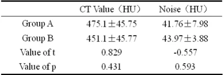
The average CT value in Group A is (475.1 ± 45.75) HU, the average CT value in Group B is (451.1 ± 45.77) HU, P>0.05. The average image noise in Group A is (41.76 ± 7.98) HU, the average image noise in Group B is (43.97 ± 3.88) HU, P> 0.05. The difference of image nose be-tween Group A and B had no statistical significance, as shown in following Table 2.
3.3. Radiation Dose
The radiation dose in the process of cardiac coronary artery examination consists of the following 3 parts: Scan, Contrast Agent Tracer Scan and Enhanced Scan. The radiation dose is the sum of these three parts. We did statistical analysis, and calculated CTDIvol, mean
dose length product (△DLP) and ED, we also compared
the results. In Group A, the average CTDIvol is (20.63 ± 2.24) mGy, in Group B the average CTDvol is (38.11 ± 10.69) mGy, it reduced (17.48 ± 8.45) mGy, and it de-creased by nearly 1 time, P <0.05. In Group A, the △
DLP is (235.75 ± 28.64)mGycm, in Group B the △DLP
is (492.59 ± 125.49) mGycm, it reduced (256.84 ± 96.85) mGycm, it decreased more than 1 time, P <0.05. In Group A the average ED is (4.01 ± 0.49) mSv, in Group B the average ED is (8.37 ± 2.13) mSv, it reduced (4.36 ± 1.64) mSv, it decreased more than 1 time, P<0.05. The radiation dose difference between two groups was statis-tically significant, as shown in Table 3.
4. DISCUSSIONS
DSCT coronary artery imaging in the diagnosis of car-diovascular diseases has unique advantages, more and more clinical cardiologist take CT as an important
[image:3.595.311.540.527.604.2]Table 2. Comparison of the image noise in Group A and B.
examination tool for non-invasive coronary artery imag-ing, it can be used as a supplement Cardiovascular Angi-ography, sometimes it can even replace Cardiovascular Angiography. In the process of the entire examination of DSCT, X-ray dose is large, the radiation hazards it pro-duces gets more and more attention. In order to follow the principle of ALARA (as low as reasonable achieva-ble), many scholars have done a lot of research in this respect, including hardware, software and optimizing programs of scan, such as Heart Bowtie, ECG Current Control, Cardiac noise subtraction filter, etc [9]. But the factors which affect the image quality are complex, some of these factors are closely related and influence each other. Some parameter settings may improve the image quality, but on the other hand, they may reduce the im-age quality in another aspect. So we must comprehen-sively consider, and separately set the parameters of scan according to the different clinical needs. But it is diffi-cult for us to balance the various factors to obtain high-quality images in order to improve the diagnostic rate. Currently, there is no standard for quality control in coronary artery imaging of Dual-Source CT in our coun-try.
So far, people have paid more and more attention to the potential hazards of ionizing radiation in CT. When ionizing radiation is working on human body, it can produce biological impacts and do harm to human body. The break of the structure of DNA double helix in hu-man body is the critical damage to the cells. Radiation induces gene mutations or the break of the double helix structure, the distortion increases, and eventually, the radiation can lead to cancer [10]. The reports pointed out that for every more radiation dose of 10mSv, the mortal-ity rate will increase by 0.04% [11]. The actual radiation dose of CT examination is 2-5 times the dose of effec-tive radiation [12]. The dose of effective radiation mainly comes from the enhanced period. In the aspect of opti-mizing scanning program, people mostly focus on con-trolling the radiation dose of enhanced period. In most cases, the image quality and the radiation dose have the inverse relationship. To balance the relationship between them, we need to change the fixed scan mode. In prac-tical application, we should pay attention to the individ-ual conditions of the different patients, such as the size of the heart, breast size of the female patients and cardi-othoracic ratio, etc. According to these conditions, we determine the appropriate scan mode and scan parame-ters. So we can achieve the personalized and reasonable scanning. A recent survey, which is an international
re-on the premise that the image quality meets the require-ments of clinical diagnosis, it becomes important for us to effectively reduce the radiation dose in the CTA ex-amination of coronary artery, and this can reduce the radiation damage caused by human factors.
Reducing the radiation dose is generally divided into two categories: new low-dose technology and optimizing scan parameters. The new scanning technology uses the ECG current modulation technique, Prospective ECG Trigger Scan Technology and noise reduction algorithms. The methods of optimizing scan parameters are lowering tube current, lowering tube voltage, reducing the scan rage of Z-axis and reducing the number of scans based on body mass or body mass index (BMI). In this article, in order to reduce the radiation dose, we used the tech-nology of regulating the dose of heart electric pulse (AUTO), and this technology belongs to retrospective cardiac-gated imaging technique. Through the study and analysis, we concluded that the regulation technology of dose of heart electric pulse (AUTO) can make the image quality completely meets the requirements of clinical diagnosis, and compared with the conventional full dose (OFF), this technology can significantly reduce the radi-ation dose of (4.36 ± 1.64) mSv, the radiradi-ation dose de-creased more than 1 time. Of course, it also has inade-quacies: 1. Data acquisition is not carried out throughout the whole cardiac cycle, and we can not analyze the car-diac function. 2. If the patient’s heart rate in unstable, it is easy for us to select the wrong full-dose regions. 3. In the non-full-dose exposure regions of the R-R interval, the image quality is a little bit worse than that of the image in full-dose regions. 4. If there are heart prema-ture beats in the scan, we can not edit the ECG.
The study has the following limitations. The individu-alized scan mode according the patient’s BMI was not adopted, the radiation dose still has some space to be reduced, and in this aspect the study should be continued. If the post-processing of image only uses one of the two periods in image reconstruction, we can only reconstruct the image of one period, therefore the radiation dose will be reduced by 1 time, and in this aspect can be studied much further.
REFERENCES
[1] Lei ZHUANG, Jihong GAN, Jianjun LIU.
Angiography. Journal of Sichuan Continuing Education College of Medical Sciences. 2009, 28 (2): 87-89.
[3] Yan Gao. Exploration of the low dose technique in
the examination of the coronary arteries angiography with dual-source CT. Sichuan Medical Journal, 2009, 30(12):1967-1969.
[4] Chiwei Wang. The current study of Dual-source CT
coronary artery imaging. Anhui Medical Journal, 2010, 31(10): 1256-1258
[5] Renwei Liu. Research Progress of Dual-source
Computed Tomography (DSCT) Low-dose Coronary Angiography. Medical Recapitulate, 2009, 15(21):3317-3320.
[6] Jiong Li. The application of optimizing scan
parameters of Dual-Source CT in coronary artery imaging for reducing radiation dose. Nei Mongol Journal of Traditional Chinese Medicine, 2012, 10:82-83.
[7] Lifeng Zhang, Maoyi Zhou, Li Yang. Influence of
coronal arterial image quality of 64 detector row CT in patients with ECG-gated scanned and optimal R-R phase imaging reconstructed. Chinese Journal of Medical Imaging Technology, 2008, 24(1):88-91.
[8] Wanshi Zhang, Jiaxing Xu. See Development of
Low-dose CT from RSNA 2008. China Medical Device Information, 2009, 7(1):12-13
[9] Aijun Wang, Jianguo Zhao, Xuejun Ping. The
comparison of two different methods of coronary artery imaging in Dual-Source CT. Journal of Ningxia Medical College, 2010, 32(3): 443-445.
[10] Zixu Yan, Zhaoqi Zhang. Low tube voltage in
reduction of radiation dose in dual-source CT coronary angiography. Chinese Journal of Medical Imaging Technology, 2009, 25(9): 1614-1616.
[11] Weiwei Qi, Xiangke Du, Ying Guo. A method to
optimize the selection of tube current for consistent image noise and dose control in 64-slice spiral helical CT cardiac imaging. Image Technology, 2008, 452(10):1026-1030.
[12] Xiaoyang Chen, Jiankun Zhai, Hui Li. Clinical
application and radiation prevention of CT scanning technology. China Modern Medicine, 2010, 17(8):61-62.
[13] Gou Wenjing, Shiyuan Liu, Hong Yu. Strategy and
research progress in coronary computed tomography angiography with low-dose scan. International Journal of Medical Radiology, 2012, 35(2): 147-150.
[14] Hausleiter J,Meyer T,Hermann F. Estimated
radiation dose associated with cardiac CT
angio-graphy. JAMA,2009, 301:500-700.
[15] Yan Qi, Shuang Qi. The application study of