ISSN Online: 2165-3410 ISSN Print: 2165-3402
DOI: 10.4236/aim.2018.89047 Sep. 17, 2018 699 Advances in Microbiology
Citrobacter rodentium
, a Gut Pathogen:
The Yin
and the Yang of Its Pathophysiology, Immunity
and Clinical Manifestation in Mice
Tania Rahman
1,2*, Md. Ferdous Seraj
3, Md. Monirul Islam
41Department of Biochemistry and Molecular Biology, Bio21 Molecular Science and Biotechnology Institute, University of
Melbourne, Melbourne, Australia
2Department of Microbiology and Immunology, University of Melbourne at the Peter Doherty Institute for Infection and
Immunity, Melbourne, Australia
3School of Civil, Environmental and Chemical Engineering, RMIT University, Melbourne, Australia 4Fiona Elsey Cancer Research Institute, Ballarat Technology Park Central, Ballarat, Australia
Abstract
Pathogenic strains of E. coli including enteropathogenic E. coli (EPEC), en-terohemorrhagic E. coli (EHEC), enterotoxigenic E. coli (ETEC) are principle
causes for diarrhoea in many parts of the globe. Citrobacter rodentium (C.
rodentium), a gram negative bacterium, is a murine pathogen that also util-izes type III secretion system and similar virulence factors to EPEC and EHEC and forms comparable attaching/effacing lesions in the intestines as
EPEC and EHEC. The infection caused by C. rodentium in mice is usually
self-limiting and results in only minor systemic effects with higher mortality in some susceptible mouse strains. All these characteristics have made the bacteria a commonly used model to study host immune responses to patho-genic E. coli infection. In this review, we focus on the impact of virulence factors of the pathogen; different immune components involved in the
im-mune response and summarize their role during C. rodentium infection.
Keywords
Citrobacter rodentium, Attaching and Effacing Pathogen, Locus of Enterocyte Effacement, Transmissible Murine Colonic Hyperplasia, Colitis, Mucosal Immune Response
1. Introduction
Escherichia coli is a frequent commensal organism of human intestine, often How to cite this paper: Rahman, T., Seraj,
Md.F. and Islam, Md.M. (2018) Citrobacter rodentium, a Gut Pathogen: The Yin and the Yang of Its Pathophysiology, Immunity and Clinical Manifestation in Mice. Advances in Microbiology, 8, 699-718.
https://doi.org/10.4236/aim.2018.89047
Received: June 15, 2018 Accepted: August 20, 2018 Published: September 17, 2018
Copyright © 2018 by authors and Scientific Research Publishing Inc. This work is licensed under the Creative Commons Attribution International License (CC BY 4.0).
http://creativecommons.org/licenses/by/4.0/
DOI: 10.4236/aim.2018.89047 700 Advances in Microbiology
colonizing immediately after birth and usually remaining for decades [1].
How-ever, some E. coli strains exhibit pathogenic potential, when they acquire certain virulence associated genes [2]. Pathogenic strains of E. coli including
enteropa-thogenic E. coli (EPEC) and enterohemorrhagic E. coli (EHEC) are the leading
causes of diarrheal outbreak in most parts of the world [3]. EPEC is a major
source of diarrhoea in children under two years of age causing deaths of a mil-lion each year in developing countries [4] [5]. EHEC causes bloody diarrhoea in children and elderly in developed countries with approximately 73,000 cases re-ported each year in the United states [6] [7] which can cause fatal diseases like
haemorrhagic colitis and haemolytic uremic syndrome [8] [9]. The hallmark of
EPEC and EHEC induced pathology is that they populate in the intestinal
epi-thelium through the development of attaching and effacing (A/E) lesions [10].
As these pathogens are a profound global health concern understanding their clinical manifestation, pathogenesis and immunity has become the focus of ex-tensive investigation. However, EPEC and EHEC elicit narrow range of host specificity and do not elicit a pertinent disease in laboratory animal genera, which makes it difficult to study EPEC and EHEC pathogenesis [11]. Citrobacter rodentium is an accepted mouse pathogen that uses comparable virulence fac-tors as EPEC and EHEC and forms analogous A/E lesions in the distal colon of mice [12] [13] [14]. Subsequently, C. rodentium has become a commonly used
animal representative to explore the immune responses to pathogenic E. coli
contamination in humans.
2.
C. rodentium
, an Attaching and Effacing Pathogen
C. rodentium (formerly known as Citrobacter freundii biotype 4280) a non-motile, gram-negative bacteria in the family of Enterobacteriaceae, is recognised as the
contributing species of transmissible murine colonic hyperplasia (TMCH) [15].
The disease is spread through the faecal-oral route and clinical symptoms occur
predominantly in weanling mice [16]. Overall, the infection is self-curing in
adult mice with higher mortality in some susceptible mouse strains [13]. In gen-eral, the infected mice exhibit decrease in body weight, excretion of soft faecal pellets with diarrhoea in severe cases and crypt hyperplasia.
DOI: 10.4236/aim.2018.89047 701 Advances in Microbiology
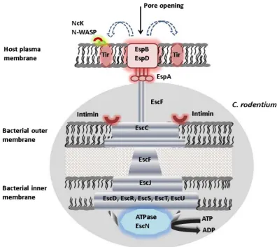
Figure 1. Illustration of type III secretion system (T3SS) in C. rodentium. The T3SS consists of a complex apparatus that actively delivers bacterial effector proteins into the cytoplasm of a host cell. The basal body spans the bacterial membranes and a needle-like syringe extends from the surface which is capped by a tip complex. Upon host cell contact, a translocon is inserted in the host cell membrane and forms a pore. Bac-terial virulence proteins are then selectively secreted through the syringe into the host cell, where they ma-nipulate host cell functions essential for subsequent pathogenicity.
2.1. Virulence Factors of
C. rodentium
The type III secretion system is programmed by a cluster of genes recognized as locus of enterocyte effacement (LEE), a conserved pathogenicity island consist-ing of 35.6 kb [24]. The LEE pathogenicity island consists of over 40 genes which are organised in to five operons including LEE1, LEE2, LEE3, LEE4 and LEE5
[24] [25] [26]. LEE encodes several structural components of T3SS, effectors, translocators and several other proteins (Figure 2) [27] [28]. One important ef-fector protein is Tir (translocated intimin receptor) a bacteria derived receptor, which following translocation binds to the host epithelial cell and interacts with intimin, the bacterial outer membrane protein, thereby facilitates anchorage of bacteria to host cell, leading to pedestal formation [29] (Figure 3). The intimins
are encoded by eae genes that are extremely conserved in N-terminal regions,
however, display substantial heterogeneity at the C-termini [30]. Five different intimin types α, β, γ, δ and ε have been recognized [31] [32]. Intimin α and in-timin β are expressed mainly by strains pertaining to EPEC clones 1 and 2,
cor-respondingly, whereas intimin γ is expressed by enterohaemorrhagic E. coli
DOI: 10.4236/aim.2018.89047 702 Advances in Microbiology
Figure 2. Genetic assembly of C. rodentium LEE. The orientation of each gene is shown by the direction of the arrow. The different locations of the rorf1 (r1) and rorf2 (espG/r2) genes in C. rodentium LEE, as well as the asso-ciation of several IS’s or IS remnants with the C. rodentium LEE. The major operons encoded by the LEE (LEE1, -2, -3, and -4, Tir, and R1/R2) and their transcriptional directions are shown and adapted from reference [25].
[image:4.595.142.539.252.605.2]DOI: 10.4236/aim.2018.89047 703 Advances in Microbiology Upon entry to the host cells, Tir is tyrosine phosphorylated, which recruits non-catalytic region of tyrosine kinase adaptor protein Nck [34] [35] [36] [37]. Nck binds to the phosphorylated tyrosine and this in turn triggers the recruit-ment of nucleation promoting factor N-WASP (neural Wiscott-Aldrich syn-drome protein) and actin-regulated protein Arp2/3 complex, resulting in host actin rearrangement [38] [39] [40]. Tir triggers localized actin polymerization by another two different pathways. After translocation, LEE and non-LEE effectors contribute to the impediment of several signalling pathways in the host cell, in-cluding actin polymerization, tight junction integrity, endosomal trafficking, apoptosis, phagocytosis and innate immune responses, as well as epithelial cell shedding and detachment [41].
Besides Tir, LEE encodes several secreted translocators: EspA, EspD, EspB, EspF, EspG, EspH, EspZ, which are entirely translocated into the host cells and are involved in modulating host cytoskeleton leading to the manifestation of
disease [4] [42] [43]. A/E pathogens secrete numerous LEE-encoded regulatory
proteins, Ler, GlrA and GlrR, which exhibit a significant role in the
transcrip-tional regulation of LEE and several non-LEE virulence determinants [44] [45]
[46]. Moreover, RegA adjusts LEE expressions through upregulating grlR/A
transcription [47]. There are several effectors that are not secreted and translo-cated by the LEE-encoded T3SS including prophages and insertion sequences. They comprise the Espl/NleA, an indispensable protein for entire virulence of C. rodentium [48] [49] [50] and binds host PDZ-domain proteins [51]; EspB, es-sential for intimate attachment and signal transduction (Figure 3) [52]; EspJ, which display a negligible part in enteric colonization [53]; and EspG, stimulates the dissociation of microtubules beneath adherent bacteria [54]. In addition, two
non-LEE encoded proteins, NleB to NleH, are found in C. rodentium and they
are mostly produced by the LEE-encoded T3SS [49] [55]. Among these, NleC
and NleD have been recognized to be translocated into host cells [56]. NleB1
binds to host cell death domain encoding proteins, diminishes the signalling of a death receptor and thereby disrupting a major antimicrobial host response [57].
Other than the genes for rorf1 and rorf2/espG and several insertion sequences
(IS) and IS remnants, both the LEE of C. rodentium and that of EPEC and
EHEC shares all 41 ORFs and the linear gene sequences (Figure 2) [25]. This
suggests that the LEE encoded pathogens has a mutual evolutionary origin and
reciprocal function which supports the use of C. rodentium as an animal model
to study A/E pathogenesis.
2.2. Disease Progression of
C. rodentium
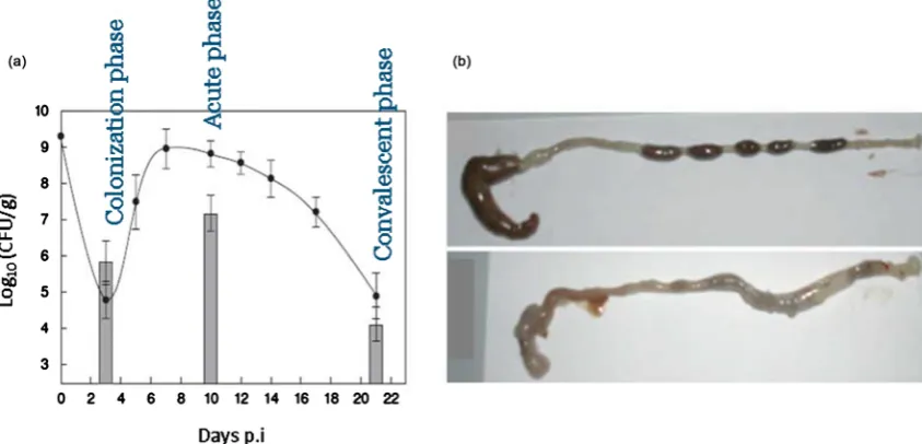
Similar to EPEC and EHEC, C. rodentium infection encompasses three distinct
DOI: 10.4236/aim.2018.89047 704 Advances in Microbiology
During the first week after inoculation, C. rodentium colonizes the brush
border microvilli and higher numbers of bacteria are seen closely adherent to the mucosal epithelial cells (Figure 4) [59] [60]. Acute phase of infection follows over the subsequent 2 weeks when the levels of bacteria peak around >109 c.f.u
per gram of tissue in the colon [16] and the bacteria induce a profound
hyper-plasia of colonic mucosa with the development of secretory diarrhoea [61]. At
the time of peak hyperplasic phase, the organism can no longer be isolated from the intestines and the infected mucosa are thickened markedly. Convalescent phase of infection comes following 4 weeks and above, for the period of which the reactive epithelial hyperplasia to clinical diarrhoea get resolved and colonic mucosa appears normal [13] [62].
2.3. Immune Defense against
C. rodentium
Most of the existing knowledge on the immune response and its relation with pathology has been expanded using mice with the targeted ablations of various immune components. Innate immune response as well as adaptive immune re-sponse appears to control mucosal defence against C. rodentium [63].
2.3.1. Role of Adaptive Mucosal Immune Responses
[image:6.595.115.537.438.641.2]The concept of exploring mice with deficiencies in immune components first came from the study that colonic mucosa of infected mice contained large infil-trates of CD4+ T cells with a helper T cell 1 cytokine response [64]. Substantial mortality was observed in mice deficient in CD4+ T cells or TCRαβ+ T cells [59]. Mice lacking CD4 showed a survival limit of two weeks and exhibited 100%
DOI: 10.4236/aim.2018.89047 705 Advances in Microbiology mortality. However, depletion of CD8+ T cells or TCRγδ+ T cells did not ad-versely affect survival of infection and played a minor role in surviving the acute phase of infection. Two studies [63] [65] [66] separately demonstrated the vital
significance of B cells for the protective immunity against C. rodentium. Mice
lacking mature B cells (μMT mice) failed to mount early inflammatory response at 2 weeks and could not lessen bacterial load or clear bacterial colonization over
a prolonged period [63] [65] [66]. RAG1-deficient mice in which both B and T
cells were absent displayed chronic intestinal colonization and more severe colonic damage and these mice were unable to clear infection and died after 3-4 weeks [59] [63] [67].
To analyse the potential of secretory antibodies in bacterial clearance, mice
with selective deficiencies for IgA, IgM or IgG were used [65]. Both IgA- and
IgM- deficient mice had been found to develop effective immunity against a secondary challenge and played a negligible role in controlling C. rodentium in-fection. However, mice deficient in IgG antibodies lost the ability to develop ro-bust protective response against secondary challenge. Thus host defense against C. rodentium was dependent on IgG antibodies but did not require secretion of IgA or IgM [65]. A comparative analysis of the specific ablation of different
adaptive immune components is summarized in Table 1.
[image:7.595.57.539.416.743.2]Simmons and co-workers investigated C. rodentium infection in IFNγ-deficient and IL-12 deficient mice. IFNγ-deficient mice had higher bacterial numbers and
Table 1. Summary of the effects of selective ablation of adaptive immune components on C. rodentium infection in mice.
Mouse models Effects of ablation of specific adaptive immune components on C. rodentium infection Refs Mice lacking CD4+ T cells or
TCRαβ+ T cells A survival limit of two weeks and exhibited 100% mortality [59] [64]
Mice lacking CD8+ T cells or
TCRγδ+ T cells Do not adversely affect survival of infection [59]
Mice without mature B cells
(μMT mice) Cannot lessen bacterial load or clear bacterial colonization over a prolonged period [63] [65] [66]
RAG1-deficient mice (both B
and T cells are absent) Develop chronic intestinal colonization and unable to clear infection and die after 3-4 weeks [59] [63] [67]
IgA- and IgM- deficient mice Develop effective immunity against a secondary challenge and play a negligible role in controlling C.
rodentium infection. [65]
Mice deficient in IgG antibodies Lose the ability to develop protective response against secondary challenge. [65]
IFNγ-deficient mice Higher bacterial numbers and enhanced mucosal thickening in colons and cannot clear infection until
day 28 [68]
IL-12 deficient mice Elicit higher bacterial numbers for the first 3 weeks of infection and eventually clear infection by day
35 [30]
Mice lacking IL-22 Display systemic bacterial load and enhanced epithelial hyperplasia and mortality range up to 100%
within the first two weeks of infection [69]
Treg deficient mice (DEREG
DOI: 10.4236/aim.2018.89047 706 Advances in Microbiology enhanced mucosal thickening in their colons and could not clear infection until day 28 [68]. Alternatively, IL-12 deficient mice had been shown to elicit higher bacterial numbers for the first 3 weeks of infection and eventually cleared infec-tion by day 35 [30]. Interleukin-22 (IL-22) has been identified as an essential
cy-tokine for mediating protection against C. rodentium infection. While compared
to wild-type mice, mice lacking IL-22 infected with C. rodentium displayed sys-temic bacterial load and enhanced epithelial hyperplasia. In multiple repeat ex-periments, the mortality of IL-22 knockout mice ranged from 80% to 100% within the first two weeks after infection [69]. Administration of Reg3γ, an an-timicrobial peptide to IL-22 knockout mice controlled infection.
During infection, CD4+ Th17 cell subsets were particularly amplified in
Peyer’s patches (PP) but were unaltered in mesenteric LNs [70]. The differentia-tion of Th17 cells in PP were dependent on the inflammatory cytokine IL-6 as treatment with anti-IL-6 antibodies reduced Th17 cells and exacerbated clinical manifestation of colitis. Moreover, following anti-IL-6 antibody treatment, there was a reduction of IL-22 mRNA expression in the small intestine during infec-tion but had no effect on IgA producinfec-tion by B cells [70].
Symonds and co-workers demonstrated an elevated FoxP3 mRNA expression in the distal colon at all stages of infection with C. rodentium. Also, C. roden-tium infection exhibited an up-regulation of IL17 mRNA expression [58]. Wang
and co-workers investigated role of Treg during C. rodentium infection using
DEREG mouse model. Depletion of Treg by diphtheria toxin led to a diminished bacterial clearance and systemic dissemination of bacteria. Also, Th17-associated immune response was compromised following Treg-depletion, with less in-flammation-associated pathology in the colons of Treg-depleted mice. Treat-ment with Anti-IL2 in depleted mice retained Th17 induction, suggesting that Treg induced a protective Th17 response by intake of local IL-2 [71]. IL-10 was found dispensable in controlling inflammation as IL-10 ablated mice resolved infection earlier than wild-type mice and had less infection associated colitis
[72]. In addition, following infection, IL-27 was produced which subsequently
suppressed Th17 in vitro and thus play role in anti-inflammatory circuit in the ab-sence of IL-10. The neutralization of IL-27 led to pronounced colitis in mice lacking IL-10 suggesting that IL-10 enacts a minor part in the bacterial clearance whereas IL-27 might be an important cytokine for attenuation of inflammation [72].
DOI: 10.4236/aim.2018.89047 707 Advances in Microbiology
with C. rodentium showed similar increases in IL-17f-/-, IL17a-/-, and
IL-17a-/-IL17f-/- mice, indicating that deficiency of just one of the IL-17 pro-teins resulted in full susceptibility to infection. However, splenomegaly and co-lon hypertrophy, which were associated with severe coco-lonic inflammation, were more pronounced in IL-17f-/- mice than in IL-17a-/- mice suggesting that IL-17f was more important than IL-17a in protecting colonic epithelial cells from the pathogenic effects of this bacterium.
2.3.2. Role of Innate Mucosal Immune Responses
There are several innate immune components which perform a vital role in mu-cosal homeostasis and in antimicrobial immunity. An augmented pathology was observed in mice lacking Toll-like receptor 2 (TLR2) due to an impaired epithe-lial barrier [75]. Mice lacking the signalling adaptor MYD88, a myeloid differen-tiation primary response protein 88, which is essential for signalling by the ma-jority of TLRs [76] [77] had greater bacterial loads both in the colon and in pe-ripheral tissues as the bacteria penetrated deeply into colonic crypts compared to WT mice. Moreover, they suffered from severe colitis and death after infection. The innate immune receptor type-I interleukin-1 receptor (IL-1R), utilizing
MyD88 signalling pathway protected mice from severe damage caused by C.
ro-dentium [78]. IL-1R deficient mice exhibited increased susceptibility to tissue damage comparable to that of MyD88 knockout mice. Yet, distinct from MyD88 knockout mice, mice deficient in IL-1R did not display amplified pathogen bur-dens in the colon. In another study, Khan and co-workers exhibited that the bacteria triggered TLR4 and prompted NF-κB nuclear translocation which was dependent on TLR4. Deficiency of TLR4 decreased tissue pathology and in-flammatory cell infiltration in gut. Unexpectedly, dissemination of bacteria through colon was hindered in mice lacking TLR4, while the extent of infection was unaffected, suggesting that TLR4-mediated responses were eventually mal-adaptive to the host [79].
Liu and co-workers demonstrated the biological function of inflammasomes
in immune response against C. rodentium. Mice deficient in inflammasome
components Nlrp3, Nlrc4, and caspase-1 were hyper susceptible to C. rodentium
induced intestinal inflammation due to impaired production of IL-1β and IL-18
[80]. However, these deficient mice exhibited only mild defects and none of
these mice died after infection, indicating that inflammasome is not essential for mice survival after C. rodentium infection. In addition, IL-1β−/− and IL-18−/− mice suffered from increased bacterial burdens and had severe histopathology.
Therefore, Nlrp3 and Nlrc4 inflammasome-mediated IL-1β and IL-18 response
contributed a significant role in host protection against C. rodentium [80] [81]. In another study, Kim and co-workers characterized the role of the intracellular
Nod-like receptor family members Nod2 in protection against C. rodentium
in-fection [82]. Nod2−/− mice displayed diminished intestinal clearance to C.
roden-tium. The enhanced bacterial load was due to impaired secretion of chemokine
Fur-DOI: 10.4236/aim.2018.89047 708 Advances in Microbiology thermore, IL-12, a cytokine produced by monocytes triggered Th1 immunity vi-tal for bacterial clearance. The adoptive transfer experiments established the sig-nificant contribution of Ly6Chi monocytes in the clearance of bacteria in vivo
[82]. Table 2 summarizes a comparative analysis of the specific ablation of dif-ferent innate immune components.
Mice lacking the p50 subunit of the NF-κB transcription factor, a nuclear factor kappa B, had reduced ability to clear C. rodentium infection [83]. Also a continued bacterial load was reported in mice deficient in p38α, a mitogen-activated pro-tein kinase (MAPK) in intestinal epithelial cells [84]. Interestingly, these animals exhibited no apparent histological lesions, however, failed to recruit CD4+ T cells
and had impaired chemokines expression. Thus, p38α in IECs by employing
immune cells and adjusting chemokine expression played a part to the host pro-tective immune responses. CXCL9, an ELR (glutamic acid-leucine-arginine)
mo-tif chemokine had direct antimicrobial potential against C. rodentium and
[image:10.595.57.535.340.735.2]de-fended crypts from bacterial dissemination. Blockade of this antimicrobial activ-ity by anti-CXCL9 antibodies escalated host exposure to C. rodentium infection
Table 2. Summary of the effects of selective ablation of innate immune components on C. rodentium infection in mice.
Mouse models Effects of ablation of specific innate immune components on C. rodentium infection Refs Mice lacking Toll-like receptor 2
(TLR2) An augmented pathology due to an impaired epithelial barrier [75]
Mice lacking MYD88 Have greater bacterial loads in colon and peripheral tissues and suffer from severe colitis and death [76] [77]
IL-1R deficient mice Increased susceptibility to tissue damage but do not display amplified pathogen burdens in colon. [78]
Deficiency of TLR4 Decreased tissue pathology and inflammatory cell infiltration in gut. While the extent of infection is unaffected, dissemination of bacteria through colon is hindered [79] Mice deficient in Nlrp3, Nlrc4, and
caspase-1 Hyper susceptible to C. rodentium induced intestinal inflammation. However, exhibit only mild defects and do not die after infection [80] [81]
IL-1β−/− and IL-18−/− mice Increased bacterial burdens and severe histopathology. [80] [81]
Nod2−/− mice Diminished intestinal clearance to C. rodentium. due to impaired secretion of CCL2 from colonic
cells [82]
Mice lacking the p50 subunit of
NF-κB Reduced ability to clear C. rodentium infection. [83]
Mice deficient in p38α A continued bacterial load with no apparent histological lesions, however, fails to recruit CD4+ T
cells and impaired chemokines expression. [84]
Ablation of specific
macrophage/monocyte compartment Neither cell type is essential to trigger immunity [85]
Mice lacking PSGL-1 and P, E and
L-selectin Mice defective in PSGL-1 and P-selectin suffer morbidity, extensive inflammatory responses and augmented bacterial burden, however, mice defective in either E or L-selectin do not exhibit severe infection
[86]
Mice lacking β7 integrin Efficiently control infection and clear bacteria 5-6 week after inoculation [59] Mice deficient Muc2 Susceptible to the C. rodentium-induced colitis and display quick weight loss and exhibit 90%
DOI: 10.4236/aim.2018.89047 709 Advances in Microbiology with noticeable bacterial dissemination, augmented bacterial titre, and
deterio-rated tissue pathology [89]. Surface lymphotoxin expression on group 3 innate
lymphoid cells (ILC3s) is critical for early immune responses against C.
roden-tium [90] [91]. LT aids in IL-22 secretion by intestinal ILCs. Blocking of LTβR signaling rapidly diminished intestinal IL-22 production after C. rodentium in-fection [91]. In addition, stimulating LTβR signaling induced IL-22 pathway in LT-deficient mice. LT-beta receptor (LTbR) signaling in intestinal epithelial cells was essential for recruitment of neutrophils to the site of infection through se-cretion of CXCL1 and CXCL2 chemokines. In contrast, surface LT produced by adaptive B and T cells was dispensable for protection against gut bacterial infec-tion [90].
The function of macrophages and monocytes during C. rodentium infection
was investigated using ablation of specific macrophage/monocyte compartment during infection. Although neither cell type was essential to trigger immunity, monocytes and macrophages played a role by secreting IL-12, which prompted
Th1 polarization and IFN-γ secretion. Thus, monocytes and macrophages
con-tribute in C. rodentium immunity by secreting cytokines that direct T cell
po-larization [85].
To outline the function of selectins and their ligands during C. rodentium in-fection, Kum and co-workers investigated infection in mice lacking PSGL-1, a P-selectin glycoprotein ligand-1 and P, E and L-selectin [86]. Mice defective in PSGL-1 and P-selectin suffered morbidity, extensive inflammatory responses and augmented bacterial burden, however, mice defective in either E or L-selectin did not exhibit severe infection. Also, intestinal inflammation and re-cruitment of inflammatory cells i.e., neutrophils and macrophages were signifi-cantly diminished in P-selectin defective mice which received blocking antibod-ies to ICAM-1 or LFA-1, suggesting that these adhesion molecules can counter-balance the defect in selectins during leucocyte recruitment [86]. Mice lacking β7 integrin efficiently controlled infection and cleared bacteria 5-6 week after in-oculation [59].
Mice deficient in main intestinal mucin, Muc2, which have an altered
intesti-nal mucus layer, were more susceptible to the C. rodentium-induced colitis and
displayed quick weight loss and exhibited about 90% mortality due to a closer interaction of intestinal microbes with the epithelial barrier [87] [88]. Muc2−/−
mice had 10 - 100 fold increased C. rodentium load, maximum of which were
closely attachded to the mucosa in colon. FITC-Dextran administration exhib-ited considerably exacerbated disruption in intestinal barrier integrity in Muc2−/− mice, with explicit bacterial translocation into the colonic mucosa [87] [88].
2.4. Role of Probiotics and Antibiotic Administration
Probiotics, a combination of live microorganisms attenuated infection with C.
rodentium in adult mice and provided a protective role in C. rodentium induced
DOI: 10.4236/aim.2018.89047 710 Advances in Microbiology mixture exhibited inhibitory role on the growth of C. rodentium. Mice that were
administered live probiotics containing a mixture of Lactobacillus rhamnosus
and L. acidophilus stayed healthy. Pretreatment of mice with probiotics restored colonic integrity and lessened both hyperplasia and inflammatory-cell infiltra-tion in colon [93]. In a recent study, Collins et al. demonstrated that probiotics such as Lactobacillus acidophilus, L. rhamnosus, and Lactobacillus helveticus
administered daily in the form of fermented dairy products (FDPs) lessened C.
rodentium induced colonic hyperplasia and stopped the loss of significant bacte-rial genera that might lead to disease pathology. However, the FDPs did not
re-sult in any noteworthy reduction in C. rodentium colonization when estimated
by bacterial load [94].
Metronidazole pretreatment augmented exposure to C. rodentium-induced
colitis compared to that of untreated mice 6 days postinfection and resulted in a
diminished number of Porphyromonadaceae and amplified population of
lacto-bacilli [95]. Metronidazole treatment resulted an impaired goblet cell function, decreased Muc2 secretion, a major component of intestinal secretory mucin and thinning of inner mucus layer, resulting in microbially induced immune activa-tion prior to disease inducactiva-tion. Perturbaactiva-tion of the microbiota with metronida-zole resulted augmented attachment of bacteria to the intestinal epithelium, re-sulting in a severe form of C. rodentium-induced colitis in mice [95].
2.5. Limitations of
C. rodentium
Model
A limitation to the study of C. rodentium infection model is the absence of anti-gen-specific tools with which to characterize the fate and function of the
patho-gen/antigen-specific response during infection [96]. The only means that are
currently available to address this limitation include transgenic strains of C. ro-dentium that express OVA or GFP [96] [97] [98]. Another probable limitation to
study this pathogen could be the loss of antibiotic sensitivity of C. rodentium
due to the development of worldwide emergence of multi-resistant strains [19].
However, the likelihood of this loss is occasional due to the germ-free condition of the animal houses.
3. Concluding Remarks
EPEC and EHEC are the leading cause of diarrhoea in human, affecting children
and adults in both developing and developed countries. C. rodentium is an
en-teric murine pathogen that mimics virulence factors of human EPEC and EHEC and forms comparable attaching and effacing lesions, as a central mechanism of tissue targeting, virulence factors and infection in mice. As a result of this asso-ciation with other important inflammatory diseases, and that there are cases of more than a million deaths each year from EPEC and EHEC, the knowledge
about the pathophysiology of C. rodentium infections and following infection
DOI: 10.4236/aim.2018.89047 711 Advances in Microbiology This review comprehensively covers the salient features of recent discoveries related to C. rodentium virulence, epithelial hyperplasia, innate and adaptive immune responses, and the pathophysiology of diarrhoea. It is acknowledged
that EPEC and EHECcan be modelled efficiently in mice.Murine C. rodentium
is a well characterised model of diarrhoeal disease as the molecular, cellular,
pathophysiological aspects of the disease have been well studied. Therefore, C.
rodentium represents an excellent model in which to study the innate and adap-tive immune components. We believe that the advances that have been included in this review will give a comprehensive insight to combat the acute diarrhoeal illness in human. Nevertheless, once again C. rodentium has been proved to be a
useful in vivo model for studying pathogenesis of secretory diarrhoeal
dis-eases/gastrointestinal pathogen and for preventive/mucosal vaccinations and therapeutic approaches.
Conflicts of Interest
The authors declare no conflict of interest that could be perceived to bias the work.
References
[1] Nataro, J.P. and Kaper, J.B. (1998) Diarrheagenic Escherichia coli.Clinical Microbi-ology Reviews, 11, 142-201.
[2] Cassels, F.J. and Wolf, M.K. (1995) Colonization Factors of Diarrheagenic E. coli
and Their Intestinal Receptors. Journal of Industrial Microbiology, 15, 214-226. https://doi.org/10.1007/BF01569828
[3] Frankel, G., et al. (1998) Enteropathogenic and Enterohaemorrhagic Escherichia coli: More Subversive Elements. Molecular Microbiology, 30, 911-921.
https://doi.org/10.1046/j.1365-2958.1998.01144.x
[4] Clarke, S.C., et al. (2003) Virulence of Enteropathogenic Escherichia coli, a Global Pathogen. Clinical Microbiology Reviews, 16, 365-378.
https://doi.org/10.1128/CMR.16.3.365-378.2003
[5] Chen, H.D. and Frankel, G. (2005) Enteropathogenic Escherichia coli: Unravelling Pathogenesis.FEMS Microbiology Reviews, 29, 83-98.
https://doi.org/10.1016/j.femsre.2004.07.002
[6] Mead, P.S., et al. (1999) Food-Related Illness and Death in the United States Reply to Dr. Hedberg. Emerging Infectious Diseases, 5, 841-842.
https://doi.org/10.3201/eid0506.990625
[7] Rangel, J.M., et al. (2005) Epidemiology of Escherichia coli O157:H7 Outbreaks, United States, 1982-2002. Emerging Infectious Diseases, 11, 603-609.
https://doi.org/10.3201/eid1104.040739
[8] Welinder-Olsson, C. and Kaijser, B. (2005) Enterohemorrhagic Escherichia coli
(EHEC). Scandinavian Journal of Infectious Diseases, 37, 405-416. https://doi.org/10.1080/00365540510038523
[9] Schmidt, H., et al. (1996) Pore-Forming Properties of the Plasmid-Encoded Hemo-lysin of Enterohemorrhagic Escherichia coli O157:H7. European Journal of Bio-chemistry, 241, 594-601. https://doi.org/10.1111/j.1432-1033.1996.00594.x
Na-DOI: 10.4236/aim.2018.89047 712 Advances in Microbiology ture Reviews Microbiology, 2, 123-140. https://doi.org/10.1038/nrmicro818
[11] Mundy, R., et al. (2006) Comparison of Colonization Dynamics and Pathology of Mice Infected with Enteropathogenic Escherichia coli, Enterohaemorrhagic E. coli
and Citrobacter rodentium.FEMS Microbiology Letters, 265, 126-132. https://doi.org/10.1111/j.1574-6968.2006.00481.x
[12] Mundy, R., et al. (2005) Citrobacter rodentium of Mice and Man. Cellular Microbi-ology, 7, 1697-1706. https://doi.org/10.1111/j.1462-5822.2005.00625.x
[13] Luperchio, S.A. and Schauer, D.B. (2001) Molecular Pathogenesis of Citrobacter rodentium and Transmissible Murine Colonic Hyperplasia. Microbes and Infection, 3, 333-340. https://doi.org/10.1016/S1286-4579(01)01387-9
[14] Savkovic, S.D., et al. (2005) Mouse Model of Enteropathogenic Escherichia coli In-fection. Infection and Immunity, 73, 1161-1170.
https://doi.org/10.1128/IAI.73.2.1161-1170.2005
[15] Schauer, D.B. and Falkow, S. (1993) Attaching and Effacing Locus of a Citrobacter freundii Biotype that Causes Transmissible Murine Colonic Hyperplasia. Infection and Immunity, 61, 2486-2492.
[16] Barthold, S.W., Coleman, G.L., Jacoby, R.O., Livestone, E.M. and Jonas, A.M. (1978) Transmissible Murine Colonic Hyperplasia. Veterinary Pathology, 15, 223-236.
https://doi.org/10.1177/030098587801500209
[17] Wiles, S., et al. (2004) Organ Specificity, Colonization and Clearance Dynamics in Vivo Following Oral Challenges with the Murine Pathogen Citrobacter rodentium.
Cellular Microbiology, 6, 963-972.https://doi.org/10.1111/j.1462-5822.2004.00414.x [18] Wiles, S., Pickard, K.M., Peng, K., MacDonald, T.T. and Frankel, G. (2006) In Vivo
Bioluminescence Imaging of the Murine Pathogen Citrobacter rodentium. Infection and Immunity, 74, 5391-5396.https://doi.org/10.1128/IAI.00848-06
[19] Buschor, S., et al. (2017) Innate Immunity Restricts Citrobacter rodentium A/E Pathogenesis Initiation to an Early Window of Opportunity. PLOS Pathogens, 13, e1006476.https://doi.org/10.1371/journal.ppat.1006476
[20] Zahavi, E.E., et al. (2011) Bundle-Forming Pilus Retraction Enhances Enteropatho-genic Escherichia coli Infectivity. Molecular Biology of the Cell, 22, 2436-2447.
https://doi.org/10.1091/mbc.e11-01-0001
[21] Vallance, B.A. and Finlay, B.B. (2000) Exploitation of Host Cells by Enteropatho-genic Escherichia coli. Proceedings of the National Academy of Sciences, 97, 8799-8806.https://doi.org/10.1073/pnas.97.16.8799
[22] Garmendia, J., Frankel, G. and Crepin, V.F. (2005) Enteropathogenic and Entero-hemorrhagic Escherichia coli Infections: Translocation, Translocation, Transloca-tion. Infection and Immunity, 73, 2573-2585.
https://doi.org/10.1128/IAI.73.5.2573-2585.2005
[23] Vallance, B.A., Deng, W., Jacobson, K. and Finlay, B.B. (2003) Host Susceptibility to the Attaching and Effacing Bacterial Pathogen Citrobacter rodentium. Infection and Immunity, 71, 3443-3453.https://doi.org/10.1128/IAI.71.6.3443-3453.2003
[24] Franzin, F.M. and Sircili, M.P. (2015) Locus of Enterocyte Effacement: A Patho-genicity Island Involved in the Virulence of Enteropathogenic and Enterohemor-ragic Escherichia coli Subjected to a Complex Network of Gene Regulation. BioMed Research International, 2015, Article ID: 534738.
https://doi.org/10.1155/2015/534738
DOI: 10.4236/aim.2018.89047 713 Advances in Microbiology
Transfer among Attaching and Effacing Pathogens. Infection and Immunity, 69, 6323-6335.https://doi.org/10.1128/IAI.69.10.6323-6335.2001
[26] Deng, W., et al. (2004) Dissecting Virulence: Systematic and Functional Analyses of a Pathogenicity Island. Proceedings of the National Academy of Sciences, 101, 3597-3602.https://doi.org/10.1073/pnas.0400326101
[27] Elliott, S.J., et al. (2000) The Locus of Enterocyte Effacement (LEE)-Encoded Regu-lator Controls Expression of Both LEE- and Non-LEE-Encoded Virulence Factors in Enteropathogenic and Enterohemorrhagic Escherichia coli. Infection and Immu-nity, 68, 6115-6126.https://doi.org/10.1128/IAI.68.11.6115-6126.2000
[28] Gaytan, M.O., Martínez-Santos, V.I., Soto, E. and González-Pedrajo, B. (2016) Type Three Secretion System in Attaching and Effacing Pathogens. Frontiers in Cellular and Infection Microbiology, 6, 129.https://doi.org/10.3389/fcimb.2016.00129 [29] Shames, S.R., Croxen, M.A., Deng, W. and Finlay, B.B. (2011) The Type III
Sys-tem-Secreted Effector EspZ Localizes to Host Mitochondria and Interacts with the Translocase of Inner Mitochondrial Membrane 17b. Infection and Immunity, 79, 4784-4790.https://doi.org/10.1128/IAI.05761-11
[30] MacDonald, T.T., Frankel, G., Dougan, G., Goncalves, N.S. and Simmons, C. (2003) Host Defences to Citrobacter rodentium. International Journal of Medical Microbi-ology, 293, 87-93.https://doi.org/10.1078/1438-4221-00247
[31] Oswald, E., Schmidt, H., Morabito, S., Karch, H., Marchès, O. and Caprioli, A. (2000) Typing of Intimin Genes in Human and Animal Enterohemorrhagic and Enteropathogenic Escherichia coli: Characterization of a New Intimin Variant. In-fection and Immunity, 68, 64-71.https://doi.org/10.1128/IAI.68.1.64-71.2000 [32] Yi, Y., et al. (2010) Crystal Structure of EHEC Intimin: Insights into the
Comple-mentarity between EPEC and EHEC. PLoS ONE, 5, e15285.
https://doi.org/10.1371/journal.pone.0015285
[33] Adu-Bobie, J., et al. (1998) Detection of Intimins Alpha, Beta, Gamma, and Delta, four Intimin Derivatives Expressed by Attaching and Effacing Microbial Pathogens.
Journal of Clinical Microbiology, 36, 662-668.
[34] Kenny, B. and Finlay, B.B. (1997) Intimin-Dependent Binding of Enteropathogenic
Escherichia coli to Host Cells Triggers Novel Signaling Events, Including Tyrosine Phosphorylation of Phospholipase C-Gamma1. Infection and Immunity, 65, 2528-2536.
[35] Kenny, B., DeVinney, R., Stein, M., Reinscheid, D.J., Frey, E.A. and Finlay, B.B. (1997) Enteropathogenic E. coli (EPEC) Transfers Its Receptor for Intimate Adher-ence into Mammalian Cells. Cell, 91, 511-520.
https://doi.org/10.1016/S0092-8674(00)80437-7
[36] Campellone, K.G., Giese, A., Tipper, D.J. and Leong, J.M. (2002) A Tyro-sine-Phosphorylated 12-Amino-Acid Sequence of Enteropathogenic Escherichia coli Tir Binds the Host Adaptor Protein Nck and Is Required for Nck Localization to Actin Pedestals. Molecular Microbiology, 43, 1227-1241.
https://doi.org/10.1046/j.1365-2958.2002.02817.x
[37] Martinez-Quiles, N., Feuerbacher, L.A., Benito-León, M. and Hardwidge, P.R. (2014) Contribution of Crk Adaptor Proteins to Host Cell and Bacteria Interactions.
BioMed Research International, 2014, Article ID: 372901.
https://doi.org/10.1155/2014/372901
DOI: 10.4236/aim.2018.89047 714 Advances in Microbiology
https://doi.org/10.1016/S0898-6568(02)00027-X
[39] Gruenheid, S., et al. (2001) Enteropathogenic E. coli Tir Binds Nck to Initiate Actin Pedestal Formation in Host Cells. Nature Cell Biology, 3, 856-859.
https://doi.org/10.1038/ncb0901-856
[40] Nieto-Pelegrin, E., Kenny, B. and Martinez-Quiles, N. (2014) Nck Adaptors, Besides Promoting N-WASP Mediated Actin-Nucleation Activity at Pedestals, Influence the Cellular Levels of Enteropathogenic Escherichia coli Tir Effector. Cell Adhesion & Migration, 8, 404-417.https://doi.org/10.4161/19336918.2014.969993
[41] Collins, J.W., et al. (2014) Citrobacter rodentium: Infection, Inflammation and the Microbiota. Nature Reviews Microbiology, 12, 612-623.
https://doi.org/10.1038/nrmicro3315
[42] Donnenberg, M.S. and Whittam, T.S. (2001) Pathogenesis and Evolution of Viru-lence in Enteropathogenic and Enterohemorrhagic Escherichia coli. Journal of Clinical Investigation, 107, 539-548.https://doi.org/10.1172/JCI12404
[43] Ugalde-Silva, P., Gonzalez-Lugo, O. and Navarro-Garcia, F. (2016) Tight Junction Disruption Induced by Type 3 Secretion System Effectors Injected by Enteropatho-genic and Enterohemorrhagic Escherichia coli. Frontiers in Cellular and Infection Microbiology, 6, 87.https://doi.org/10.3389/fcimb.2016.00087
[44] Abe, H., et al. (2008) Global Regulation by Horizontally Transferred Regulators Es-tablishes the Pathogenicity of Escherichia coli. DNA Research, 15, 25-38.
https://doi.org/10.1093/dnares/dsm033
[45] Torres, A.G., et al. (2007) Ler and H-NS, Regulators Controlling Expression of the Long Polar Fimbriae of Escherichia coli O157:H7. Journal of Bacteriology, 189, 5916-5928.https://doi.org/10.1128/JB.00245-07
[46] Holmes, A., Lindestam Arlehamn, C.S., Wang, D., Mitchell, T.J., Evans, T.J. and Roe, A.J. (2012) Expression and Regulation of the Escherichia coli O157:H7 Effector Proteins NleH1 and NleH2. PLoS ONE, 7, e33408.
https://doi.org/10.1371/journal.pone.0033408
[47] Yang, J., Tauschek, M., Hart, E., Hartland, E.L. and Robins-Browne, R.M. (2010) Virulence Regulation in Citrobacter rodentium: The Art of Timing. Microbial Bio-technology, 3, 259-268.https://doi.org/10.1111/j.1751-7915.2009.00114.x
[48] Gruenheid, S., et al. (2004) Identification and Characterization of NleA, a Non-LEE-Encoded Type III Translocated Virulence Factor of Enterohaemorrhagic
Escherichia coli O157:H7. Molecular Microbiology, 51, 1233-1249.
https://doi.org/10.1046/j.1365-2958.2003.03911.x
[49] Kelly, M., et al. (2006) Essential Role of the Type III Secretion System Effector NleB in Colonization of Mice by Citrobacter rodentium. Infection and Immunity, 74, 2328-2337.https://doi.org/10.1128/IAI.74.4.2328-2337.2006
[50] Thanabalasuriar, A., et al. (2012) Sec24 Interaction Is Essential for Localization and Virulence-Associated Function of the Bacterial Effector Protein NleA. Cellular Mi-crobiology, 14, 1206-1218.https://doi.org/10.1111/j.1462-5822.2012.01789.x
[51] Lee, S.F., et al. (2008) A C-Terminal Class I PDZ Binding Motif of EspI/NleA Modulates the Virulence of Attaching and Effacing Escherichia coli and Citrobacter rodentium. Cellular Microbiology, 10, 499-513.
[52] Newman, J.V., Zabel, B.A., Jha, S.S. and Schauer, D.B. (1999) Citrobacter rodentium
espB Is Necessary for Signal Transduction and for Infection of Laboratory Mice.
Infection and Immunity, 67, 6019-6025.
At-DOI: 10.4236/aim.2018.89047 715 Advances in Microbiology
taching and Effacing Pathogens That Modulates Infection Dynamics. Infection and Immunity, 73, 679-686.https://doi.org/10.1128/IAI.73.2.679-686.2005
[54] Shaw, R.K., et al. (2005) Enteropathogenic Escherichia coli Type III Effectors EspG and EspG2 Disrupt the Microtubule Network of Intestinal Epithelial Cells. Infection and Immunity, 73, 4385-4390.https://doi.org/10.1128/IAI.73.7.4385-4390.2005 [55] Newton, H.J., et al. (2010) The Type III Effectors NleE and NleB from
Enteropa-thogenic E. coli and OspZ from Shigella Block Nuclear Translocation of NF-kappaB p65. PLOS Pathogens, 6, e1000898.https://doi.org/10.1371/journal.ppat.1000898 [56] Yen, H., Ooka, T., Iguchi, A., Hayashi, T., Sugimoto, N. and Tobe, T. (2010) NleC, a
Type III Secretion Protease, Compromises NF-kappaB Activation by Targeting p65/RelA. PLOS Pathogens, 6, e1001231.
https://doi.org/10.1371/journal.ppat.1001231
[57] Pearson, J.S., et al. (2013) A Type III Effector Antagonizes Death Receptor Signal-ling during Bacterial Gut Infection. Nature, 501, 247-251.
https://doi.org/10.1038/nature12524
[58] Symonds, E.L., Riedel, C.U., O’Mahony, D., Lapthorne, S., O’Mahony, L. and Shanahan, F. (2009) Involvement of T Helper Type 17 and Regulatory T Cell Activ-ity in Citrobacter rodentium Invasion and Inflammatory Damage. Clinical & Ex-perimental Immunology, 157, 148-154.
https://doi.org/10.1111/j.1365-2249.2009.03934.x
[59] Bry, L. and Brenner, M.B. (2004) Critical Role of T Cell-Dependent Serum Anti-body, But Not the Gut-Associated Lymphoid Tissue, for Surviving Acute Mucosal Infection with Citrobacter rodentium, an Attaching and Effacing Pathogen. The Journal of Immunology, 172, 433-441.https://doi.org/10.4049/jimmunol.172.1.433 [60] Shen-Tu, G., Schauer, D.B., Jones, N.L. and Sherman, P.M. (2010)
Deter-gent-Resistant Microdomains Mediate Activation of Host Cell Signaling in Re-sponse to Attaching-Effacing Bacteria. Laboratory Investigation, 90, 266-281.
https://doi.org/10.1038/labinvest.2009.131
[61] Umar, S., Scott, J., Sellin, J.H., Dubinsky, W.P. and Morris, A.P. (2000) Murine Colonic Mucosa Hyperproliferation. I. Elevated CFTR Expression and Enhanced cAMP-Dependent Cl(-) Secretion. American Journal of Physiology-Gastrointestinal and Liver Physiology, 278, G753-G764.
https://doi.org/10.1152/ajpgi.2000.278.5.G753
[62] Goosney, D.L., Gruenheid, S. and Finlay, B.B. (2000) Gut Feelings: Enteropatho-genic E. coli (EPEC) Interactions with the Host. Annual Review of Cell and Devel-opmental Biology, 16, 173-189.https://doi.org/10.1146/annurev.cellbio.16.1.173 [63] Simmons, C.P., et al. (2003) Central Role for B Lymphocytes and CD4+ T Cells in
Immunity to Infection by the Attaching and Effacing Pathogen Citrobacter roden-tium. Infection and Immunity, 71, 5077-5086.
https://doi.org/10.1128/IAI.71.9.5077-5086.2003
[64] Higgins, L.M., Frankel, G., Douce, G., Dougan, G. and MacDonald, T.T. (1999)
Citrobacter rodentium Infection in Mice Elicits a Mucosal Th1 Cytokine Response and Lesions Similar to Those in Murine Inflammatory Bowel Disease. Infection and Immunity, 67, 3031-3039.
[65] Maaser, C., et al. (2004) Clearance of Citrobacter rodentium Requires B Cells But Not Secretory Immunoglobulin A (IgA) or IgM Antibodies. Infection and Immu-nity, 72, 3315-3324.https://doi.org/10.1128/IAI.72.6.3315-3324.2004
DOI: 10.4236/aim.2018.89047 716 Advances in Microbiology
of an Enteric Citrobacter rodentium Infection Is Enhanced by Deficiencies in the Antioxidants Selenium and Vitamin E. Infection and Immunity, 79, 1471-1478.
https://doi.org/10.1128/IAI.01017-10
[67] Vallance, B.A., Deng, W., Knodler, L.A. and Finlay, B.B. (2002) Mice Lacking T and B Lymphocytes Develop Transient Colitis and Crypt Hyperplasia Yet Suffer Im-paired Bacterial Clearance during Citrobacter rodentium Infection. Infection and Immunity, 70, 2070-2081.https://doi.org/10.1128/IAI.70.4.2070-2081.2002
[68] Simmons, C.P., et al. (2002) Impaired Resistance and Enhanced Pathology during Infection with a Noninvasive, Attaching-Effacing Enteric Bacterial Pathogen,
Citrobacter rodentium, in Mice Lacking IL-12 or IFN-Gamma. The Journal of Im-munology, 168, 1804-1812.https://doi.org/10.4049/jimmunol.168.4.1804
[69] Zheng, Y., et al. (2008) Interleukin-22 Mediates Early Host Defense against Attach-ing and EffacAttach-ing Bacterial Pathogens. Nature Medicine, 14, 282-289.
https://doi.org/10.1038/nm1720
[70] Li, L., et al. (2014) Cytokine IL-6 Is Required in Citrobacter rodentium Infec-tion-Induced Intestinal Th17 Responses and Promotes IL-22 Expression in Inflam-matory Bowel Disease. Molecular Medicine Reports, 9, 831-836.
https://doi.org/10.3892/mmr.2014.1898
[71] Wang, Z., et al. (2014) Regulatory T Cells Promote a Protective Th17-Associated Immune Response to Intestinal Bacterial Infection with C. rodentium. Mucosal Immunology, 7, 1290-1301.https://doi.org/10.1038/mi.2014.17
[72] Dann, S.M., et al. (2014) Attenuation of Intestinal Inflammation in Inter-leukin-10-Deficient Mice Infected with Citrobacter rodentium. Infection and Im-munity, 82, 1949-1958.https://doi.org/10.1128/IAI.00066-14
[73] Basu, R., et al. (2012) Th22 Cells Are an Important Source of IL-22 for Host Protec-tion against Enteropathogenic Bacteria. Immunity, 37, 1061-1075.
https://doi.org/10.1016/j.immuni.2012.08.024
[74] Ishigame, H., et al. (2009) Differential Roles of Interleukin-17A and -17F in Host Defense against Mucoepithelial Bacterial Infection and Allergic Responses. Immu-nity, 30, 108-119.https://doi.org/10.1016/j.immuni.2008.11.009
[75] Gibson, D.L., et al. (2008) Toll-Like Receptor 2 Plays a Critical Role in Maintaining Mucosal Integrity during Citrobacter rodentium-Induced Colitis. Cellular Microbi-ology, 10, 388-403.
[76] Gibson, D.L., Ma, C., Bergstrom, K.S., Huang, J.T., Man, C. and Vallance, B.A. (2008) MyD88 Signalling Plays a Critical Role in Host Defence by Controlling Pathogen Burden and Promoting Epithelial Cell Homeostasis during Citrobacter rodentium-Induced Colitis. Cellular Microbiology, 10, 618-631.
https://doi.org/10.1111/j.1462-5822.2007.01071.x
[77] Lebeis, S.L., Bommarius, B., Parkos, C.A., Sherman, M.A. and Kalman, D. (2007) TLR Signaling Mediated by MyD88 Is Required for a Protective Innate Immune Response by Neutrophils to Citrobacter rodentium. The Journal of Immunology, 179, 566-577.https://doi.org/10.4049/jimmunol.179.1.566
[78] Lebeis, S.L., Powell, K.R., Merlin, D., Sherman, M.A. and Kalman, D. (2009) Inter-leukin-1 Receptor Signaling Protects Mice from Lethal Intestinal Damage Caused by the Attaching and Effacing Pathogen Citrobacter rodentium. Infection and Im-munity, 77, 604-614.https://doi.org/10.1128/IAI.00907-08
DOI: 10.4236/aim.2018.89047 717 Advances in Microbiology and Immunity, 74, 2522-2536.https://doi.org/10.1128/IAI.74.5.2522-2536.2006 [80] Liu, Z., et al. (2012) Role of Inflammasomes in Host Defense against Citrobacter
rodentium Infection. The Journal of Biological Chemistry, 287, 16955-16964.
https://doi.org/10.1074/jbc.M112.358705
[81] Nordlander, S., Pott, J. and Maloy, K.J. (2014) NLRC4 Expression in Intestinal Epithelial Cells Mediates Protection against an Enteric Pathogen. Mucosal Immu-nology, 7, 775-785.https://doi.org/10.1038/mi.2013.95
[82] Kim, Y.G., et al. (2011) The Nod2 Sensor Promotes Intestinal Pathogen Eradication via the Chemokine CCL2-Dependent Recruitment of Inflammatory Monocytes.
Immunity, 34, 769-780.https://doi.org/10.1016/j.immuni.2011.04.013
[83] Dennis, A., et al. (2008) The p50 Subunit of NF-kappaB Is Critical for in Vivo
Clearance of the Noninvasive Enteric Pathogen Citrobacter rodentium. Infection and Immunity, 76, 4978-4988.https://doi.org/10.1128/IAI.00736-08
[84] Kang, Y.J., et al. (2010) Epithelial p38 Alpha Controls Immune Cell Recruitment in the Colonic Mucosa. PLOS Pathogens, 6, e1000934.
https://doi.org/10.1371/journal.ppat.1000934
[85] Schreiber, H.A., et al. (2013) Intestinal Monocytes and Macrophages Are Required for T Cell Polarization in Response to Citrobacter rodentium. The Journal of Ex-perimental Medicine, 210, 2025-2039.https://doi.org/10.1084/jem.20130903 [86] Kum, W.W., Lo, B.C., Deng, W., Ziltener, H.J. and Finlay, B.B. (2010) Impaired
In-nate Immune Response and Enhanced Pathology during Citrobacter rodentium In-fection in Mice Lacking Functional P-Selectin. Cellular Microbiology, 12, 1250-1271.
https://doi.org/10.1111/j.1462-5822.2010.01466.x
[87] Bergstrom, K.S., et al. (2010) Muc2 Protects against Lethal Infectious Colitis by Disassociating Pathogenic and Commensal Bacteria from the Colonic Mucosa.
PLOS Pathogens, 6, e1000902.https://doi.org/10.1371/journal.ppat.1000902 [88] Van der Sluis, M., et al. (2006) Muc2-Deficient Mice Spontaneously Develop Colitis,
Indicating That MUC2 Is Critical for Colonic Protection. Gastroenterology, 131, 117-129.https://doi.org/10.1053/j.gastro.2006.04.020
[89] Reid-Yu, S.A., Tuinema, B.R., Small, C.N., Xing, L. and Coombes, B.K. (2015) CXCL9 Contributes to Antimicrobial Protection of the Gut during Citrobacter ro-dentium Infection Independent of Chemokine-Receptor Signaling. PLOS Patho-gens, 11, e1004648.https://doi.org/10.1371/journal.ppat.1004648
[90] Wang, Y., et al. (2010) Lymphotoxin Beta Receptor Signaling in Intestinal Epithelial Cells Orchestrates Innate Immune Responses against Mucosal Bacterial Infection.
Immunity, 32, 403-413.https://doi.org/10.1016/j.immuni.2010.02.011
[91] Tumanov, A.V., et al. (2011) Lymphotoxin Controls the IL-22 Protection Pathway in Gut Innate Lymphoid Cells during Mucosal Pathogen Challenge. Cell Host & Microbe, 10, 44-53.https://doi.org/10.1016/j.chom.2011.06.002
[92] Gareau, M.G., Wine, E., Reardon, C. and Sherman, P.M. (2010) Probiotics Prevent Death Caused by Citrobacter rodentium Infection in Neonatal Mice. The Journal of Infectious Diseases, 201, 81-91.https://doi.org/10.1086/648614
[93] Johnson-Henry, K.C., et al. (2005) Amelioration of the Effects of Citrobacter roden-tium Infection in Mice by Pretreatment with Probiotics. The Journal of Infectious Diseases, 191, 2106-2117.https://doi.org/10.1086/430318
DOI: 10.4236/aim.2018.89047 718 Advances in Microbiology
https://doi.org/10.1093/infdis/jiu205
[95] Wlodarska, M., et al. (2011) Antibiotic Treatment Alters the Colonic Mucus Layer and Predisposes the Host to Exacerbated Citrobacter rodentium-Induced Colitis.
Infection and Immunity, 79, 1536-1545.https://doi.org/10.1128/IAI.01104-10 [96] Silberger, D.J., Zindl, C.L. and Weaver, C.T. (2017) Citrobacter rodentium: A Model
Enteropathogen for Understanding the Interplay of Innate and Adaptive Compo-nents of Type 3 Immunity. Mucosal Immunology, 10, 1108-1117.
https://doi.org/10.1038/mi.2017.47
[97] Shiomi, H., et al. (2010) Gamma Interferon Produced by Antigen-Specific CD4+ T Cells Regulates the Mucosal Immune Responses to Citrobacter rodentium Infection.
Infection and Immunity, 78, 2653-2666.https://doi.org/10.1128/IAI.01343-09 [98] Yang, Y., et al. (2014) Focused Specificity of Intestinal TH17 Cells towards
Com-mensal Bacterial Antigens. Nature, 510, 152-156.