ISOLATION, CHARACTERIZATION AND PURIFICATION OF AN EXTRACELLULAR ENZYME
AGARASE FROM AGAROLYTIC BACTERIA
1
Aanshi,
1
Institute of Applied Medicines and Research, Ghaziabad
2
Institute of Environment and Development Studies, Bundelkhand University, Jhansi
ARTICLE INFO ABSTRACT
In the present study the bacteria
degrading, were found to contain agarolytic bacteria. In each sample, various patterns of agar degradation were seen indicating different bacteria present in the sample. Different isolate found all are gram negative they were checked for reproducibility of the agarolytic activity by culturing on the capex dox media. They were identified to be rod shaped. The strain was susceptible to penicillin, kanamycin and reactive to cloistin. A
oligosaccharide units, but not free galactose, from agar. Therefore, it is evident that the agarase specifically cleaves β
structur
activity of 0.1 % galactose gradually increases as purification step and the enzyme activity of 1 % agar gradually decreases as purification step.
Copyright © 2015 Aanshi et al.This is an open access article distributed under the Creative Commons Att distribution, and reproduction in any medium, provided the original work is properly cited.
INTRODUCTION
Agar is a jelly like substance obtains from algae and dipolysaccharide agarose which form the supporting structure in the cell wall of certain species of alga in culture media. An agar degrading bacterium having guanine: cytosine content has been isolated from soil this gram negative grew well in a media of agar and agarose. It was cell bound in growing culture but was released into the medium at stationary phase Agar is the
trivial name for a complex group of gel
polysaccharides derived from red algae. Agar is derived from the polysaccharide agarose which forms the supporting structures is the cell wall of certain species of algae which is released on boiling these algae are known as agraophytes and belong to rhodophytes. Agar is actually the resulting mixture of two components the linear polysaccharides agarose and heterogeneous mixture of smaller molecules called agar pectin. The gelling agent is agar is a branched polysaccharides obtained from the cell wall of some species of red alg
the genera gelidium and gracileria for commercial purpose. Agar is a polymer made up of subunits of galctose. It can be fractionated into components which differ in molecular weight, charge and gelling properties (Duckworth & Yaphe, 1971a; Izumi, 1972).
*Corresponding author: Bhupendra
Institute of Environment and Development Studies, Bundelkhand University, Jhansi.
ISSN: 0975-833X
Article History:
Received 28th May, 2015
Received in revised form
15th June, 2015
Accepted 25th July, 2015
Published online 31st August,2015
Key words:
Agarolytic bacteria, Colistin, Penicillin, Spartcamycin and Agarase.
Citation: Aanshi, Kiran and Bhupendra, 2015. “
Agarolytic bacteria”, International Journal of Current Research
RESEARCH ARTICLE
ISOLATION, CHARACTERIZATION AND PURIFICATION OF AN EXTRACELLULAR ENZYME
AGARASE FROM AGAROLYTIC BACTERIA
Aanshi,
2Kiran and *
,2Bhupendra
Institute of Applied Medicines and Research, Ghaziabad
Institute of Environment and Development Studies, Bundelkhand University, Jhansi
ABSTRACT
In the present study the bacteria are isolate from soil samples. On screening of the samples for agar degrading, were found to contain agarolytic bacteria. In each sample, various patterns of agar degradation were seen indicating different bacteria present in the sample. Different isolate found all are gram negative they were checked for reproducibility of the agarolytic activity by culturing on the capex dox media. They were identified to be rod shaped. The strain was susceptible to penicillin, kanamycin and reactive to cloistin. An enzyme, agarase has purified appears to release oligosaccharide units, but not free galactose, from agar. Therefore, it is evident that the agarase specifically cleaves β- I, 4 linkages between D-galactose and 3, 6
structure. The enzyme had a specific agarase activity with 1% agar and 0.1% galactose. The enzyme activity of 0.1 % galactose gradually increases as purification step and the enzyme activity of 1 % agar gradually decreases as purification step.
is an open access article distributed under the Creative Commons Attribution License, which distribution, and reproduction in any medium, provided the original work is properly cited.
Agar is a jelly like substance obtains from algae and dipolysaccharide agarose which form the supporting structure in the cell wall of certain species of alga in culture media. An agar degrading bacterium having guanine: cytosine content has been isolated from soil this gram negative grew well in a media of agar and agarose. It was cell bound in growing culture but was released into the medium at stationary phase Agar is the
trivial name for a complex group of gel-forming
ed from red algae. Agar is derived from the polysaccharide agarose which forms the supporting structures is the cell wall of certain species of algae which is released on boiling these algae are known as agraophytes and lly the resulting mixture of two components the linear polysaccharides agarose and heterogeneous mixture of smaller molecules called agar pectin. The gelling agent is agar is a branched polysaccharides obtained from the cell wall of some species of red algae from the genera gelidium and gracileria for commercial purpose. Agar is a polymer made up of subunits of galctose. It can be fractionated into components which differ in molecular weight, charge and gelling properties (Duckworth & Yaphe, 1971a;
Institute of Environment and Development Studies, Bundelkhand
All these components are composed mainly of alternating residues of 3-linked P-D-galactopyranose and linked anhydro-a-~-galactopyranos residues the essentially neutral components of agar are called agarose; other fractions contain sulphate and pyruvate residues in varying amounts. Several groups of bacteria can degrade agar (Araki & Arai, 1954) and some agarases which hydrolyse specific residues have been partially purified and used in studies of the structure of agar. Agarolytic bacteria it is the type of bacteria that has been found in marine water and soil also it is the bacteria that has the special ability of degrading agar from culturing plates. An Agaraolytic bacterium breaks the agar in the plates or liquefies the agar in the plates. Plate’s turns to white in color depressions are also seen in the plates as the plates are degraded by the agaraolytic bacteria. As the degradation across by the agarolytic bacteria it’s when the enzyme name agarase has been released by agarolytic bacteria in the cultures plates.
Large amount of waste agar is generated in plant tissue culture units. Several isolates has been obtain by isolation of agarolytic bacteria from soil and all the isolates are gram negative rod and caucus shaped divided into two groups which only softened agar and liquefaction of the agar. Agar degrading agarolytic bacteria is a physiological class o
agar as a sole carbon source the ability is made available by the use of agarase enzyme which break down agarose into
Available online at http://www.journalcra.com
International Journal of Current Research Vol. 7, Issue, 08, pp.19345-19349, August, 2015
INTERNATIONAL
2015. “Isolation, Characterization and Purification of an Extracellular Enzyme Agarase from
International Journal of Current Research, 7, (8), 19345-19349.
ISOLATION, CHARACTERIZATION AND PURIFICATION OF AN EXTRACELLULAR ENZYME
Institute of Applied Medicines and Research, Ghaziabad
Institute of Environment and Development Studies, Bundelkhand University, Jhansi
are isolate from soil samples. On screening of the samples for agar-degrading, were found to contain agarolytic bacteria. In each sample, various patterns of agar degradation were seen indicating different bacteria present in the sample. Different isolates were found all are gram negative they were checked for reproducibility of the agarolytic activity by culturing on the capex dox media. They were identified to be rod shaped. The strain was susceptible n enzyme, agarase has purified appears to release oligosaccharide units, but not free galactose, from agar. Therefore, it is evident that the agarase galactose and 3, 6-anhydro-lactose of the agarose e. The enzyme had a specific agarase activity with 1% agar and 0.1% galactose. The enzyme activity of 0.1 % galactose gradually increases as purification step and the enzyme activity of 1 %
ribution License, which permits unrestricted use,
All these components are composed mainly of alternating galactopyranose and linked
3,6-residues the essentially neutral components of agar are called agarose; other fractions contain sulphate and pyruvate residues in varying amounts. Several groups of bacteria can degrade agar (Araki & Arai, 1954) and ases which hydrolyse specific residues have been partially purified and used in studies of the structure of agar. Agarolytic bacteria it is the type of bacteria that has been found in marine water and soil also it is the bacteria that has the ty of degrading agar from culturing plates. An Agaraolytic bacterium breaks the agar in the plates or liquefies the agar in the plates. Plate’s turns to white in color depressions are also seen in the plates as the plates are degraded by the cteria. As the degradation across by the agarolytic bacteria it’s when the enzyme name agarase has been released by agarolytic bacteria in the cultures plates.
Large amount of waste agar is generated in plant tissue culture n obtain by isolation of agarolytic bacteria from soil and all the isolates are gram negative rod and caucus shaped divided into two groups which only softened agar and liquefaction of the agar. Agar degrading agarolytic bacteria is a physiological class of bacteria capable of utilising agar as a sole carbon source the ability is made available by the use of agarase enzyme which break down agarose into INTERNATIONAL JOURNAL OF CURRENT RESEARCH
oligosaccharides. Agarase enzymes are classified alpha agarase and beta agarase based upon whether they degrade alpha or beta linkage breaking them into oligosaccharides. Alpha agarase yield oligosaccharides with 3,6anhydro L-galactose at the reducing end where as beta-agarase results in D-galactose residues optimal pH of agarases is 5.5. Agarases enzyme has wide application in food industry, cosmetics remarkable activities. Bacterium characteristics that were gram negative rods are a group of bacteria that do not retain the crystal violet stain used in the Gram staining method of bacterial
differentiationmaking positive identification possible.
Agarases are classified as either-agarases or-agarases based upon whether they degrade or linkages in agarose, breaking them into oligosaccharides. Agarases yield oligosaccharides with 3.6 anhydro-L-galactose at the reducing end whereas agarases result in D-galactose residues. While the optimal pH of agarase is 5.5, it is stable at a tolerant range, from 4.0 to 9.0. Enzyme activity which agarase (as an enzyme) should behave similarly too. While not an exact representation, it does deliver a rough approximation suitable for demonstrating the enzymatic activity where agarose (green) is broken down into oligosaccharides (blue and red). The agarase is not affected by the catalysis reaction, merely resuming its original state. Due to this the same molecule of agarase can catalyse many reactions. However, radically differing temperatures or pH values may denature the enzyme permanently, destroying its capacity to catalyse reactions.
MATERIALS AND METHODS
Isolation & identification of agarolytic bacteria from soil
Ten sample of soil from varies place of Ghaziabad were collected. Soil sample was taken and dilution of soil has done. Czapex dox media was used for the isolation of agaraolytic bacteria. After pouring and spreading the sample over the
media plate keep the pates upside down in the incubator at 370c
for 48 hours. The plates are now identified by Lugol’s iodine solution and also observed under UV light by UV transilluminator.
Lugol’s Iodine Solutions
Lugol’s iodine solution is used for the confirmatory test of agaraolytic bacteria isolates. Screening of agaraolytic bacterial isolates done by Lugol’s iodine test Lugol’s iodine solution was poured on to media plates which are at least 48 hours.
Gram staining
Gram’s staining was performed by picking up each of the colonies from all samples to find out the nature of organism. The Gram staining method was done by Hans Christian Gram 1882.
Antibiotic activity of agarolytic bacteria
Agaraolytic bacteria antibiotic activity test has done against three antibiotic Colistin, Penicillin, and Spartcamycin. The pour plating method has been used for antibiotic sensitivity test. After making wells in the plates pour the antibiotic in the
wells by the micropipettes. When the pouring of antibiotic has completed then pour the media that has the growth of agaraolytic bacterial isolates. When the pouring of antibiotics and the broth media has done keep the plates in the incubator at
370C for 48 hours.
Production of extra cellular enzyme agarases
The isolated potential strain producing agarases were pick up and inoculated into the culture media. The culture media to find out the enzyme activity on 1% agar and 0.1% galactose after inoculation into the culture media the inoculums were kept in
shaker at 140 rpm, 48hrs and 350C.
Agarase Purification
All the procedure was carried out at below 40C. The cultures
were centrifuge at 9000 rpm for 20 minutes and 100ml of the
supernatant was obtained. The culture supernatant was
obtained. The culture supernatant was adjusted to 70% saturation with solid ammonium sulphate and stirred over night. The precipitate collected by centrifugation at 15,000 g 30 mintues. The collected precipitate was dissolved in 50 ml of 20Mm tris HCL buffer (pH 7.5) containing 0.1 Mm EDTA.
Column Chromatography
The dialysate was applied onto DEAE- cellulose column equilibrated with buffer A, and the column was washed with 70ml of the same buffer with 0.13M NaCl. The active fraction were eluted with a linear gradient of 0.13M NaCl to 0.4 M NaCl in buffer A. Active fraction were pooled and concentrated
to 5 ml and stored at 40c.
SDS-PAGE
SDS –PAGE was performed on an 8% accrylamide gel by the method of (lammeli) protein standard (geni lab Bangalore) were used as molecular marker standard. The proteins were stained with coomassie brilliant blue.
RESULTS AND DISCUSSION
All the samples were screened agarolytic organism after plating them onto cepax dox agar media. When plated onto the cepax dox agar medium, gradual degradation of agar was for seen. The white colour media was converted into colourless pin point colonies was seen. Depressions were formed around the colonies indicating agar degradation. (Figure 1) Colonies that formed a characteristic of agarolytic bacteria were observed when the UV light passes through them. A thin layer of growth of organism was observed that caused softening of agar and the agar become sunken. (Figure 2) Similar result also found by Srinivasan 2012. Now lugol’s iodine test done for the confirmation. Lugol’s solution was poured over the plates the appearance of pale yellow zones around the colonies of bacteria on plates was observed.
(Saraswathi et al., 2011, Figure 3). The observed result was
compared to other agar-degrading bacterial agarase from
Bacillus sp. which had an optimal pH at 7.6 (Suzuki et al.,
2003) According to the report Lakshmikanth et al. 2006 most
Fig.1. Isolates of agarolytic bacteria from soil Fig.2. Examination under UV light for agarolytic bacteria
[image:3.595.102.511.60.219.2]Fig.3. Lugol’s iodine test for agarolytic bacteria Fig.4. Gram staining for agarolytic bacteria
Fig. 5. Effect of antibiotic coliitin on agarolytic bacteria
[image:3.595.108.508.255.409.2]Pseudomonas aeruginosa grew at the broad pH range of 5.0–
11. Hosoya et al., 2009 repoted P. Agarivorans as a slightly
alkali tolerant with pH growth range of 6 to 9 and with an optimum pH of 8 to 9. With comparison of these results indicated that B. subtilis as an alkali-tolerant has a specific requirement of sodium chloride for growth and production of extracellular agarases just like most of agarolytic bacteria isolated from marine sources. Gram staining was performed by picking up the colonies of agaraolytic bacteria gram staining proofed that the agaraolytic bacteria is gram negative. Bacteria and the agaraolytic bacteria is Bacillus and Crocus species. Figure 4. The strain was Gram-negative rods, non-fermentative and highly motile. Gram's Method is a method of differentiating bacterial species into two large groups (gram-positive and gram-negative). Gram staining differentiates bacteria by the chemical and physical properties of their cell walls by detecting peptidoglycan, which is present in a thick layer in positive bacteria. In a Gram stain test, gram-positive bacteria retain the crystal violet dye, while a counterstain (commonly safranin added after the crystal violet gives all Gram-negative bacteria a red or pink coloring. gram staining also done by Srinivasan Thulasidas 2012.
Antibiotic test of agarolytic bacteria strain was done against
antibiotic penicillin, clostin and spartamycine and it is found that the bacterial strain was susceptible to penicillin and
spartamycine and reactive to colistin. Figure 5. As the bacterial strain is reactive to colistin depression are seen in the form of zone. Similar result of antibiotic test was also found by
Yu-fong et al. 2009. Agarase was purified from the 0.1 % galactose
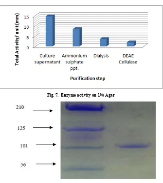
[image:4.595.134.469.56.431.2]and 1% agar culture supernatant of organism by the Ammonium sulphate precipitation, dialysis and column chromatography. The Figure 6 & & summarizes the results of each step. The activity of 0.1 % galactose maximum found in DEAE cellulose step and the activity of 1% agar maximum found in culture supernatant step. I.e. the activity of 0.1 % galactose gradually increases as purification step and in 1% Agra the activity of gradullay decreses as purification step. Similar result also found by Srinivasan 2012. The purified agarase had a molecular mass of approximately 90-105KD estimated by SDS-PAGE shown in Figure 8. The agarase secreted into the liquid culture and the enzyme was purified to homogeneity on the SDS-PAGE. The enzyme respectively indicating that the enzyme is a monomer. The enzyme cleaves β, 1- 4 linkage of agarose to produce neoagarotetrose as the predominant product. As India has a very rich soil resource, intensive research on the agar-digesters will help production of the important enzyme, agarase, in a commercial scale and in a cost-effective manner so that India no longer needs to import this enzyme thereby saving the valuable foreign exchange to a greater extent. Its widely useful properties can then be applied by the molecular biologists at lesser cost and made beneficial to the common people.
Fig. 7. Enzyme activity on 1% Agar
Conclusion
Agar-degrading agarolytic bacteria were isolated from the soil. All were Gram-negative rods and coccus shape. It could be divided into two groups. Those which only softened the agar and those which caused extensive liquefaction of agar the strains of agarolytic bacteria produced at least two enzyme complexes, one cell-free and the other cell-bound and hydrolysed agar with the formation of oligosaccharides. The strains released ‘agarase’ into the medium yielding monosaccharide as major end-products. The agar-degrading enzymes of both groups were inducible, not only by agar, but also by other galactans and polysaccharides associated with plants. The strain was Gram-negative rods, non-fermentative and highly motile. Agarases enzyme has highly commercial value agarases enzyme has used in Food, cosmetic and medical
industry (Kobayashi et al., 1997), Protoplast isolation from
seaweeds (Araki et al., 1998) and Production of agar-derived
oligosaccharides
REFERENCES
Araki, C. and Arai, K. 1954. Studies on agar-digesting bacteria. The isolation of agar-digesting bacteria and their enzymatic activities. Memoirs of the Faculty of Industrial
Arts, Kyoto Technical University, Science and Technology,
3B, 7-23
Araki, T., Hayakawa, M., Lu, Z., Karita, S. and Morishita, T. (1998). Purification and characterization of agarases from a
marine bacterium, Vibrio sp.PO-303. J. Mar. Biotechnol.,
6: 260-265.
Duckworth, M. and Yaphe, W. 1971a. The structure of agar. I. Fractionation of a complex mixture of poly saccharides.
Carbohydrate Research, 16, 189-197.
Hosoya, S., Jang, J.H., Yasumoto, H.M., Matsuda, S. and
Kasai, H. 2009. Psychromonas agarivorans sp. nov., a
novel agarolytic bacterium. Int. J. Syst. Evol. Microbiol.,
59: 1262-1266.
Izumi, K. 1972. Chemical heterogeneity of the agar from
Gracilaria verrucosa. Journal of Biochemistry, 72, 135-140.
Kobayashi, R., Takisada, M., Suzuki, T., Kirimura, K. and Usami, S. 1997. Neoagarobiose as a novel moisturizer with
whitening effect. Biosci. Biotechnol. Biochem., 61:
162-163.
Lakshmikanth, M., Manohar, S., Souche, Y. and Lalitha, J.
2006. Extracellular agarase LSL-1 producing
neoagarobiose from a newly isolated agar-liquefying soil
bacterium, Acinetobacter sp. AG LSL-1. World J.
Microbiol. Biotechnol., 22: 1087-1094.
Saraswathi, S., Vasanthabharathi, V., Kalaiselvi, V. and Jayalakshmi, S. 2011 Characterization and optimization of
agarase from an estuarine Bacillus subtilis African Journal
of Microbiology Research, Vol. 5(19), pp. 2960-2968, 23
September
Srinivasan Thulasidas 2012. Isolation and Characterization of Agarolytic Microorganisms and Purification of an
Extracellular Enzyme Agarase. International Journal of
Pharmaceutical & Biological Archives, 3(4):965-968.
Suzuki, H., Sawai, Y., Suzuki, T. and Kawai, K. 2003. Purification and characterization of an extracellular
beta-agarase from Bacillus sp. MK03. J. Biosci. Bioeng., 95:
328-334.
Yu-Fong Siea, Hui-Chun Yangb, and Yen Leea. 2009. The Discoveery of Agarolytic Bacterium with Agrarse Gene Containing Plasmid, and Sone Enzymology Characteristics.
International Journal of Applied Science and Engineering,
7, 1: 25-41