Microglia in steady state
Katrin Kierdorf, Marco Prinz
J Clin Invest.
2017;
127(9)
:3201-3209.
https://doi.org/10.1172/JCI90602
.
Microglial cells are the resident tissue macrophages of the CNS and are widely recognized
for their immune surveillance of the healthy CNS. In addition to this well-accepted function,
recent findings point to major roles for microglia in instructing and regulating the proper
function of the neuronal networks in the adult CNS, but these cells are also involved in
creating neuronal networks by orchestrating construction of the whole network during
development. In this Review, we highlight recent findings about the steady-state functions of
microglial cells, the factors that are important for physiological microglial function, and how
microglia help to maintain tissue homeostasis in the CNS.
Review Series
Find the latest version:
Introduction
The adult CNS consists of billions of neurons, with each neuron creating up to 15,000 interconnections with other neurons (1). This immense neuronal network is delicate and can be only par-tially replaced; therefore, it is necessary to maintain, support, and guard this fragile system (2–5). In the adult CNS, neuronal main-tenance and support are performed by different cell types: glial cells, including astrocytes and oligodendrocytes, often referred to as macroglia, and the resident tissue macrophages of the CNS parenchyma, including the microglia and macrophages of the CNS interfaces such as perivascular, meningeal, and choroid plexus macrophages (6, 7). Historically, the name “glia” is derived from the Greek word for glue, and for a long time, glial cells were just seen as the “cement” that maintains the structure of the CNS. Interestingly, Rudolf Virchow, who first named and described the neuroglia as a connective material between the neurons in 1856, pointed out that understanding the function and behavior of the nervous system requires characterization of the neuroglial biolo-gy (8). Following Virchow’s observations, more and more studies were performed to determine the identity of glial cells. Following the characterization of astrocytes, Müller cells, and radial glia, Santiago Ramón y Cajal discovered a new element of the neurog-lia, which he claimed was of mesodermal origin (9, 10). In 1919, Pío del Río Hortega showed for the first time that this newly dis-covered “third element” included both oligodendrocytes and microglia (11). He described the microglial cells as wandering his-tiocytes with a mesodermal origin, separating them from the other “classical” glial cells with an epidermal origin (12, 13). Nowadays, it is still accepted that microglia are resident tissue macrophages with a mesodermal origin.
Nearly every tissue in the body contains specialized tissue macrophages; however, microglia are a special subset of immune cells, as they are located within an organ that is considered to be immune-privileged (14). During the last hundred years, the aims in the field were to obtain a detailed understanding of
microg-lia under physiological and pathological conditions in the CNS. Microglia were long considered to be in a resting state in the healthy CNS, while inflammation or an infection in the CNS resulted in activation of the microglia to a professional phagocyte that either resolves the inflammation or infection or potentially promotes a pathological process. Nowadays, mounting evidence indicates that microglia are not in a resting state under healthy conditions, but instead play an essential role in maintaining tissue homeostasis under physiological conditions.
In this Review, we summarize the current state of knowledge about the physiological role and function of microglia during brain development and in the healthy adult CNS. We aim to give a detailed view of the physiological tasks microglia cells have in the healthy CNS and further highlight how microglia senescence is created and maintained during development to fulfill these essen-tial tasks in the parenchyma.
Microglial development: from an erythromyeloid
progenitor to a specialized tissue macrophage
To fully understand microglial function and properties under steady-state conditions in the CNS, it is important to under-stand the origins and developmental pathways of these cells. As proposed by Pío del Río Hortega, microglia are of mesodermal origin. In recent decades, it has become accepted that a hema-topoietic progenitor enters the CNS early on during embryonic development from a single wave of mesodermal progenitors (6, 15, 16). After several decades of debate as to whether microglia are derived from a single wave of progenitors that enter the CNS only once during development and locally expand and maintain themselves within the CNS throughout an individual’s life, or whether microglial hematopoietic progenitors invade the CNS during early embryogenesis and are also continuously exchanged from a circulating hematopoietic progenitor pool throughout life (17). A number of studies in the last several years have pro-vided detailed insights into microglial development and proven that microglial cells are derived from one single embryonic wave of progenitors (18–21). The first microglia enter the developing neuroectoderm in the mouse embryo at 9.5 days postconception (dpc) (18, 21, 22). These early embryonic microglia are derivedMicroglial cells are the resident tissue macrophages of the CNS and are widely recognized for their immune surveillance of the healthy CNS. In addition to this well-accepted function, recent findings point to major roles for microglia in instructing and regulating the proper function of the neuronal networks in the adult CNS, but these cells are also involved in creating neuronal networks by orchestrating construction of the whole network during development. In this Review, we highlight recent findings about the steady-state functions of microglial cells, the factors that are important for physiological microglial function, and how microglia help to maintain tissue homeostasis in the CNS.
Microglia in steady state
Katrin Kierdorf1 and Marco Prinz2
1Department of Life Sciences and MRC Centre for Molecular Bacteriology and Infection, Imperial College London, London, United Kingdom. 2Institute of Neuropathology, Faculty of Medicine,
University of Freiburg and BIOSS Centre for Biological Signalling Studies, University of Freiburg, Freiburg, Germany.
Conflict of interest: The authors have declared that no conflict of interest exists.
and differentiation of microglia during development are highly dependent on colony-stimulating factor 1 receptor (Csf1r) expres-sion (18, 29). Csf1r-deficient mice lack microglia and are devoid of the yolk sac–derived EMPs that give rise to embryonic microg-lia. Interestingly, mice deficient in one of the two known Csf1r ligands, colony-stimulating factor 1 (Csf1) or interleukin-34 (Il34), show only reduced numbers of microglia. Il34-deficient animals have decreased numbers of microglia in several brain regions (30, 31), whereas Csf1-deficient animals are reported to have either no microglia phenotype or a slightly decreased number of microglia (29, 32–34). In a study comparing microRNA expres-sion in adult microglia and other myeloid cells, it was found that microglia are highly dependent on TGF-β signaling during their differentiation. Microglia are absent in the CNS of TGF-β –defi-cient mice, and they develop defects in glutamate homeostasis and synaptic plasticity (35). It is not yet clear how microglia are distributed throughout the CNS, and several different mecha-nisms have been proposed for their distribution, such as migra-tion along radial glial cells toward different brain regions or the recruitment of microglia via guidance cues supplied by different brain regions (36). Undisturbed microglial development is tightly coupled to normal physiological function of adult microglia in the CNS, and a slight disturbance in the maturation and expansion of the cell population can have severe effects on the functional prop-erties of the CNS (37). Therefore, it is still necessary to uncover more details of microglial cell development to completely under-stand the process that establishes a network of adult steady-state microglia during development.
[image:3.585.54.526.60.325.2]from erythromyeloid progenitors (EMPs), which are generated from extraembryonic hematogenic endothelium positive for the angiopoietin receptor Tie2 in the blood islands of the yolk sac around 8.0 dpc. The EMPs leave the yolk sac soon after their generation and colonize the developing fetal liver, eventual-ly giving rise to a plethora of other tissue macrophages such as Langerhans cells in the epidermis or Kupffer cells in the liver (19). EMPs are c-Kit+CD45+CSF1R+CX3CR1–F4/80– progenitor cells that differentiate further into immature macrophage interme-diates and then to yolk sac–derived macrophages, which invade the CNS and give rise to the first embryonic microglia (18, 19, 21, 23). Interestingly, the CNS seems to be one of the first tissues to be colonized with tissue macrophages derived from the yolk sac EMPs (18). The differentiation of embryonic microglia from yolk sac EMPs is dependent on two myeloid transcription factors, the ETS-domain transcription factor Pu.1 and IFN regulatory factor 8 (Irf8). However, the yolk sac EMPs and embryonic microglia are independent of the transcription factor c-myb, which is a key transcription factor for the maintenance of hematopoietic stem cells (HSCs) derived from embryonic definitive hematopoiesis microglia as well as perivascular and meningeal macrophages in the CNS. It is also well accepted that adult microglia are derived from a single wave of yolk sac EMPs, with no further contribution of myeloid progenitors from another hematopoietic origin, such as circulating HSC-derived myeloid progenitors from adult bone marrow (20, 21, 23–27). Upon entering the developing neuroec-toderm, the embryonic microglial population expands via pro-liferation throughout development (21, 22, 28). The expansion
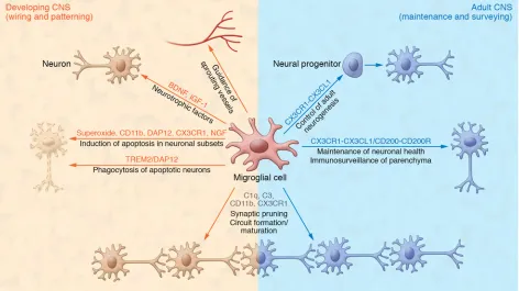
Figure 1. Homeostatic function of microglia in the developing and adult CNS. In addition to their function as a resident immune cell in the CNS
binding to low-affinity neuronal growth factor receptor p75 (52). Several in vitro studies showing the effects of microglia-secreted factors on neuronal survival indicate that there may be a variety of factors that can promote or reduce neuronal survival during development. Microglia are also involved in controlling the size of the neural precursor pool in the subventricular zone (SVZ), by eliminating T-box brain protein 2–positive (TBR2+) and paired box protein-6–positive (Pax6+) neural precursors (53). Microglia acti-vation by LPS or microglia inactiacti-vation by doxycycline or proges-terone modulates neural precursor cell number (53, 54). Overacti-vation of microglia leads to a decreased number of precursors and a thinning of the SVZ, whereas a decrease in microglial activity results in an increased number of neural precursors (54). Howev-er, it should be mentioned here that the treatments used in these studies were not specific to microglia. Also, Pu.1-deficient animals show a decreased number of proliferating cortical progenitors in ex vivo cultures (55). Thus, microglia seem to play a major part in the establishment of the correct number of neurons by regulating cell death and survival in a region-specific manner through the orchestration of prodeath and prosurvival signals.
Guidance of the developing vasculature. One important event
during the development of the CNS from a few neuronal layers to billions of neurons organized in different circuits within distinct brain regions is the vascularization of the CNS to provide nutri-ents and oxygen. The sprouting of blood vessels into the neuro-ectoderm starts around 10 dpc in mice (56), shortly after the first microglial progenitors enter the CNS. Microglia in the developing neuroectoderm play an essential role in vascular networking by serving as “tip macrophages” that connect sprouting vessels. In mice and zebrafish, microglia promote the branching of invad-ing sproutinvad-ing vessels, and a reduction in microglia leads to a decreased number of vascular branching points (57). Moreover,
Csf1-deficient animals, which do not have retinal microglia,
exhib-it decreased branching of the vascular plexus compared wexhib-ith their WT counterparts (58). Vessel growth and density were reduced after microglia depletion, but could be restored after intravitreal injection of microglia (59). Microglia guide the sprouting vessel tip by secretion of soluble guidance factors rather than by direct contact, with bidirectional communication between the sprouting vessel and the microglia (60).
Maturation and refinement of neuronal circuits. Recently,
microglia were shown to play an important role in the maturation and formation of neuronal circuits, especially during postnatal development. Electron microscopy data revealed that microglia in the healthy CNS are in constant contact with neuronal and astro-cytic elements, and it was therefore suggested that microglia are located close to synapses in the healthy brain (61, 62). It was also shown that microglial cells make direct contact with pre- and post-synaptic structures, and these cells have been seen to continuously interact for 4–5 minutes in the healthy CNS (63). However, this con-tact is greatly prolonged during the status epilepticus in the isch-emic CNS (63), and neuronal hyperactivity results in an increase in microglial processes that are recruited toward the active synapse (64). One can conclude from these and other studies that microglia seem to sense synaptic activity, prolong their contact with actively signaling synapses, and also modulate the neuronal activity at the synapse (63–65). Notably, it has not been shown that interactions
Physiological function of microglia during
embryonic and postnatal development
While the first microglial progenitors are entering the neuroec-toderm, the first neurons are born in the mouse CNS (38). There is also some evidence that microglial cells appear in CNS regions with the onset of functional neuronal networks (39). This correla-tive event might indicate that during the development of neurons and neuronal circuit formation, microglia are required in the devel-oping neuroectoderm. Csf1r-deficient animals, which do not have any microglia, exhibit severe defects in brain development with marked structural abnormalities in brain architecture, including olfactory bulb atrophy, expansion of the lateral ventricles, and dra-matic thinning of the neocortex (29, 40). Microglia seem to serve several different functions during CNS development particularly as “architects,” orchestrating and coordinating the patterning and wiring in the developing CNS. In the following sections we will fur-ther discuss four main functions of microglia during development: the phagocytosis of dead cells, the trophic support of developing neurons in the CNS, the guidance of developing vasculature in the CNS, and the support and refinement of developing neural circuits by synaptic pruning (Figure 1).
Phagocytosis of apoptotic neurons during development. As
pro-fessional phagocytes, microglia play a major role in removing apoptotic neurons from the developing CNS. In some regions up to half of all neurons born during development undergo apoptosis prior to adulthood (41, 42). Microglia are found in close proximity to apoptotic neurons in the developing brain, where they remove apoptotic debris (43–45), as well as induce programmed cell death. Microglia induce apoptosis in Purkinje cells of the cerebellum, but also in other brain regions including hippocampus, via release of superoxide ion, similar to the respiratory bursts seen in neutro-phils (46–48). Release of superoxide ion is induced by the integrin CD11b and the immune receptor DNAX activation protein of 12 kDa (DAP12) after microglial interaction with the target neuron (48). DAP12 and its receptor, triggering receptor expressed on myeloid cells 2 (TREM2), have been shown to induce removal of apoptotic neurons in vitro and indicate that microglia play a critical role in eliminating apoptotic neurons during development without inducing an inflammatory phenotype (49, 50). Interestingly, mice lacking microglia, such as Pu.1- or Csf1r-deficient animals, have not been found to have major defects in removal of apoptotic neu-rons during development, suggesting that other cells in the CNS might be involved in the removal of apoptotic neurons.
Trophic support of developing neurons. In addition to the
altered sensory response to nociceptive and thermal stimuli that results from a defect of interneurons in the dorsal spinal cord (80). HOXB8 in the CNS is expressed by a subpopulation of microglia, and transplantation of WT bone marrow into Hoxb8-mutant mice rescued pathological grooming behavior, but did not rescue the sensory phenotype (81).
Several studies in the last decade have revealed that microg-lia contribute to the formation and development of the CNS. As summarized above, this contribution includes regulation of circuit maturation processes and the guidance of developing vessels in the brain. Moreover, microglia enter the CNS at an early time point during development and function both as innate immune cells to protect the developing brain and to guide and construct the develop-ing nervous system. These finddevelop-ings suggest we are just beginndevelop-ing to understand the important roles of microglia in CNS development.
Physiological function of microglia
in the adult brain
Microglia are distributed over the entire CNS parenchyma and can be found in every region in the healthy adult CNS. The density of microglia differs between anatomical regions and ranges from 5% in the cortex to 12% in the substantia nigra (82). Interestingly, microglia seem to be more abundant in gray matter than in white matter (83). In several studies it was suggested that microglial cells maintain their own population in the adult CNS via endog-enous proliferation (20, 27, 28, 84), but only a limited amount of experimental evidence supporting this hypothesis is available so far. A recent investigation revealed that microglial cell number is tightly controlled by temporal and spatial coupling of apoptosis and proliferation within the microglial population, which provides evidence for tight control of microglial cell numbers (85). Using a novel microglia-fate mapping system, it was shown that in the healthy brain regional differences in microglia self-renewal exist and that microglia expansion is a random process during homeo-stasis that can shift to clonality upon pathology (86).
In each CNS region, microglia occupy distinct nonoverlapping territories. Therefore, each microglial cell has a highly specialized morphology under physiological conditions with a small cell soma containing few organelles and surrounding elongated processes with secondary branching and lamellipodia (87, 88). This delicate morphology was already recognized by Pío del Río Hortega, who described the radial ramified processes of microglia in 1919 (11). Compared with other tissue macrophages, microglia have a large cell surface area; for example, the perimeter of a hippocampal microglial cell is seven times that of a Kupffer cell in the liver (82). While the phenotype of microglia under physiological conditions has long been described as resting and downregulated, there is accumulating evidence that microglia are important for the main-tenance of tissue homeostasis and regulate numerous processes in the adult CNS (6, 89). New imaging techniques and genetic tools have allowed visualization of living microglia in vivo and ex vivo. It was also shown that the elongated processes of microglia move continuously, scanning the surrounding area (62, 90). Several ex vivo studies have monitored physiological microglial motility in hippocampal or cortical slice cultures (90, 91). Even though these studies do not reflect the overall physiological behavior of microglia in vivo, they showed that ramified microglia are motile between microglial processes and synaptic boutons are correlated
with the disappearance of these boutons after contact occurs. In the developing brain, the creation of the synapse architecture in neuro-nal circuits is a critical aspect of CNS maturation (66). Tremblay and colleagues used detailed light and electron microscopy to show that during learning and sensory experiences, microglia are in con-tact with different synaptic elements, including dendritic spines, axonal terminals, astrocytic processes, and the synaptic cleft (67). Microglia also engulf and remove synaptic elements during senso-ry experience and are involved in the plasticity of the synapse (67). It was later shown that microglia take up postsynaptic material, as indicated by the presence of postsynaptic density protein 95–posi-tive (PSD95+) puncta in the microglial processes, as well as synap-tosomal-associated protein 25–positive (SNAP25+) material, which is derived from presynaptic elements (68). This study further revealed a dynamic role of microglia in synaptic pruning through removal of unused dendritic spines during postnatal phases. Fur-thermore, synaptic pruning was dependent on microglia number during the postnatal phase. Cx3cr1 deficiency reduces the number of microglia during the postnatal phase, leading to reduced synap-tic pruning and impaired neuronal circuit maturation (68). Microg-lial synaptic pruning is also dependent on the neuronal activity of the dendritic spines as well as the labeling of developing synapses with complement component C3, which leads to phagocytosis via the microglial complement receptor CD11b (also known as CR3) (69, 70). Several studies indicate that microglia play a major role in synaptic pruning of neurons, and we are only starting to understand the detailed mechanisms of this process. More research is needed to further elucidate the role of the synaptic pruning performed by microglial cells compared with synaptic pruning seen in other glial cells such as astrocytes and self-pruning of neurons (70–73).
The developing CNS has many more synapses than the mature adult CNS; thus, the removal of unused synapses and the mainte-nance of functional interconnections is an important process. Dis-ruptions in this process are thought to be associated with autism spectrum disorders or other behavior defects and learning dis-orders (74, 75). Methyl CpG-binding protein 2–deficient (Mecp2- deficient) animals or animals with a myeloid-specific deletion of
Mecp2 show Rett syndrome–like phenotypes, which can be
res-cued by reintroduction of WT bone marrow–derived macrophages to the CNS (76). Interestingly, it has been shown that phagocytic activity of the WT bone marrow–derived cells is necessary to res-cue the Rett syndrome phenotype. In contrast, other studies inves-tigating the function of microglia in the development of Mecp2 deficiency–induced Rett syndrome revealed enhanced phagocyto-sis of dendritic spines during synaptic pruning that is not rescued by reintroduction of Mecp2 into microglia (77, 78). These studies suggest a contribution of microglia in disease pathology, but other mechanisms of disease could also be involved. Further investiga-tion needs to be carried out to reveal the precise contribuinvestiga-tion of microglia to the development of Rett syndrome.
Microglia as modulators of synaptic plasticity. Microglia play a
critical role in maintaining synaptic plasticity under physiological conditions and in regulating synaptic properties, especially during learning and circuit maturation (98), which particularly occur during postnatal development and early adulthood. However, it is not yet clear whether microglia play a role in the maintenance of synapse properties in the healthy adult CNS. Depletion of microg-lia in the postnatal CNS or deletion of brain-derived neurotrophic factor (BDNF) in CX3CR1+ cells, which include microglia, resulted in reduced synaptic structural plasticity associated with learning (99). Depletion of microglia in different postnatal phases led to a decrease in both synapse elimination and formation during learn-ing, indicating that microglia play a role in dendritic spine remod-eling during motor skill learning. Interestingly, microglia deletion in early adulthood still resulted in reduced synapse elimination and decreased motor learning (99). Therefore, microglia retain the capacity to regulate learning-induced synapse modification in young adult animals. Surprisingly, microglia deletion via adminis-tration of the CSF1R inhibitor PLX3397 in adult animals (2 months old) did not result in behavioral or cognitive defects or changes in synaptic properties, even after 3 weeks of treatment (100).
Adult resident microglia are in continuous contact with neu-rons to monitor their functional status. This interaction and com-munication between neurons and microglia are maintained by a variety of receptors and ligands such as CX3CR1/fractalkine (CX3CL1) or CD200/CD200R (Figure 2). The detailed inter-action network between microglia and neurons and how this network maintains the physiological status of microglia and the appropriate function of the neuronal network are reviewed in detail elsewhere (101–103).
Regulation of adult neurogenesis by microglia. Adult microglia
regulate postnatal neurogenesis in the hippocampus and SVZ. Whereas aging seems to have a negative effect on microglia, resulting in a decrease in neurogenesis, exercise and environ-mental enrichment physiologically prime microglia to support adult neurogenesis (104–106). Exercise enhances hippocampal microglial release of the chemokine CX3CL1, which protects neuronal precursor cells (106). Additionally, microglia help main-tain the hippocampal neurogenic niche in the dentate gyrus via expression of CX3CR1, which mediates the communication and interaction between microglia and neurons in adult mice (103). In several studies, microglial CX3CR1 was also shown to play a major role in supporting and regulating adult neurogenesis. Defi-ciency in CX3CR1 or interruption of CX3CR1 signaling resulted in an upregulation of proinflammatory signaling in microglia and a dramatic reduction of adult hippocampal neurogenesis (107, 108). Neuronal progenitors continuously proliferate in the sub-granular zone of the dentate gyrus, but only a few of them become integrated in functional neuronal circuits; all other cells undergo apoptosis. Ramified microglia in the hippocampus phagocytose apoptotic cells via terminal branches, building characteristic ultrastructural formations that can be described as “ball and chain” structures (109). Because of their role in the maintenance of the hippocampal neurogenic niche during adulthood, microg-lia are essential components in learning and memory formation. A recent study also showed that microglia serve important func-tions in phagocytosing apoptotic cells from the neurogenic niche and seem to retract and extend their processes while mediating
pinocytosis (90–92). Two-photon microscopy imaging revealed a strict territorial organization of microglia in vivo, with distances of 50–60 μm between each cell soma. The somata of physiolog-ical microglia are stable and nonmotile; however, the processes of each microglial cell are highly dynamic, and retract and extend to sample the surrounding CNS (61, 62). The sampling is highly random and has a high turnover rate, such that each microglia can sample the CNS parenchyma once in a few hours. The ramified morphology as well as the motility of microglial processes allows them to serve different physiological functions in the adult CNS.
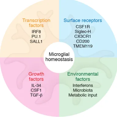
[image:6.585.68.260.62.254.2]Based on gene expression and surface protein expression profiles, a microglial cell can easily be identified as a resident tis-sue macrophage derived from yolk sac EMPs, while all yolk sac EMP-derived tissue macrophages share a core signature with each other (20, 27, 37). However, as a result of their adaptation to the CNS, microglia exhibit decreased expression of hundreds of tran-scripts compared with other tissue macrophage populations (93). Using the microglial surface marker transmembrane protein 119 (TMEM119), Bennett and colleagues performed gene expression profiling of microglia at different developmental time points and found that these cells undergo a defined maturation process that stabilizes around postnatal day 14 (94). In addition to TMEM119, several other genes were found to be highly expressed by microg-lia and could potentially be used to separate them from other tissue macrophage populations and bone marrow–derived myeloid cells. Potential microglia-specific markers are the cell surface protein Siglec-H and the transcriptional regulator Sal-like 1 (SALL1) (refs. 93, 95–97, and Figure 2). However, whether these surface markers are shared by nonmicroglia CNS macrophages in the perivascular and meningeal space is currently unclear.
Figure 2. Factors regulating microglial homeostasis. Microglial
in the SVZ (110). The Tyro3, Axl, and Mertk (TAM) RTKs Mer and Axl expressed on microglial cells were shown to be detrimental for phagoptosis. Phagoptosis is the phagocytosis of neuronal pro-genitors that are still viable but already upregulated first apop-totic markers leading to recognition by phagocytes. Therefore, microglia regulate cellular density of neuronal progenitors in the SVZ during adult neurogenesis (110).
Mutations affecting microglial function in the adult CNS. The
findings discussed above indicate that microglia play an essen-tial role in maintaining tissue homeostasis in the healthy CNS and therefore the undisturbed function of the neuronal net-work; however, the entire repertoire of microglial function has not been fully elucidated. Newly developed genetic tools and techniques will help to identify and verify more physiological functions in the future. We would now like to highlight a few examples demonstrating the detrimental role of microglial cells for a physiological and healthy tissue homeostasis in the CNS. The induced deficiency of the microglial signature transcription factor SALL1 in adult microglia resulted in a loss of microglia- specific genes and the expression of genes associated with oth-er tissue macrophages. SALL1 deficiency in microglia and othoth-er CNS tissue macrophages resulted in an increase in the number of microglia and a reactive microglia phenotype that was accompa-nied by a decrease in differentiating doublecortin-positive neu-roblasts in the hippocampus (95). Loss of the transcription factor IRF8, which is involved in microglial development, resulted in drastic morphological changes in adult microglia and a matura-tion defect accompanied by marked changes in the gene expres-sion profile of microglia under physiological conditions (20). Further, IRF8-deficient patients exhibited severe immunodefi-ciency; however, the impact of IRF8 deficiency on their cognitive function was not studied (111).
Ubiquitin-specific protease 18 (USP18) was recently found to be a critical negative regulator of microglial activation, especially in white matter microglia (112). Loss of USP18 induces an overac-tivated phenotype in resting microglia and severe CNS pathology.
Usp18-deficient microgliopathy resulted from prolonged STAT1
activation and expression of IFN-induced genes in microglial cells. Absence of type 1 IFN receptor α chain (IFNAR1) rescued the hyperactivated microglial phenotype, indicating that there is a tonic IFN signal under physiological conditions in the CNS, as well as an inability of Usp18-deficient microglia to terminate IFN sig-naling. Therefore, dysregulation of IFN signaling results in a loss of microglial homeostasis in the adult CNS (Figure 2). Notably, the recent identification of Usp18-deficient patients with severe brain abnormalities underscores the essential role of USP18 for normal CNS homeostasis (113).
Microglial interaction with peripheral processes. Under
phys-iological conditions, microglia are meant to serve as resident immune cells in a highly immune-privileged organ with little con-tact with the periphery. However, it is now clear that microglia may interact directly or indirectly (through perivascular macrophages and endothelial cells) with the periphery, including with the cir-culating blood. Microglia are highly sensitive to peripheral meta-bolic changes, with excess dietary lipids resulting in an immuno-reactive phenotype (114). It has also been shown that microglial cells in the hypothalamus exhibit a proinflammatory gene
expres-sion profile and activated morphology upon high-fat-diet feeding. Another study demonstrated that microglia are highly dependent on the presence of the gut microbiota and associated metabolites to develop into fully functional mature microglia in the adult CNS (115) (Figure 2). Microglia from germ-free mice or adult mice with depleted gut microbiota showed an immature microglial pheno-type with a concomitant impaired immunological response and a drastic change in gene expression (110), as short-chain fatty acids, which are metabolites produced from resident gut microbiota, trigger maturation of adult microglia. These recent data demon-strate an important and unexpected role of the gut microbiota in the maturation and physiological status of microglia in the CNS and suggest studies to further analyze the interaction of the gut microbiota and microglia and the consequences of these interac-tions for CNS homeostasis.
Summary
The findings summarized in this Review highlight a plethora of novel microglia functions in the CNS under physiological condi-tions, from the guidance of developing neuronal networks to the maintenance of neuronal circuits in the adult. Unlike a resting immune cell, microglia are responsible for multiple tasks in the physiological CNS to maintain homeostatic function. Further-more, these cells resemble a dynamic and highly specialized tissue macrophage, equipped with a gene signature imprinted from their yolk sac EMP origin and a highly specialized gene signature acquired upon entry into the CNS. Although we have a good idea where microglial cells originate and when they pop-ulate the CNS, it still remains unclear how microglial cells con-trol their cell number and distribution throughout the adult CNS. Based on the recent literature, we can conclude that if microglia cannot develop into their physiological ramified state, the tissue homeostasis of the adult CNS is highly disturbed. During devel-opment, microglia are integrated in a complex signaling network and highly adapted to the CNS environment; even small alter-ations during this time can disturb these cellular interactions and result in dramatic short- or long-term changes. This signaling net-work of microglial cells with neurons, but also with other neigh-boring microglial cells, is not fully understood, and we are just beginning to understand the communication and interactions of microglial cells in the healthy CNS. Recent studies have only started to demonstrate the complexity of microglial involvement in neuronal network formation and wiring, and it is still uncer-tain how many behavioral and psychiatric disorders may result from microglial dysfunction or mutations affecting microglial cells. Therefore, there are still many questions about microgli-al behavior under steady-state conditions. We believe that microgli-all of the findings described in this Review mark only the start of a full understanding of how microglial cells function under steady-state conditions, and that physiological microglial behavior will be uncovered in future studies that employ newly developed tools to study microglia in the healthy CNS.
Acknowledgments
Bundesministe-rium für Bildung und Forschung-funded Competence Network on Multiple Sclerosis (KKNMS), the European Union’s Sev-enth Framework Program FP7 under grant agreement 607962 (nEURO inflammation), the Sobek-Stiftung and the DFG (SFB 992, SFB1140, SFB/TRR167, Reinhart-Koselleck-Grant), and the Ministry of Science, Research and the Arts, Baden-Wuerttemberg (Sonderlinie “Neuroinflammation”).
Address correspondence to: Katrin Kierdorf, MRC Centre for Molec-ular Bacteriology and Infection, Department of Life Sciences, Flow-ers Building, South Kensington Campus, Imperial College London, London SW7 2AZ, United Kingdom. Email: k.kierdorf@imperial. ac.uk. Or to: Marco Prinz, Institute of Neuropathology, University of Freiburg, Breisacher Str. 64, D-79106 Freiburg, Germany. Phone: 49.761.270.51050; Email: [email protected].
1. Tang Y, Nyengaard JR, De Groot DM, Gundersen HJ. Total regional and global number of syn-apses in the human brain neocortex. Synapse. 2001;41(3):258–273.
2. Gage FH. Neurogenesis in the adult brain. J Neu-rosci. 2002;22(3):612–613.
3. Hagg T. Molecular regulation of adult CNS neu-rogenesis: an integrated view. Trends Neurosci. 2005;28(11):589–595.
4. Ming GL, Song H. Adult neurogenesis in the mammalian central nervous system. Annu Rev Neurosci. 2005;28:223–250.
5. Nottebohm F. Neuronal replacement in adult-hood. Ann N Y Acad Sci. 1985;457:143–161. 6. Prinz M, Priller J. Microglia and brain
macro-phages in the molecular age: from origin to neuropsychiatric disease. Nat Rev Neurosci. 2014;15(5):300–312.
7. Prinz M, Erny D, Hagemeyer N. Ontogeny and homeostasis of CNS myeloid cells. Nat Immunol. 2017;18(4):385–392.
8. Virchow R. Die Cellularpathologie in ihrer Begründung auf physiologische und pathol-ogische Gewebelehre. Berlin, Germany(20): August Hirschwald; 1858.
9. Kettenmann H, Verkhratsky A. Neuroglia: the 150 years after. Trends Neurosci. 2008;31(12):653–659. 10. Somjen GG. Nervenkitt: notes on the history of
the concept of neuroglia. Glia. 1988;1(1):2–9. 11. Del Rio-Hortega P. El “tercer elemento” de los
centros nerviosus. I. La microglia en estado normal. II. Intervencion de la microglia en los procesos patologicos (Celulas en bastoncito y cuerpos granuloadiposos). III. Naturaleza proba-ble de la microglia. 1919;9:68–120.
12. Del Rio-Hortega P. Microglia. In: Wilder P, ed. Cytology and Cellular Pathology of the Nervous Sys-tem. New York, New York, USA: 1932:483–534. 13. Rio-Hortega P. The microglia. Lancet.
1939;233(6036):1023–1026.
14. Carson MJ, Doose JM, Melchior B, Schmid CD, Ploix CC. CNS immune privilege: hiding in plain sight. Immunol Rev. 2006;213:48–65.
15. Ginhoux F, Lim S, Hoeffel G, Low D, Huber T. Origin and differentiation of microglia. Front Cell Neurosci. 2013;7:45.
16. Gomez Perdiguero E, Schulz C, Geissmann F. Development and homeostasis of “resident” myeloid cells: the case of the microglia. Glia. 2013;61(1):112–120.
17. Chan WY, Kohsaka S, Rezaie P. The origin and cell lineage of microglia: new concepts. Brain Res Rev. 2007;53(2):344–354.
18. Ginhoux F, et al. Fate mapping analysis reveals that adult microglia derive from primitive macro-phages. Science. 2010;330(6005):841–845. 19. Gomez Perdiguero E, et al. Tissue-resident
mac-rophages originate from yolk-sac-
derived erythro-myeloid progenitors. Nature. 2015;518(7540):547–551.
20. Hagemeyer N, et al. Transcriptome-based pro-filing of yolk sac-derived macrophages reveals a role for Irf8 in macrophage maturation. EMBO J. 2016;35(16):1730–1744.
21. Kierdorf K, et al. Microglia emerge from erythro-myeloid precursors via Pu.1- and Irf8-dependent pathways. Nat Neurosci. 2013;16(3):273–280. 22. Alliot F, Godin I, Pessac B. Microglia derive from
progenitors, originating from the yolk sac, and which proliferate in the brain. Brain Res Dev Brain Res. 1999;117(2):145–152.
23. Hoeffel G, et al. C-Myb(+) erythro-myeloid progenitor-derived fetal monocytes give rise to adult tissue-resident macrophages. Immunity. 2015;42(4):665–678.
24. Ajami B, Bennett JL, Krieger C, Tetzlaff W, Rossi FM. Local self-renewal can sustain CNS microg-lia maintenance and function throughout adult life. Nat Neurosci. 2007;10(12):1538–1543. 25. Goldmann T, et al. Origin, fate and dynamics of
macrophages at central nervous system interfac-es. Nat Immunol. 2016;17(7):797–805. 26. Mildner A, et al. Microglia in the adult brain
arise from Ly-6ChiCCR2+ monocytes only
under defined host conditions. Nat Neurosci. 2007;10(12):1544–1553.
27. Schulz C, et al. A lineage of myeloid cells inde-pendent of Myb and hematopoietic stem cells. Science. 2012;336(6077):86–90.
28. Hashimoto D, et al. Tissue-resident macrophages self-maintain locally throughout adult life with minimal contribution from circulating mono-cytes. Immunity. 2013;38(4):792–804. 29. Erblich B, Zhu L, Etgen AM, Dobrenis K, Pollard
JW. Absence of colony stimulation factor-1 recep-tor results in loss of microglia, disrupted brain development and olfactory deficits. PLoS One. 2011;6(10):e26317.
30. Greter M, et al. Stroma-derived interleukin-34 controls the development and maintenance of langerhans cells and the maintenance of microg-lia. Immunity. 2012;37(6):1050–1060. 31. Wang Y, et al. IL-34 is a tissue-restricted ligand
of CSF1R required for the development of Langerhans cells and microglia. Nat Immunol. 2012;13(8):753–760.
32. Blevins G, Fedoroff S. Microglia in colony-stim-ulating factor 1-deficient op/op mice. J Neurosci Res. 1995;40(4):535–544.
33. Kondo Y, Lemere CA, Seabrook TJ. Osteopetrotic (op/op) mice have reduced microglia, no Abeta deposition, and no changes in dopaminergic neu-rons. J Neuroinflammation. 2007;4:31. 34. Luo J, et al. Colony-stimulating factor 1
recep-tor (CSF1R) signaling in injured neurons facilitates protection and survival. J Exp Med.
2013;210(1):157–172.
35. Butovsky O, et al. Identification of a unique
TGF-β-dependent molecular and functional
signa-ture in microglia. Nat Neurosci. 2014;17(1):131–143. 36. Pont-Lezica L, Béchade C, Belarif-Cantaut
Y, Pascual O, Bessis A. Physiological roles of microglia during development. J Neurochem. 2011;119(5):901–908.
37. Matcovitch-Natan O, et al. Microglia development follows a stepwise program to regulate brain homeostasis. Science. 2016;353(6301):aad8670.
38. Martynoga B, Drechsel D, Guillemot F. Molecular control of neurogenesis: a view from the mam-malian cerebral cortex. Cold Spring Harb Perspect Biol. 2012;4(10):a008359.
39. Rigato C, Buckinx R, Le-Corronc H, Rigo JM, Legendre P. Pattern of invasion of the embryonic mouse spinal cord by microglial cells at the time of the onset of functional neuronal networks. Glia. 2011;59(4):675–695.
40. Nandi S, et al. The CSF-1 receptor ligands IL-34 and CSF-1 exhibit distinct developmental brain expression patterns and regulate neural progen-itor cell maintenance and maturation. Dev Biol. 2012;367(2):100–113.
41. Dekkers MP, Barde YA. Developmental biology. Programmed cell death in neuronal develop-ment. Science. 2013;340(6128):39–41. 42. Dekkers MP, Nikoletopoulou V, Barde YA. Cell
biology in neuroscience: Death of developing neurons: new insights and implications for con-nectivity. J Cell Biol. 2013;203(3):385–393. 43. Ashwell K. Microglia and cell death in the
devel-oping mouse cerebellum. Brain Res Dev Brain Res. 1990;55(2):219–230.
44. Brockhaus J, Möller T, Kettenmann H. Phago-cytozing ameboid microglial cells studied in a mouse corpus callosum slice preparation. Glia. 1996;16(1):81–90.
45. Witting A, Müller P, Herrmann A, Kettenmann H, Nolte C. Phagocytic clearance of apoptotic neu-rons by microglia/brain macrophages in vitro: involvement of lectin-, integrin-, and phospha-tidylserine-mediated recognition. J Neurochem. 2000;75(3):1060–1070.
46. Marín-Teva JL, Dusart I, Colin C, Gervais A, van Rooijen N, Mallat M. Microglia promote the death of developing Purkinje cells. Neuron. 2004;41(4):535–547.
47. Peri F, Nüsslein-Volhard C. Live imaging of neu-ronal degradation by microglia reveals a role for v0-ATPase a1 in phagosomal fusion in vivo. Cell. 2008;133(5):916–927.
J Neurosci. 2008;28(32):8138–8143.
49. Takahashi K, Rochford CD, Neumann H. Clear-ance of apoptotic neurons without inflammation by microglial triggering receptor expressed on myeloid cells-2. J Exp Med. 2005;201(4):647–657. 50. Takahashi K, Prinz M, Stagi M, Chechneva O,
Neumann H. TREM2-transduced myeloid pre-cursors mediate nervous tissue debris clearance and facilitate recovery in an animal model of multiple sclerosis. PLoS Med. 2007;4(4):e124. 51. Ueno M, et al. Layer V cortical neurons require
microglial support for survival during postnatal development. Nat Neurosci. 2013;16(5):543–551. 52. Frade JM, Barde YA. Microglia-derived nerve
growth factor causes cell death in the developing retina. Neuron. 1998;20(1):35–41.
53. Cunningham CL, Martínez-Cerdeño V, Noctor SC. Microglia regulate the number of neural precursor cells in the developing cerebral cortex. J Neurosci. 2013;33(10):4216–4233.
54. Tronnes AA, Koschnitzky J, Daza R, Hitti J, Ramirez JM, Hevner R. Effects of lipopolysac-charide and progesterone exposures on embry-onic cerebral cortex development in mice. Reprod Sci. 2016;23(6):771–778.
55. Antony JM, Paquin A, Nutt SL, Kaplan DR, Miller FD. Endogenous microglia regulate development of embryonic cortical precursor cells. J Neurosci Res. 2011;89(3):286–298.
56. Risau W. Development and differentiation of endothelium. Kidney Int Suppl. 1998;67:S3–S6. 57. Fantin A, et al. Tissue macrophages act as cellular
chaperones for vascular anastomosis down-stream of VEGF-mediated endothelial tip cell induction. Blood. 2010;116(5):829–840. 58. Kubota Y, et al. M-CSF inhibition selectively
targets pathological angiogenesis and lymphan-giogenesis. J Exp Med. 2009;206(5):1089–1102. 59. Checchin D, Sennlaub F, Levavasseur E, Leduc
M, Chemtob S. Potential role of microglia in reti-nal blood vessel formation. Invest Ophthalmol Vis Sci. 2006;47(8):3595–3602.
60. Rymo SF, Gerhardt H, Wolfhagen Sand F, Lang R, Uv A, Betsholtz C. A two-way communication between microglial cells and angiogenic sprouts regulates angiogenesis in aortic ring cultures. PLoS One. 2011;6(1):e15846.
61. Davalos D, et al. ATP mediates rapid microglial response to local brain injury in vivo. Nat Neuros-ci. 2005;8(6):752–758.
62. Nimmerjahn A, Kirchhoff F, Helmchen F. Resting microglial cells are highly dynamic sur-veillants of brain parenchyma in vivo. Science. 2005;308(5726):1314–1318.
63. Wake H, Moorhouse AJ, Jinno S, Kohsaka S, Nabekura J. Resting microglia directly monitor the functional state of synapses in vivo and deter-mine the fate of ischemic terminals. J Neurosci. 2009;29(13):3974–3980.
64. Eyo UB, Peng J, Swiatkowski P, Mukherjee A, Bispo A, Wu LJ. Neuronal hyperactivity recruits microglial processes via neuronal NMDA recep-tors and microglial P2Y12 receprecep-tors after status epilepticus. J Neurosci. 2014;34(32):10528–10540. 65. Li Y, Du XF, Liu CS, Wen ZL, Du JL. Reciprocal
regulation between resting microglial dynam-ics and neuronal activity in vivo. Dev Cell. 2012;23(6):1189–1202.
66. Katz LC, Shatz CJ. Synaptic activity and the construction of cortical circuits. Science. 1996;274(5290):1133–1138.
67. Tremblay MÈ, Lowery RL, Majewska AK. Microg-lial interactions with synapses are modulated by visual experience. PLoS Biol. 2010;8(11):e1000527. 68. Paolicelli RC, et al. Synaptic pruning by microglia is necessary for normal brain development. Sci-ence. 2011;333(6048):1456–1458.
69. Schafer DP, et al. Microglia sculpt postnatal neu-ral circuits in an activity and complement-depen-dent manner. Neuron. 2012;74(4):691–705. 70. Stevens B, et al. The classical complement
cas-cade mediates CNS synapse elimination. Cell. 2007;131(6):1164–1178.
71. Allen NJ, Barres BA. Signaling between glia and neurons: focus on synaptic plasticity. Curr Opin Neurobiol. 2005;15(5):542–548.
72. Stettler DD, Yamahachi H, Li W, Denk W, Gilbert CD. Axons and synaptic boutons are highly dynamic in adult visual cortex. Neuron. 2006;49(6):877–887.
73. Tasdemir-Yilmaz OE, Freeman MR. Astrocytes engage unique molecular programs to engulf pruned neuronal debris from distinct subsets of neurons. Genes Dev. 2014;28(1):20–33. 74. Marín O. Interneuron dysfunction in psychiatric
disorders. Nat Rev Neurosci. 2012;13(2):107–120. 75. Schafer DP, Lehrman EK, Stevens B. The
“quad-partite” synapse: microglia-synapse inter-actions in the developing and mature CNS. Glia. 2013;61(1):24–36.
76. Derecki NC, et al. Wild-type microglia arrest pathology in a mouse model of Rett syndrome. Nature. 2012;484(7392):105–109.
77. Schafer DP, et al. Microglia contribute to circuit defects in Mecp2 null mice independent of microglia-specific loss of Mecp2 expression. Elife. 2016;5:e15224.
78. Wang J, et al. Wild-type microglia do not reverse pathology in mouse models of Rett syndrome. Nature. 2015;521(7552):E1–E4.
79. Greer JM, Capecchi MR. Hoxb8 is required for normal grooming behavior in mice. Neuron. 2002;33(1):23–34.
80. Holstege JC, et al. Loss of Hoxb8 alters spinal dor-sal laminae and sensory responses in mice. Proc Natl Acad Sci U S A. 2008;105(17):6338–6343. 81. Chen SK, et al. Hematopoietic origin of patho-logical grooming in Hoxb8 mutant mice. Cell. 2010;141(5):775–785.
82. Lawson LJ, Perry VH, Dri P, Gordon S. Hetero-geneity in the distribution and morphology of microglia in the normal adult mouse brain. Neu-roscience. 1990;39(1):151–170.
83. Perry VH, Hume DA, Gordon S. Immunohisto chemical localization of macrophages and microglia in the adult and developing mouse brain. Neuroscience. 1985;15(2):313–326. 84. Lawson LJ, Perry VH, Gordon S. Turnover of
res-ident microglia in the normal adult mouse brain. Neuroscience. 1992;48(2):405–415.
85. Askew K, et al. Coupled proliferation and apop-tosis maintain the rapid turnover of microglia in the adult brain. Cell Rep. 2017;18(2):391–405. 86. Tay TL, et al. A new fate mapping system reveals
context-dependent random or clonal expansion of microglia. Nat Neurosci. 2017;20(6):793–803.
87. Perry VH, Gordon S. Macrophages and microg-lia in the nervous system. Trends Neurosci. 1988;11(6):273–277.
88. Perry VH, Gordon S. Macrophages and the ner-vous system. Int Rev Cytol. 1991;125:203–244. 89. Prinz M, Priller J. The role of peripheral immune
cells in the CNS in steady state and disease. Nat Neurosci. 2017;20(2):136–144.
90. Stence N, Waite M, Dailey ME. Dynamics of microglial activation: a confocal time-lapse analysis in hippocampal slices. Glia. 2001;33(3):256–266.
91. Glenn JA, Booth PL, Thomas WE. Pinocytotic activity in ramified microglia. Neurosci Lett. 1991;123(1):27–31.
92. Booth PL, Thomas WE. Evidence for motility and pinocytosis in ramified microglia in tissue culture. Brain Res. 1991;548(1–2):163–171. 93. Gautier EL, et al. Gene-expression profiles and
transcriptional regulatory pathways that underlie the identity and diversity of mouse tissue macro-phages. Nat Immunol. 2012;13(11):1118–1128. 94. Bennett ML, et al. New tools for studying
microg-lia in the mouse and human CNS. Proc Natl Acad Sci U S A. 2016;113(12):E1738–E1746.
95. Buttgereit A, et al. Sall1 is a transcriptional reg-ulator defining microglia identity and function. Nat Immunol. 2016;17(12):1397–1406. 96. Koso H, et al. Conditional rod photoreceptor
ablation reveals Sall1 as a microglial marker and regulator of microglial morphology in the retina. Glia. 2016;64(11):2005–2024.
97. Lavin Y, et al. Tissue-resident macrophage enhancer landscapes are shaped by the local microenvironment. Cell. 2014;159(6):1312–1326. 98. Tremblay MÈ, Stevens B, Sierra A, Wake
H, Bessis A, Nimmerjahn A. The role of microglia in the healthy brain. J Neurosci. 2011;31(45):16064–16069.
99. Parkhurst CN, et al. Microglia promote learning- dependent synapse formation through brain- derived neurotrophic factor. Cell.
2013;155(7):1596–1609.
100. Elmore MR, et al. Colony-stimulating factor 1 receptor signaling is necessary for microglia via-bility, unmasking a microglia progenitor cell in the adult brain. Neuron. 2014;82(2):380–397. 101. Biber K, Neumann H, Inoue K, Boddeke HW.
Neuronal ‘On’ and ‘Off’ signals control microg-lia. Trends Neurosci. 2007;30(11):596–602. 102. Hanisch UK, Kettenmann H. Microglia:
active sensor and versatile effector cells in the normal and pathologic brain. Nat Neurosci. 2007;10(11):1387–1394.
103. Kierdorf K, Prinz M. Factors regulating microglia activation. Front Cell Neurosci. 2013;7:44. 104. Choi SH, et al. Non-cell-autonomous effects of
presenilin 1 variants on enrichment-mediated hippocampal progenitor cell proliferation and differentiation. Neuron. 2008;59(4):568–580. 105. Gebara E, Sultan S, Kocher-Braissant J, Toni N.
Adult hippocampal neurogenesis inversely cor-relates with microglia in conditions of voluntary running and aging. Front Neurosci. 2013;7:145. 106. Vukovic J, Colditz MJ, Blackmore DG, Ruitenberg
107. Bachstetter AD, et al. Fractalkine and CX 3 CR1 regulate hippocampal neurogenesis in adult and aged rats. Neurobiol Aging. 2011;32(11):2030–2044. 108. Sellner S, et al. Microglial CX3CR1 promotes
adult neurogenesis by inhibiting Sirt 1/p65 sig-naling independent of CX3CL1. Acta Neuropathol Commun. 2016;4(1):102.
109. Sierra A, et al. Microglia shape adult hippocam-pal neurogenesis through apoptosis-coupled phagocytosis. Cell Stem Cell. 2010;7(4):483–495.
110. Fourgeaud L, et al. TAM receptors regulate mul-tiple features of microglial physiology. Nature. 2016;532(7598):240–244.
111. Hambleton S, et al. IRF8 mutations and human dendritic-cell immunodeficiency. N Engl J Med. 2011;365(2):127–138.
112. Goldmann T, et al. USP18 lack in microglia causes destructive interferonopathy of the mouse brain. EMBO J. 2015;34(12):1612–1629.
113. Meuwissen ME, et al. Human USP18 deficiency
underlies type 1 interferonopathy leading to severe pseudo-TORCH syndrome. J Exp Med. 2016;213(7):1163–1174.
114. Baufeld C, Osterloh A, Prokop S, Miller KR, Hep-pner FL. High-fat diet-induced brain region- specific phenotypic spectrum of CNS resident microglia. Acta Neuropathol. 2016;132(3):361–375. 115. Erny D, et al. Host microbiota constantly control