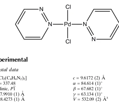
trans
-Dichloridobis(pyridazine-
j
N
)-palladium(II)
Baptiste Larame´e* and Garry S. Hanan
De´partement de Chimie, Universite´ de Montre´al, Pavillon J.-A. Bombardier, 5155 Decelles Avenue, Montre´al, Que´bec, H3T 2B1, Canada
Correspondence e-mail: baptiste.laramee-milette@umontreal.ca
Received 25 September 2013; accepted 2 December 2013
Key indicators: single-crystal X-ray study;T= 150 K; mean(C–C) = 0.004 A˚; Rfactor = 0.023;wRfactor = 0.068; data-to-parameter ratio = 14.2.
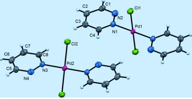
The title compound, [PdCl2(C4H4N2)2], contains two crystal-lographically unique complexes; the PdII atom lies on an inversion center in both cases. The two pyridazine units bonded to the PdIIatom are thus coplanar although dihedral angles within each complex are different. In one complex, the angle between the ring plane and Pd—Cl bond is almost perpendicular [89.4 (1)], while the other is tilted with an angle of 60.0 (1). In the crystal, weak C H—N hydrogen bonds and C H—Cl interactions connect the two indepen-dent complex molecules.
Related literature
For related pyridazine copper, nickel, silver and rhenium metal complexes, see: Otienoet al.(1995); Canoet al.(2000); Degtyarenko et al. (2008) and Raimondi et al. (2012), respectively.
Experimental
Crystal data
[PdCl2(C4H4N2)2]
Mr= 337.48 Triclinic,P1
a= 7.9910 (1) A˚
b= 8.4273 (1) A˚
c= 9.6172 (2) A˚
= 84.614 (1)
= 67.682 (1)
= 63.134 (1) V= 532.09 (2) A˚3
Z= 2
CuKradiation
= 18.45 mm1
T= 150 K
0.080.060.06 mm
Data collection
Bruker APEXII CCD diffractometer
Absorption correction: multi-scan (SADABS; Sheldrick, 1996)
Tmin= 0.216,Tmax= 0.260
13678 measured reflections 1971 independent reflections 1957 reflections withI> 2(I)
Rint= 0.018
Refinement
R[F2> 2(F2)] = 0.023
wR(F2) = 0.068
S= 1.07 1971 reflections
139 parameters
H-atom parameters constrained
max= 0.97 e A˚
3
min=0.75 e A˚
[image:1.610.58.256.524.697.2]3
Table 1
Hydrogen-bond geometry (A˚ ,).
D—H A D—H H A D A D—H A
C4—H4 N4i 0.95 2.55 3.438 (3) 155
C3—H3 Cl2ii
0.95 2.94 3.569 (3) 125
C1—H1 Cl2iii
0.95 2.92 3.787 (3) 153
C8—H8 Cl1iv 0.95 2.82 3.529 (3) 132
Symmetry codes: (i)xþ1;yþ1;zþ2; (ii) x;yþ1;zþ1; (iii)x;yþ1;z; (iv)
x;y;z1.
Data collection:APEX2(Bruker, 2011); cell refinement:SAINT (Bruker, 2011); data reduction:SAINT; program(s) used to solve structure:SHELXS97(Sheldrick, 2008); program(s) used to refine structure: SHELXL97 (Sheldrick, 2008); molecular graphics: SHELXTL(Sheldrick, 2008); software used to prepare material for publication:publCIF(Westrip, 2010).
The authors thank the Department of Chemistry of the Universite´ de Montre´al for access to the CCD facility. We thank Thierry Maris for useful crystallographic discussions. We are grateful to the Universite´ de Montre´al for financial assistance.
Supplementary data and figures for this paper are available from the IUCr electronic archives (Reference: NK2214).
References
Bruker (2011).APEX2andSAINT. Bruker AXS Inc., Madison, Wisconsin, USA.
Cano, J., De Munno, G., Lloret, F. & Julve, M. (2000).Inorg. Chem.39, 1611– 1614.
Degtyarenko, A. S., Solntsev, P. V., Krautscheid, H., Rusanov, E. B., Chernega, A. N. & Domasevitch, K. V. (2008).New J. Chem.32, 1910–1918. Otieno, T., Rettig, S. J., Thompson, R. C. & Trotter, J. (1995).Inorg. Chem.34,
1718–1725.
Raimondi, A., Panigati, M., Maggioni, D., D’Alfonso, L., Mercandelli, P., Mussini, P. & D’Alfonso, G. (2012).Inorg. Chem.51, 2966–2975. Sheldrick, G. M. (1996).SADABS. University of Go¨ttingen, Germany. Sheldrick, G. M. (2008).Acta Cryst.A64, 112–122.
Westrip, S. P. (2010).J. Appl. Cryst.43, 920–925.
Acta Crystallographica Section E
Structure Reports
Online
supporting information
Acta Cryst. (2014). E70, m17 [https://doi.org/10.1107/S1600536813032716]
trans
-Dichloridobis(pyridazine-
κ
N
)palladium(II)
Baptiste Laram
é
e and Garry S. Hanan
S1. Comment
In the present work, a square planar trans-bis(chloro)-bis(pyridazine-κN) palladium(II) metal complex has been
synthesized. Similar metal complexes are already known in coordination polymer chemistry (Degtyarenko et al., 2008)).
The molecular structure of the title compound is illustrated in Fig. 1, where two molecules are found in the asymmetric
unit. The bond distances are unexceptional. In one complex the plane of the pyridazyl ring is perpendicular with respect
to the Cl–Pd–Cl axis, while in the second molecules the ring is slightly tilted with an angle of 60 (1)°, which may be due
to the presence of weak hydrogen bonds.
S2. Experimental
trans-bis(chloro)-bis(pyridazine-κN)palladium(II). Pyridazine (0.12 mg, 0.0015 mmol) is added into a nitromethane
solution (1.0 mL) of PdCl2(MeCN)2 (0.39 mg, 0.0015 mmol), and heated to 80 °C for 12 hours. After 3 hours, a yellow
precipitate started to form. The precipitate was isolated by filtration and redissolved in a minimum amount of dimethyl
sulfoxide. Clear bronze crystals were obtained by slow diffusion of THF into the DMSO solution over 2 weeks. 1H NMR
(400 MHz, CD3NO2) delta ppm 9.15-9.13 (t, J=3.5 Hz. 4 H) 8.80 (t, J=3.2 Hz, 4 H).
S3. Refinement
H atoms were positioned geometrically (C—H 0.95 Å) and included in the refinement in the riding model approximation;
their temperature displacement parameters were set to 1.2 times the equivalent isotropic temperature factors of the parent
Figure 1
The molecular structure of trans-bis(chloro)-bis(pyridazine-κN)palladium(II), with atom labels and displacement
ellipsoids drawn at the 80% probability level. The two halves of both complexes are related by inversion symmetry.
trans-Dichloridobis(pyridazine-κN)palladium(II)
Crystal data
[PdCl2(C4H4N2)2]
Mr = 337.48 Triclinic, P1 Hall symbol: -P 1
a = 7.9910 (1) Å
b = 8.4273 (1) Å
c = 9.6172 (2) Å
α = 84.614 (1)°
β = 67.682 (1)°
γ = 63.134 (1)°
V = 532.09 (2) Å3
Z = 2
F(000) = 328
Dx = 2.106 Mg m−3
Cu Kα radiation, λ = 1.54178 Å Cell parameters from 9992 reflections
θ = 5.9–70.9°
µ = 18.45 mm−1
T = 150 K Block, brown
0.08 × 0.06 × 0.06 mm
Data collection
Bruker APEXII CCD diffractometer
Radiation source: fine-focus sealed tube Graphite monochromator
φ and ω scans
Absorption correction: multi-scan (SADABS; Sheldrick, 1996)
Tmin = 0.216, Tmax = 0.260
13678 measured reflections 1971 independent reflections 1957 reflections with I > 2σ(I)
Rint = 0.018
θmax = 70.9°, θmin = 5.0°
h = −9→9
k = −9→10
l = −11→11
Refinement
Refinement on F2
Least-squares matrix: full
R[F2 > 2σ(F2)] = 0.023
wR(F2) = 0.068
S = 1.07 1971 reflections
Primary atom site location: structure-invariant direct methods
Secondary atom site location: difference Fourier map
w = 1/[σ2(F
o2) + (0.0456P)2 + 0.8794P]
where P = (Fo2 + 2Fc2)/3
(Δ/σ)max < 0.001
Δρmax = 0.97 e Å−3
Δρmin = −0.75 e Å−3
Special details
Experimental. X-ray crystallographic data for I were collected from a single-crystal sample, which was mounted on a loop fiber. Data were collected using a Bruker Platform diffractometer, equipped with a Bruker SMART 4 K Charged-Coupled Device (CCD) Area Detector using the program APEX2 and a Nonius FR591 rotating anode equiped with a Montel 200 optics The crystal-to-detector distance was 5.0 cm, and the data collection was carried out in 512 x 512 pixel mode. The initial unit-cell parameters were determined by a least-squares fit of the angular setting of strong reflections, collected by a 10.0 degree scan in 33 frames over four different parts of the reciprocal space (132 frames total). One complete sphere of data was collected, to better than 0.80 Å resolution.
Geometry. All e.s.d.'s (except the e.s.d. in the dihedral angle between two l.s. planes) are estimated using the full covariance matrix. The cell e.s.d.'s are taken into account individually in the estimation of e.s.d.'s in distances, angles and torsion angles; correlations between e.s.d.'s in cell parameters are only used when they are defined by crystal symmetry. An approximate (isotropic) treatment of cell e.s.d.'s is used for estimating e.s.d.'s involving l.s. planes.
Refinement. Refinement of F2 against ALL reflections. The weighted R-factor wR and goodness of fit S are based on F2,
conventional R-factors R are based on F, with F set to zero for negative F2. The threshold expression of F2 > σ(F2) is used
only for calculating R-factors(gt) etc. and is not relevant to the choice of reflections for refinement. R-factors based on F2
are statistically about twice as large as those based on F, and R- factors based on ALL data will be even larger.
Fractional atomic coordinates and isotropic or equivalent isotropic displacement parameters (Å2)
x y z Uiso*/Ueq
Pd1 0.5000 0.5000 1.0000 0.01106 (11)
Cl1 0.18605 (9) 0.51044 (8) 1.13317 (7) 0.01691 (15)
N1 0.3652 (3) 0.7675 (3) 1.0036 (3) 0.0132 (5)
N2 0.3068 (3) 0.8322 (3) 0.8878 (3) 0.0170 (5)
C1 0.2179 (4) 1.0087 (4) 0.8859 (3) 0.0162 (5)
H1 0.1750 1.0556 0.8050 0.019*
C2 0.1834 (4) 1.1295 (4) 0.9954 (3) 0.0171 (6)
H2 0.1207 1.2548 0.9892 0.021*
C3 0.2440 (4) 1.0592 (4) 1.1123 (3) 0.0186 (6)
H3 0.2238 1.1347 1.1906 0.022*
C4 0.3358 (4) 0.8747 (4) 1.1130 (3) 0.0166 (5)
H4 0.3785 0.8237 1.1932 0.020*
Pd2 0.5000 0.0000 0.5000 0.01214 (11)
Cl2 0.20050 (9) 0.03267 (8) 0.49612 (7) 0.01848 (16)
N3 0.3747 (3) 0.2644 (3) 0.5494 (3) 0.0141 (5)
N4 0.3653 (3) 0.3203 (3) 0.6799 (3) 0.0169 (5)
C5 0.2860 (4) 0.4951 (4) 0.7117 (3) 0.0175 (5)
H5 0.2774 0.5357 0.8042 0.021*
C6 0.2145 (4) 0.6235 (4) 0.6188 (4) 0.0191 (6)
H6 0.1618 0.7473 0.6452 0.023*
C7 0.2239 (4) 0.5624 (4) 0.4878 (3) 0.0196 (6)
H7 0.1769 0.6429 0.4197 0.023*
C8 0.3034 (4) 0.3806 (4) 0.4574 (3) 0.0156 (5)
Atomic displacement parameters (Å2)
U11 U22 U33 U12 U13 U23
Pd1 0.01260 (16) 0.00575 (16) 0.01452 (16) −0.00218 (11) −0.00736 (11) 0.00139 (10)
Cl1 0.0152 (3) 0.0133 (3) 0.0216 (3) −0.0055 (2) −0.0080 (2) 0.0037 (2)
N1 0.0136 (10) 0.0093 (11) 0.0164 (11) −0.0043 (9) −0.0067 (9) 0.0027 (9)
N2 0.0188 (11) 0.0152 (12) 0.0160 (11) −0.0060 (9) −0.0078 (9) 0.0023 (9)
C1 0.0147 (12) 0.0135 (13) 0.0184 (12) −0.0041 (10) −0.0077 (10) 0.0048 (10)
C2 0.0139 (13) 0.0115 (14) 0.0226 (14) −0.0038 (11) −0.0063 (11) 0.0029 (11)
C3 0.0201 (14) 0.0147 (15) 0.0219 (14) −0.0060 (11) −0.0105 (11) −0.0007 (11)
C4 0.0187 (13) 0.0126 (13) 0.0194 (13) −0.0050 (11) −0.0108 (11) 0.0018 (11)
Pd2 0.01237 (16) 0.00725 (17) 0.01663 (16) −0.00237 (11) −0.00801 (11) 0.00169 (11)
Cl2 0.0156 (3) 0.0139 (3) 0.0278 (3) −0.0051 (2) −0.0121 (3) 0.0025 (2)
N3 0.0125 (10) 0.0092 (11) 0.0197 (12) −0.0032 (9) −0.0069 (9) 0.0000 (9)
N4 0.0168 (11) 0.0162 (12) 0.0179 (11) −0.0063 (9) −0.0082 (9) 0.0022 (9)
C5 0.0157 (12) 0.0131 (13) 0.0214 (13) −0.0040 (10) −0.0065 (11) −0.0039 (10)
C6 0.0143 (13) 0.0102 (14) 0.0266 (14) −0.0031 (11) −0.0041 (11) −0.0006 (11)
C7 0.0164 (13) 0.0173 (15) 0.0214 (14) −0.0048 (11) −0.0077 (11) 0.0048 (12)
C8 0.0157 (12) 0.0135 (13) 0.0169 (12) −0.0043 (10) −0.0085 (10) 0.0025 (10)
Geometric parameters (Å, º)
Pd1—N1 2.009 (2) Pd2—N3 2.003 (2)
Pd1—N1i 2.009 (2) Pd2—N3ii 2.004 (2)
Pd1—Cl1 2.3072 (6) Pd2—Cl2 2.2969 (6)
Pd1—Cl1i 2.3073 (6) Pd2—Cl2ii 2.2969 (6)
N1—C4 1.333 (4) N3—C8 1.335 (4)
N1—N2 1.344 (3) N3—N4 1.346 (3)
N2—C1 1.329 (4) N4—C5 1.327 (3)
C1—C2 1.396 (4) C5—C6 1.393 (4)
C1—H1 0.9500 C5—H5 0.9500
C2—C3 1.370 (4) C6—C7 1.369 (4)
C2—H2 0.9500 C6—H6 0.9500
C3—C4 1.388 (4) C7—C8 1.377 (4)
C3—H3 0.9500 C7—H7 0.9500
C4—H4 0.9500 C8—H8 0.9500
N1—Pd1—N1i 180.0 N3—Pd2—N3ii 180.000 (1)
N1—Pd1—Cl1 89.26 (7) N3—Pd2—Cl2 89.44 (7)
N1i—Pd1—Cl1 90.74 (7) N3ii—Pd2—Cl2 90.56 (7)
N1—Pd1—Cl1i 90.74 (7) N3—Pd2—Cl2ii 90.56 (7)
N1i—Pd1—Cl1i 89.26 (7) N3ii—Pd2—Cl2ii 89.44 (7)
Cl1—Pd1—Cl1i 179.999 (1) Cl2—Pd2—Cl2ii 180.0
C4—N1—N2 121.9 (2) C8—N3—N4 121.2 (2)
C4—N1—Pd1 122.5 (2) C8—N3—Pd2 121.78 (19)
N2—C1—H1 117.9 N4—C5—H5 117.7
C2—C1—H1 117.9 C6—C5—H5 117.7
C3—C2—C1 117.0 (3) C7—C6—C5 116.8 (3)
C3—C2—H2 121.5 C7—C6—H6 121.6
C1—C2—H2 121.5 C5—C6—H6 121.6
C2—C3—C4 118.1 (3) C6—C7—C8 118.1 (3)
C2—C3—H3 120.9 C6—C7—H7 121.0
C4—C3—H3 120.9 C8—C7—H7 121.0
N1—C4—C3 121.5 (3) N3—C8—C7 122.1 (3)
N1—C4—H4 119.3 N3—C8—H8 118.9
C3—C4—H4 119.3 C7—C8—H8 118.9
N1i—Pd1—N1—C4 37 (100) N3ii—Pd2—N3—C8 −88 (32)
Cl1—Pd1—N1—C4 90.8 (2) Cl2—Pd2—N3—C8 −60.0 (2)
Cl1i—Pd1—N1—C4 −89.2 (2) Cl2ii—Pd2—N3—C8 120.0 (2)
N1i—Pd1—N1—N2 −143 (100) N3ii—Pd2—N3—N4 92 (32)
Cl1—Pd1—N1—N2 −89.38 (18) Cl2—Pd2—N3—N4 120.08 (18)
Cl1i—Pd1—N1—N2 90.62 (18) Cl2ii—Pd2—N3—N4 −59.92 (18)
C4—N1—N2—C1 0.1 (4) C8—N3—N4—C5 −1.2 (4)
Pd1—N1—N2—C1 −179.70 (18) Pd2—N3—N4—C5 178.73 (18)
N1—N2—C1—C2 0.5 (4) N3—N4—C5—C6 −0.7 (4)
N2—C1—C2—C3 −0.8 (4) N4—C5—C6—C7 1.5 (4)
C1—C2—C3—C4 0.4 (4) C5—C6—C7—C8 −0.4 (4)
N2—N1—C4—C3 −0.4 (4) N4—N3—C8—C7 2.4 (4)
Pd1—N1—C4—C3 179.4 (2) Pd2—N3—C8—C7 −177.6 (2)
C2—C3—C4—N1 0.1 (4) C6—C7—C8—N3 −1.5 (4)
Symmetry codes: (i) −x+1, −y+1, −z+2; (ii) −x+1, −y, −z+1.
Hydrogen-bond geometry (Å, º)
D—H···A D—H H···A D···A D—H···A
C4—H4···N4i 0.95 2.55 3.438 (3) 155
C3—H3···Cl2iii 0.95 2.94 3.569 (3) 125
C1—H1···Cl2iv 0.95 2.92 3.787 (3) 153
C8—H8···Cl1v 0.95 2.82 3.529 (3) 132