946
https://doi.org/10.1107/S2056989019007758 Acta Cryst.(2019). E75, 946–950research communications
Received 7 May 2019 Accepted 28 May 2019
Edited by C. Massera, Universita` di Parma, Italy
Keywords:crystal structure; N,N,N-trimethyl-1-(4-vinylphenyl)methanaminium cation; 4-vinyl-benzenesulfonate anion; hydrogen bonds; Hirshfeld surface analysis.
CCDC reference:1919325
Supporting information:this article has supporting information at journals.iucr.org/e
Structure and Hirshfeld surface analysis of the salt
N
,
N
,
N
-trimethyl-1-(4-vinylphenyl)methanaminium
4-vinylbenzenesulfonate
C. John McAdam, Lyall R. Hanton, Stephen C. Moratti, Jim Simpson* and Ravindra N. Wickramasinhage
Department of Chemistry, University of Otago, PO Box 56, Dunedin, New Zealand. *Correspondence e-mail: jsimpson@alkali.otago.ac.nz
In the title compound, the asymmetric unit comprises anN,N,N -trimethyl-1-(4-vinylphenyl)methanaminium cation and a 4-vinylbenzenesulfonate anion, C12H18N
+
C8H7O3S
. The salt has a polymerizable vinyl group attached to both the cation and the anion. The methanaminium and vinyl substituents on the benzene ring of the cation subtend angles of 86.6 (3) and 10.5 (9) to the ring plane, while the anion is planar excluding the sulfonate O atoms. The vinyl substituent on the benzene ring of the cation is disordered over two sites with a refined occupancy ratio of 0.542 (11):0.458 (11). In the crystal, C—H O hydrogen bonds dominate the packing and combine with a C—H (ring) contact to stack the cations and anions along the a-axis direction. Hirshfeld surface analysis of the salt and of the individual cation and anion components is also reported.
1. Chemical context
Hydrogels continue to be the subject of intense study, parti-cularly with regard to biomedical applications and new tech-nologies (Van Vlierberghe et al., 2011; Sun et al., 2015; Goswami et al., 2017; Pushparajan et al., 2018). Limiting development has been the poor mechanical strength of conventional hydrogel formulations. Numerous strategies, singly and in combination, have been utilized in efforts to improve toughness and stretchability, and the results have been extensively reviewed (Naficyet al., 2011; Peaket al., 2013; Zhao, 2014). Our current approach is to build in capacity for self-healing, and exploits polyampholytes (Zurick & Bernards, 2014), polymers formed from the covalent cross-linking of mixed cationic and anionic monomers. The title compound is one such set of ion-pair co-monomers, simply prepared from commercially available trimethylammonium cation and sulfonate anion salts.
2. Structural commentary
The asymmetric unit of the title salt, (I), comprises anN,N,N -trimethyl-1-(4-vinylphenyl)methanaminium cation and a 4-vinylbenzenesulfonate anion, linked by a C14—H14B O3 hydrogen bond (Table 1) between a methyl group of the tri-methylmethanaminium unit and a sulfonate oxygen, Fig. 1. The vinyl substituent on the benzene ring of the cation is disordered over two sites with a refined occupancy ratio of 0.542 (11):0.458 (11). In the cation, the C7/C13/N1 and C10/ C101/C102 planes of the methanaminium and major vinyl substituents on the benzene ring subtend angles of 86.6 (3) and 10.5 (9), respectively, to the ring plane. In contrast, excluding the sulfonate O atoms, the S and ordered vinyl substituents lie close to the benzene ring plane in the anion with an r.m.s. deviation of 0.0753 A˚ from the S1/C1–C6/C41/ C42 plane.
3. Supramolecular features
Packing in this salt is dominated by an extensive number of C—H O hydrogen bonds, Table 1. O2 acts as a trifurcated acceptor forming C14—H14A O2i, C15—H15A O2i and C16—H16C O2ihydrogen bonds [symmetry code: (i)x1,
y, z]. C14 and C15 are bifurcated donors with the C15— H15A O1i and C15—H15A O2i contacts forming R2
1(4)
research communications
Acta Cryst.(2019). E75, 946–950 McAdamet al. C
[image:2.610.314.566.70.234.2]12H18N+C8H7O3S
947
Table 1
Hydrogen-bond geometry (A˚ ,).
Cg1 is the centroid of the C1–C6 benzene ring.
D—H A D—H H A D A D—H A
C14—H14B O3 0.98 2.32 3.264 (5) 161
C14—H14A O2i 0.98 2.48 3.346 (5) 147
C15—H15A O1i 0.98 2.63 3.544 (4) 155
C15—H15A O2i 0.98 2.49 3.348 (4) 147
C13—H13B O3ii 0.99 2.56 3.466 (5) 152
C15—H15B O2ii 0.98 2.60 3.477 (4) 149
C16—H16B O1iii 0.98 2.61 3.365 (4) 134
C16—H16C O2i 0.98 2.52 3.370 (5) 146
C41—H41 O2iv 0.95 2.58 3.481 (4) 157
C42—H42B O1v 0.95 2.63 3.494 (4) 151
C5—H5 Cg1iv 0.95 2.93 3.837 (4) 161
Symmetry codes: (i)x1;y;z; (ii)xþ1;yþ1 2;zþ
1
2; (iii)xþ1;y 1 2;zþ
1 2; (iv)
x1 2;yþ
1
2;z; (v)xþ 3
[image:2.610.44.295.109.229.2] [image:2.610.316.563.275.472.2]2;yþ1;z 1 2.
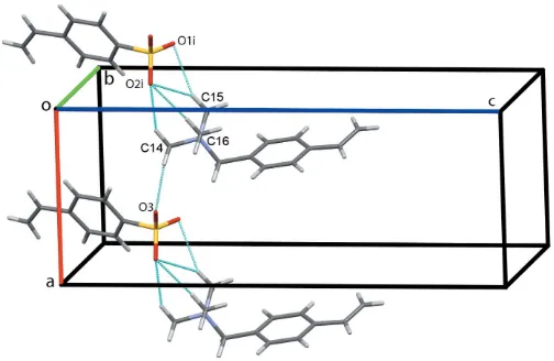
Figure 2
Chains of cations and anions of (I) along theaaxis. Hydrogen bonds are shown as cyan dotted lines [symmetry code: (i)x1,y,z].
Figure 3
Double chains of cation–anion dimers along b. Hydrogen bonds are shown as cyan dotted lines [symmetry codes: (ii) 1x,1
2+y, 1 2z; (iii)
1x,1 2+y,
[image:2.610.313.566.532.707.2]1 2z].
Figure 1
[image:2.610.43.298.612.689.2]The asymmetric unit of the title compound showing the atom numbering with ellipsoids drawn at the 50% probability level. The C—H O hydrogen bond linking the two components is drawn as a dotted black line. For clarity, only the major disorder component of the vinyl substituent on the benzene ring of the cation is shown.
Figure 4
Chains of anions alonga. Hydrogen bonds and C—H interactions are shown as cyan and green dotted lines, respectively [symmetry code: (iv)
x1 2,
1
ring motifs. C14—H14B O3 contacts link the cation–anion pairs into chains along the a-axis direction, Fig. 2. Cation– anion dimers are generated by C13—H13B O3iiand C15— H15B O2ii contacts with adjacent dimers linked into columns along b by C16—H16B O1iii hydrogen bonds [symmetry codes: (ii) 1x,1
2+y, 1
2z; (iii) 1x, 1 2+y,
1 2z]. Additional C14—H14B O3 hydrogen bonds form double columns alongbwith the vinyl substituents of the proximate cations and anions pointing in opposite directions, Fig. 3. Chains of anions form along a through C41—H41 O2iv hydrogen bonds augmented by C5—H5 Cg1ivcontacts, Fig. 4 [symmetry code: (iv) x1
2, 1
2y,z]. Finally, weak C42— H42B O1vhydrogen bonds link the anions in a head-to-tail fashion into zigzag chains alongc, Fig. 5 [symmetry code: (v) 3
2x, 1y,z 1
2]. This extensive series of contact combines to assemble an extended network structure with the cations and anions stacked along thea-axis direction, Fig. 6.
4. Hirshfeld surface analysis
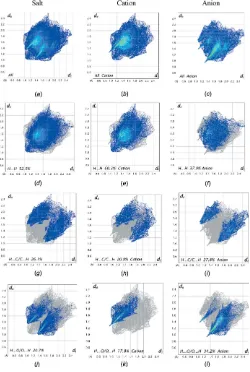
Further details of the intermolecular architecture of this salt were obtained using Hirshfeld surface analysis (Spackman & Jayatilaka, 2009) with surfaces and two-dimensional finger-print plots generated byCrystalExplorer(Turneret al., 2017).
Hirshfeld surfaces viewed for opposite faces of the complete salt are shown in Fig. 7. Both disorder components are included in these surface calculations. The red circles on the Hirshfeld surfaces correspond to the numerous C—H O contacts that play a significant role in stabilizing the packing in this structure. Fingerprint plots of the principal contacts on the Hirshfeld surface of the salt are shown in Fig. 8. These comprise H H, H C/C H, and H O/O H contacts. The much less significant C C and H S/S H contribu-tions are not shown in the figure but are detailed in Table 2.
It is also instructive to investigate the differences in contacts for the discrete cation and anion components of (I) by recording fingerprint plots of the cation and anion individu-ally. All of the surface contributions for the cation and anion are also shown in Table 2, with fingerprint plots for principal contacts found in the individual cation and anion also
948
McAdamet al. C12H18N+C8H7O3S Acta Cryst.(2019). E75, 946–950
[image:3.610.45.306.68.182.2]research communications
Figure 5
Zigzag chains of anions along c. Hydrogen bonds are shown as cyan dotted lines [symmetry code: (v)3
[image:3.610.315.560.74.301.2]2x, 1y,z 1 2].
Figure 6
Overall packing for (I) viewed along thea-axis direction.
Figure 7
[image:3.610.75.528.550.725.2]displayed in Fig. 8. The most notable differences between the values for the salt and its components are that the H H van der Waals interactions increase significantly for the cation, while the anion shows considerable increases in the H O/ O H and H C/C H contacts. These differences reflect the fact that, whereas the contacts for the cations are limited to cation–anion interactions, the anions are also involved in distinct anion–anion contacts, vide supra. The C C and
H S/S H contributions to all of the surfaces are very weak but are included in Table 2 for completeness.
5. Database survey
A search of the Cambridge Structural Database (Version 5.40 November 2018 with one update; Groomet al., 2016) reveals the fact that the salt reported here is quite unusual. Only two structures involving the N,N,N -trimethyl(4-vinylphenyl)-methylammonium cation acting as counter-ions to poly-molybdate (QAJXEH) and poly-tungstate (QAJXAD) anions were found (Vorotnikovet al., 2015). Structures of salts of the 4-vinylbenzenesulfonate anion are slightly more abun-dant, with organic methylquinolinium (RUMGAJ; Leeet al., 2015) and 4-{2-[4-(dimethylamino)phenyl]vinyl}-1-methyl-pyridinium (SAPDAR; Vijay et al., 2012) cations and hexa-aqua manganese, cobalt and nickel complex cations (SUVBOA, SUVBUG and SUVCAN; Leonardet al., 1999).
6. Synthesis and crystallization
The title compound was preparedviaan argentometric mixing approach (Li et al., 2010) from the silver salt of 4-vinyl-benzenesulfonic acid, Ag-VBS (Woesteet al., 1993; Sikkemaet al., 2007) and (vinylbenzyl)trimethylammonium chloride, VBT-Cl (Sigma Aldrich). A suspension of Ag-VBS in water and equimolar amount of VBT-Cl were stirred 30 minutes. After filtration of the AgCl precipitate, the solution was freeze-dried and the ion-pair co-monomers recrystallized from chloroform as irregular colourless blocks.
ESI MS +ve (m/z): 176.14 [C12H18N] +
; -ve: 183.01 [C8H7SO3]
.1H NMR (400 MHz, DMSO-d6): 5.95 (dd,J= 18, 1 Hz, 1H, VBT CH2), 5.38 (dd, J = 11, 1 Hz, 1H, VBT CH2), 6.80 (dd,J= 18, 11 Hz, 1H, VBT –CH ), 7.61 & 7.50 [2(d, J= 8 Hz, 2H, VBT benzene H)], 4.51 (s, 2H, VBT CH2), 4.51 (s, 2H, VBT CH2), 3.02 (s, 9H, VBT CH3). 5.84 (dd,
J= 18, 1 Hz, 1H, VBS CH2), 5.27 (dd,J= 11, 1 Hz, 1H, VBS CH2), 6.73 (dd,J= 18, 11 Hz, 1H, VBS –CH ), 7.57 & 7.42 [2(d,J= 8 Hz, 2H, VBS benzene H)]
7. Refinement
Crystal data, data collection and structure refinement details are summarized in Table 3. All H atoms were refined using a riding model withd(C—H) = 0.95 A˚ andUiso(H) = 1.2Ueq(C) for aromatic and vinyl H atoms,d(C—H) = 0.99 A˚ andUiso(H) = 1.2Ueq(C) for methylene andd(C—H) = 0.98 A˚ andUiso(H) = 1.5Ueq(C) for methyl H atoms. The vinyl substituent on the benzene ring of the cation is disordered over two sites (C101=C102 and C103=C104) with a refined occupancy ratio of 0.542 (11):0.458 (11).
Funding information
We thank the NZ Ministry of Business, Innovation and Employment Science Investment Fund (grant No. UOO-X1206) for support of this work and the University of Otago
research communications
Acta Cryst.(2019). E75, 946–950 McAdamet al. C
[image:4.610.46.295.103.178.2]12H18N+C8H7O3S
949
Table 2
Percentage contributions of interatomic contacts to the Hirshfeld surface for (I).
Contacts Included surface area
Salt Cation Anion
H H 52.5 60.3 37.9
H C/C H 26.1 20.8 27.8
H O/O H 20.7 17.8 34.2
C C 0.5 0.9 0.0
H S/S H 0.1 0.1 0.1
Figure 8
[image:4.610.46.296.325.697.2]for the purchase of the diffractometer. JS also thanks the Department of Chemistry, University of Otago for support of his work.
References
Allen, F. H., Johnson, O., Shields, G. P., Smith, B. R. & Towler, M. (2004).J. Appl. Cryst.37, 335–338.
Farrugia, L. J. (2012).J. Appl. Cryst.45, 849–854.
Goswami, S. K., McAdam, C. J., Hanton, L. R. & Moratti, S. C. (2017).
Macromol. Rapid Commun.38, 1700103.
Groom, C. R., Bruno, I. J., Lightfoot, M. P. & Ward, S. C. (2016).Acta Cryst.B72, 171–179.
Hunter, K. A. & Simpson, J. (1999). TITAN2000. University of Otago, New Zealand.
Lee, S.-H., Yoo, B.-W., Yun, H., Jazbinsek, M. & Kwon, O.-P. (2015).J. Mol. Struct.1100, 359–365.
Leonard, M. A., Squattrito, P. J. & Dubey, S. N. (1999).Acta Cryst.
C55, 35–39.
Li, G., Xue, H., Gao, C., Zhang, F. & Jiang, S. (2010).Macromolecules,
43, 14–16.
Macrae, C. F., Bruno, I. J., Chisholm, J. A., Edgington, P. R., McCabe, P., Pidcock, E., Rodriguez-Monge, L., Taylor, R., van de Streek, J. & Wood, P. A. (2008).J. Appl. Cryst.41, 466–470.
Naficy, S., Brown, H. R., Razal, J. M., Spinks, G. M. & Whitten, P. G. (2011).Aust. J. Chem.64, 1007–1025.
Parsons, S., Flack, H. D. & Wagner, T. (2013).Acta Cryst.B69, 249– 259.
Peak, C. W., Wilker, J. J. & Schmidt, G. (2013).Colloid Polym. Sci. 291, 2031–2047.
Pushparajan, C., Goswami, S. K., McAdam, C. J., Hanton, L. R., Dearden, P. K., Moratti, S. C. & Cridge, A. G. (2018). Electrophor-esis,39, 824–832.
Rigaku OD (2018).CrysAlis PRO. Rigaku Oxford Diffraction Ltd, Yarnton, England.
Sheldrick, G. M. (2015a).Acta Cryst.A71, 3–8. Sheldrick, G. M. (2015b).Acta Cryst.C71, 3–8.
Sikkema, F. D., Comellas-Aragone`s, M., Fokkink, R. G., Verduin, B. J. M., Cornelissen, J. J. L. M. & Nolte, R. J. M. (2007). Org. Biomol. Chem.5, 54–57.
Spackman, M. A. & Jayatilaka, D. (2009).CrystEngComm,11, 19–32. Spek, A. L. (2009).Acta Cryst.D65, 148–155.
Sun, Z., Lv, F., Cao, L., Liu, L., Zhang, Y. & Lu, Z. (2015).Angew. Chem. Int. Ed.54, 7944–7948.
Turner, M. J., McKinnon, J. J., Wolff, S. K., Grimwood, D. J., Spackman, P. R., Jayatilaka, D. & Spackman, M. A. (2017).
CrystalExplorer17. University of Western Australia, Nedlands, Western Australia; http://hirshfeldsurface.net.
Van Vlierberghe, S., Dubruel, P. & Schacht, E. (2011). Biomacromo-lecules,12, 1387–1408.
Vijay, R. J., Melikechi, N., Thomas, T., Gunaseelan, R., Arockiaraj, M. A. & Sagayaraj, P. (2012).J. Cryst. Growth,338, 170–176. Vorotnikov, Y. A., Mikhailov, M. A., Brylev, K. A., Piryazev, D. A.,
Kuratieva, N. V., Sokolov, M. N., Mironov, Y. V. & Shestopalov, M. A. (2015). Izv. Akad. Nauk SSSR, Ser. Khim. (Russ. Chem. Bull.),64, 2591–2596.
Westrip, S. P. (2010).J. Appl. Cryst.43, 920–925.
Woeste, G., Meyer, W. H. & Wegner, G. (1993).Makromol. Chem. 194, 1237–1248.
950
McAdamet al. C12H18N+C8H7O3S Acta Cryst.(2019). E75, 946–950
[image:5.610.44.294.95.412.2]research communications
Table 3
Experimental details.
Crystal data
Chemical formula C12H18N+C8H7O3S
Mr 359.47
Crystal system, space group Orthorhombic,P212121
Temperature (K) 100
a,b,c(A˚ ) 8.3344 (3), 10.5937 (4), 21.1228 (8)
V(A˚3) 1864.98 (12)
Z 4
Radiation type CuK
(mm1) 1.69
Crystal size (mm) 0.200.180.08
Data collection
Diffractometer Rigaku Oxford Diffraction
Super-Nova, Dual, Cu at home/near, Atlas
Absorption correction Multi-scan (CrysAlis PRO; Rigaku OD, 2018)
Tmin,Tmax 0.911, 1.000
No. of measured, independent and observed [I> 2(I)] reflections
4767, 3103, 2784
Rint 0.029
(sin/)max(A˚1) 0.620
Refinement
R[F2> 2(F2)],wR(F2),S 0.040, 0.103, 1.04
No. of reflections 3103
No. of parameters 248
No. of restraints 10
H-atom treatment H-atom parameters constrained
max,min(e A˚3) 0.37,0.29
Absolute structure Flackxdetermined using 870 quotients [(I+)(I
)]/[(I+)+(I )] (Parsonset al., 2013)
Absolute structure parameter 0.040 (19)
Computer programs:CrysAlis PRO(Rigaku OD, 2018),SHELXT(Sheldrick, 2015a),
SHELXL2018(Sheldrick, 2015b),TITAN(Hunter & Simpson, 1999),Mercury(Macrae
supporting information
sup-1
Acta Cryst. (2019). E75, 946-950
supporting information
Acta Cryst. (2019). E75, 946-950 [https://doi.org/10.1107/S2056989019007758]
Structure and Hirshfeld surface analysis of the salt
N
,
N
,
N
-trimethyl-1-(4-vinyl-phenyl)methanaminium 4-vinylbenzenesulfonate
C. John McAdam, Lyall R. Hanton, Stephen C. Moratti, Jim Simpson and Ravindra N.
Wickramasinhage
Computing details
Data collection: CrysAlis PRO (Rigaku OD, 2018); cell refinement: CrysAlis PRO (Rigaku OD, 2018); data reduction:
CrysAlis PRO (Rigaku OD, 2018); program(s) used to solve structure: SHELXT (Sheldrick, 2015a); program(s) used to
refine structure: SHELXL2018 (Sheldrick, 2015b) and TITAN (Hunter & Simpson, 1999); molecular graphics: Mercury
(Macrae et al., 2008); software used to prepare material for publication: SHELXL2014 (Sheldrick, 2015b), enCIFer
(Allen et al., 2004), PLATON (Spek, 2009), publCIF (Westrip, 2010) and WinGX (Farrugia, 2012).
N,N,N-Trimethyl-1-(4-vinylphenyl)methanaminium 4-vinylbenzenesulfonate
Crystal data
C12H18N+·C8H7O3S− Mr = 359.47
Orthorhombic, P212121 a = 8.3344 (3) Å b = 10.5937 (4) Å c = 21.1228 (8) Å V = 1864.98 (12) Å3
Z = 4
F(000) = 768
Dx = 1.280 Mg m−3
Cu Kα radiation, λ = 1.54184 Å Cell parameters from 2591 reflections θ = 4.2–72.3°
µ = 1.69 mm−1 T = 100 K
Irregular block, colourless 0.20 × 0.18 × 0.08 mm
Data collection
Rigaku Oxford Diffraction SuperNova, Dual, Cu at home/near, Atlas
diffractometer
Radiation source: micro-focus sealed X-ray tube Detector resolution: 5.1725 pixels mm-1
ω scans
Absorption correction: multi-scan (CrysAlis PRO; Rigaku OD, 2018) Tmin = 0.911, Tmax = 1.000
4767 measured reflections 3103 independent reflections 2784 reflections with I > 2σ(I) Rint = 0.029
θmax = 72.8°, θmin = 4.2° h = −6→10
k = −12→12 l = −25→24
Refinement
Refinement on F2
Least-squares matrix: full R[F2 > 2σ(F2)] = 0.040 wR(F2) = 0.103 S = 1.04 3103 reflections
248 parameters 10 restraints
Hydrogen site location: inferred from neighbouring sites
supporting information
sup-2
Acta Cryst. (2019). E75, 946-950 w = 1/[σ2(F
o2) + (0.0465P)2 + 0.5842P]
where P = (Fo2 + 2Fc2)/3
(Δ/σ)max < 0.001
Δρmax = 0.37 e Å−3
Δρmin = −0.29 e Å−3
Absolute structure: Flack x determined using 870 quotients [(I+)-(I-)]/[(I+)+(I-)] (Parsons et al.,
2013)
Absolute structure parameter: −0.040 (19)
Special details
Geometry. All esds (except the esd in the dihedral angle between two l.s. planes) are estimated using the full covariance matrix. The cell esds are taken into account individually in the estimation of esds in distances, angles and torsion angles; correlations between esds in cell parameters are only used when they are defined by crystal symmetry. An approximate (isotropic) treatment of cell esds is used for estimating esds involving l.s. planes.
Refinement. The vinyl substituent on the benzene ring of the cation is disordered over two sites with a refined occupancy ratio of 0.542 (11):0.458 (11).
Fractional atomic coordinates and isotropic or equivalent isotropic displacement parameters (Å2)
x y z Uiso*/Ueq Occ. (<1)
O1 0.7129 (3) 0.3773 (2) 0.23060 (11) 0.0340 (6)
O2 0.9047 (3) 0.2234 (2) 0.19192 (11) 0.0324 (6)
O3 0.6241 (3) 0.1666 (2) 0.20070 (11) 0.0316 (6)
S1 0.73918 (10) 0.26835 (7) 0.19052 (3) 0.0240 (2)
C1 0.7098 (4) 0.3210 (3) 0.11156 (14) 0.0213 (7)
C2 0.8167 (4) 0.4059 (3) 0.08477 (16) 0.0254 (7)
H2 0.901987 0.439140 0.109530 0.030*
C3 0.8000 (4) 0.4426 (3) 0.02186 (16) 0.0267 (8)
H3 0.873464 0.501524 0.004257 0.032*
C4 0.6771 (4) 0.3943 (3) −0.01563 (16) 0.0259 (7)
C41 0.6602 (5) 0.4228 (3) −0.08413 (17) 0.0304 (8)
H41 0.567919 0.390300 −0.104880 0.036*
C42 0.7607 (5) 0.4885 (3) −0.11868 (16) 0.0351 (8)
H42A 0.854786 0.522910 −0.100025 0.042*
H42B 0.739494 0.501740 −0.162365 0.042*
C5 0.5676 (4) 0.3129 (3) 0.01217 (17) 0.0302 (8)
H5 0.480488 0.281673 −0.012223 0.036*
C6 0.5829 (4) 0.2760 (3) 0.07539 (16) 0.0283 (8)
H6 0.506511 0.220260 0.093620 0.034*
C7 0.3486 (4) 0.3494 (4) 0.39999 (17) 0.0282 (8)
C8 0.2924 (5) 0.4592 (4) 0.4279 (2) 0.0391 (10)
H8 0.270082 0.530860 0.402275 0.047*
C9 0.2687 (6) 0.4656 (5) 0.4924 (2) 0.0577 (14)
H9 0.228496 0.541594 0.510296 0.069*
C10 0.3017 (5) 0.3649 (6) 0.5319 (2) 0.0600 (15)
C101 0.2819 (10) 0.3361 (8) 0.6023 (3) 0.038 (2) 0.542 (11)
H101 0.324589 0.260969 0.620146 0.046* 0.542 (11)
C102 0.2045 (14) 0.4175 (8) 0.6374 (4) 0.066 (4) 0.542 (11)
H10A 0.162395 0.492277 0.618983 0.080* 0.542 (11)
H10B 0.190496 0.401869 0.681348 0.080* 0.542 (11)
C103 0.2724 (12) 0.4160 (9) 0.5973 (3) 0.033 (2) 0.458 (11)
supporting information
sup-3
Acta Cryst. (2019). E75, 946-950
C104 0.2771 (11) 0.3345 (9) 0.6444 (4) 0.038 (3) 0.458 (11)
H10C 0.298320 0.247947 0.636175 0.045* 0.458 (11)
H10D 0.259356 0.362379 0.686556 0.045* 0.458 (11)
C11 0.3597 (5) 0.2564 (6) 0.5043 (2) 0.0538 (13)
H11 0.384283 0.185948 0.530359 0.065*
C12 0.3830 (5) 0.2473 (4) 0.43947 (19) 0.0385 (9)
H12 0.422829 0.171040 0.421795 0.046*
C13 0.3803 (4) 0.3451 (4) 0.32982 (17) 0.0329 (8)
H13A 0.468168 0.284452 0.321549 0.039*
H13B 0.417035 0.429445 0.315753 0.039*
C14 0.2826 (5) 0.3111 (5) 0.22225 (17) 0.0528 (13)
H14A 0.190237 0.290203 0.195519 0.079*
H14B 0.368715 0.249929 0.214829 0.079*
H14C 0.320828 0.396094 0.211787 0.079*
C15 0.0986 (4) 0.3955 (3) 0.30061 (19) 0.0330 (8)
H15A 0.010714 0.374402 0.271685 0.050*
H15B 0.134872 0.481981 0.292401 0.050*
H15C 0.061038 0.388818 0.344442 0.050*
N1 0.2336 (4) 0.3069 (3) 0.29050 (13) 0.0289 (7)
C16 0.1808 (5) 0.1749 (3) 0.3060 (2) 0.0444 (10)
H16A 0.148219 0.170607 0.350491 0.067*
H16B 0.269731 0.116280 0.298539 0.067*
H16C 0.089814 0.151749 0.278940 0.067*
Atomic displacement parameters (Å2)
U11 U22 U33 U12 U13 U23
O1 0.0421 (16) 0.0313 (13) 0.0287 (12) 0.0004 (12) 0.0005 (11) −0.0081 (10)
O2 0.0250 (12) 0.0454 (14) 0.0269 (12) 0.0102 (11) −0.0034 (10) −0.0010 (13)
O3 0.0370 (14) 0.0303 (13) 0.0275 (13) −0.0076 (12) −0.0008 (11) 0.0046 (11)
S1 0.0258 (4) 0.0259 (4) 0.0203 (3) 0.0004 (4) −0.0009 (3) −0.0022 (3)
C1 0.0243 (17) 0.0198 (14) 0.0198 (14) 0.0036 (14) −0.0016 (13) −0.0010 (12)
C2 0.0218 (16) 0.0237 (16) 0.0307 (17) −0.0006 (15) −0.0001 (14) −0.0042 (14)
C3 0.0259 (18) 0.0232 (16) 0.0311 (17) −0.0003 (15) 0.0064 (15) 0.0011 (14)
C4 0.0283 (18) 0.0244 (16) 0.0249 (17) 0.0043 (15) −0.0011 (14) −0.0019 (14)
C41 0.035 (2) 0.0286 (18) 0.0280 (18) 0.0035 (17) −0.0043 (16) 0.0003 (15)
C42 0.035 (2) 0.0423 (19) 0.0275 (17) 0.005 (2) 0.0014 (18) 0.0049 (15)
C5 0.0280 (18) 0.0302 (18) 0.0324 (19) −0.0023 (16) −0.0100 (15) 0.0016 (15)
C6 0.0283 (18) 0.0257 (17) 0.0308 (18) −0.0034 (16) −0.0057 (14) 0.0030 (16)
C7 0.0211 (17) 0.0344 (19) 0.0292 (18) −0.0060 (16) −0.0032 (14) −0.0010 (16)
C8 0.034 (2) 0.038 (2) 0.046 (2) 0.0006 (18) −0.0138 (18) −0.0090 (18)
C9 0.034 (2) 0.088 (4) 0.051 (3) 0.008 (3) −0.013 (2) −0.036 (3)
C10 0.031 (2) 0.116 (5) 0.034 (2) −0.018 (3) −0.0053 (18) −0.012 (3)
C101 0.045 (5) 0.032 (5) 0.038 (5) −0.016 (4) −0.016 (4) 0.009 (4)
C102 0.126 (11) 0.043 (5) 0.030 (5) −0.009 (6) −0.009 (6) 0.005 (4)
C103 0.040 (5) 0.031 (5) 0.028 (5) −0.003 (5) −0.002 (4) −0.001 (4)
C104 0.044 (6) 0.045 (6) 0.023 (5) −0.010 (5) 0.002 (4) 0.002 (4)
supporting information
sup-4
Acta Cryst. (2019). E75, 946-950
C12 0.036 (2) 0.033 (2) 0.046 (2) −0.007 (2) −0.0098 (17) 0.0038 (18)
C13 0.0223 (17) 0.042 (2) 0.0347 (19) −0.0019 (17) −0.0027 (15) 0.0019 (17)
C14 0.039 (2) 0.094 (4) 0.0250 (19) 0.000 (3) 0.0010 (17) −0.007 (2)
C15 0.0293 (18) 0.0291 (18) 0.041 (2) 0.0064 (16) −0.0063 (17) −0.0055 (17)
N1 0.0267 (15) 0.0331 (15) 0.0271 (14) 0.0018 (14) −0.0027 (13) −0.0027 (11)
C16 0.056 (2) 0.0216 (17) 0.055 (3) −0.0022 (18) −0.019 (2) −0.0020 (19)
Geometric parameters (Å, º)
O1—S1 1.448 (2) C10—C101 1.527 (7)
O2—S1 1.460 (2) C101—C102 1.307 (9)
O3—S1 1.459 (2) C101—H101 0.9500
S1—C1 1.776 (3) C102—H10A 0.9500
C1—C2 1.387 (4) C102—H10B 0.9500
C1—C6 1.389 (5) C103—C104 1.317 (9)
C2—C3 1.392 (5) C103—H103 0.9500
C2—H2 0.9500 C104—H10C 0.9500
C3—C4 1.392 (5) C104—H10D 0.9500
C3—H3 0.9500 C11—C12 1.387 (6)
C4—C5 1.386 (5) C11—H11 0.9500
C4—C41 1.485 (5) C12—H12 0.9500
C41—C42 1.311 (5) C13—N1 1.533 (5)
C41—H41 0.9500 C13—H13A 0.9900
C42—H42A 0.9500 C13—H13B 0.9900
C42—H42B 0.9500 C14—N1 1.499 (4)
C5—C6 1.397 (4) C14—H14A 0.9800
C5—H5 0.9500 C14—H14B 0.9800
C6—H6 0.9500 C14—H14C 0.9800
C7—C8 1.386 (5) C15—N1 1.481 (4)
C7—C12 1.395 (5) C15—H15A 0.9800
C7—C13 1.506 (5) C15—H15B 0.9800
C8—C9 1.380 (6) C15—H15C 0.9800
C8—H8 0.9500 N1—C16 1.501 (4)
C9—C10 1.382 (8) C16—H16A 0.9800
C9—H9 0.9500 C16—H16B 0.9800
C10—C11 1.376 (8) C16—H16C 0.9800
C10—C103 1.504 (7)
O1—S1—O3 113.77 (15) C10—C101—H101 120.8
O1—S1—O2 113.04 (15) C101—C102—H10A 120.0
O3—S1—O2 112.16 (16) C101—C102—H10B 120.0
O1—S1—C1 106.15 (14) H10A—C102—H10B 120.0
O3—S1—C1 106.25 (15) C104—C103—C10 116.9 (8)
O2—S1—C1 104.59 (15) C104—C103—H103 121.5
C2—C1—C6 119.2 (3) C10—C103—H103 121.5
C2—C1—S1 119.9 (2) C103—C104—H10C 120.0
C6—C1—S1 120.9 (3) C103—C104—H10D 120.0
supporting information
sup-5
Acta Cryst. (2019). E75, 946-950
C1—C2—H2 119.8 C10—C11—C12 121.7 (5)
C3—C2—H2 119.8 C10—C11—H11 119.1
C2—C3—C4 120.9 (3) C12—C11—H11 119.1
C2—C3—H3 119.5 C11—C12—C7 120.5 (4)
C4—C3—H3 119.5 C11—C12—H12 119.8
C5—C4—C3 118.2 (3) C7—C12—H12 119.8
C5—C4—C41 118.5 (3) C7—C13—N1 113.7 (3)
C3—C4—C41 123.3 (3) C7—C13—H13A 108.8
C42—C41—C4 126.1 (4) N1—C13—H13A 108.8
C42—C41—H41 116.9 C7—C13—H13B 108.8
C4—C41—H41 116.9 N1—C13—H13B 108.8
C41—C42—H42A 120.0 H13A—C13—H13B 107.7
C41—C42—H42B 120.0 N1—C14—H14A 109.5
H42A—C42—H42B 120.0 N1—C14—H14B 109.5
C4—C5—C6 121.3 (3) H14A—C14—H14B 109.5
C4—C5—H5 119.4 N1—C14—H14C 109.5
C6—C5—H5 119.4 H14A—C14—H14C 109.5
C1—C6—C5 119.9 (3) H14B—C14—H14C 109.5
C1—C6—H6 120.0 N1—C15—H15A 109.5
C5—C6—H6 120.0 N1—C15—H15B 109.5
C8—C7—C12 117.8 (4) H15A—C15—H15B 109.5
C8—C7—C13 120.1 (4) N1—C15—H15C 109.5
C12—C7—C13 121.9 (4) H15A—C15—H15C 109.5
C9—C8—C7 120.6 (4) H15B—C15—H15C 109.5
C9—C8—H8 119.7 C15—N1—C16 109.7 (3)
C7—C8—H8 119.7 C15—N1—C14 109.1 (3)
C8—C9—C10 122.0 (5) C16—N1—C14 108.5 (3)
C8—C9—H9 119.0 C15—N1—C13 111.1 (3)
C10—C9—H9 119.0 C16—N1—C13 111.2 (3)
C11—C10—C9 117.4 (4) C14—N1—C13 107.2 (3)
C11—C10—C103 138.3 (6) N1—C16—H16A 109.5
C9—C10—C103 104.1 (6) N1—C16—H16B 109.5
C11—C10—C101 106.5 (6) H16A—C16—H16B 109.5
C9—C10—C101 136.0 (6) N1—C16—H16C 109.5
C102—C101—C10 118.3 (8) H16A—C16—H16C 109.5
C102—C101—H101 120.8 H16B—C16—H16C 109.5
O1—S1—C1—C2 67.5 (3) C7—C8—C9—C10 1.0 (7)
O3—S1—C1—C2 −171.1 (2) C8—C9—C10—C11 −0.1 (7)
O2—S1—C1—C2 −52.3 (3) C8—C9—C10—C103 175.4 (6)
O1—S1—C1—C6 −114.1 (3) C8—C9—C10—C101 −175.7 (6)
O3—S1—C1—C6 7.3 (3) C11—C10—C101—C102 −169.3 (7)
O2—S1—C1—C6 126.1 (3) C9—C10—C101—C102 6.6 (12)
C6—C1—C2—C3 −1.9 (5) C11—C10—C103—C104 −13.7 (13)
S1—C1—C2—C3 176.5 (3) C9—C10—C103—C104 172.3 (8)
C1—C2—C3—C4 −0.7 (5) C9—C10—C11—C12 −0.6 (7)
C2—C3—C4—C5 2.9 (5) C103—C10—C11—C12 −174.0 (7)
supporting information
sup-6
Acta Cryst. (2019). E75, 946-950
C5—C4—C41—C42 −173.8 (4) C10—C11—C12—C7 0.2 (6)
C3—C4—C41—C42 4.5 (6) C8—C7—C12—C11 0.7 (5)
C3—C4—C5—C6 −2.5 (5) C13—C7—C12—C11 176.9 (4)
C41—C4—C5—C6 175.9 (3) C8—C7—C13—N1 −88.7 (4)
C2—C1—C6—C5 2.3 (5) C12—C7—C13—N1 95.2 (4)
S1—C1—C6—C5 −176.2 (3) C7—C13—N1—C15 59.1 (4)
C4—C5—C6—C1 −0.1 (6) C7—C13—N1—C16 −63.3 (4)
C12—C7—C8—C9 −1.3 (6) C7—C13—N1—C14 178.2 (3)
C13—C7—C8—C9 −177.6 (4)
Hydrogen-bond geometry (Å, º)
Cg1 is the centroid of the C1–C6 benzene ring.
D—H···A D—H H···A D···A D—H···A
C14—H14B···O3 0.98 2.32 3.264 (5) 161
C14—H14A···O2i 0.98 2.48 3.346 (5) 147
C15—H15A···O1i 0.98 2.63 3.544 (4) 155
C15—H15A···O2i 0.98 2.49 3.348 (4) 147
C13—H13B···O3ii 0.99 2.56 3.466 (5) 152
C15—H15B···O2ii 0.98 2.60 3.477 (4) 149
C16—H16B···O1iii 0.98 2.61 3.365 (4) 134
C16—H16C···O2i 0.98 2.52 3.370 (5) 146
C41—H41···O2iv 0.95 2.58 3.481 (4) 157
C42—H42B···O1v 0.95 2.63 3.494 (4) 151
C5—H5···Cg1iv 0.95 2.93 3.837 (4) 161