2-(Prop-2-enyloxy)benzamide
Bernhard Bugenhagen,aYosef Al Jasem,bFarah Barkhad,c Bassam al Hindawid and Thies Thiemannd*
a
Institute of Inorganic Chemistry, University of Hamburg, Hamburg, Germany,
b
Department of Chemical Engineering, UAE University, AL Ain, Abu Dhabi, United Arab Emirates,cDepartment of Petroleum Engineering, UAE University, AL Ain, Abu
Dhabi, United Arab Emirates, anddDepartment of Chemistry, UAE University,
AL Ain, Abu Dhabi, United Arab Emirates Correspondence e-mail: [email protected]
Received 22 September 2012; accepted 9 October 2012
Key indicators: single-crystal X-ray study;T= 100 K; mean(C–C) = 0.003 A˚; Rfactor = 0.033;wRfactor = 0.087; data-to-parameter ratio = 8.6.
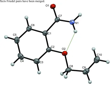
In the title molecule, C10H11NO2, the benzene ring forms
dihedral angles of 33.15 (2) and 6.20 (2)with the mean planes of the amide and propenoxy groups, respectively. The amide – NH2group is oriented toward the propenoxy substituent and
forms a weak intramolecular N—H O hydrogen bond to the propenoxy O atom. The conformation of the propenoxy group at the Csp2—Csp3 and Csp3—O bonds is synperiplanarand
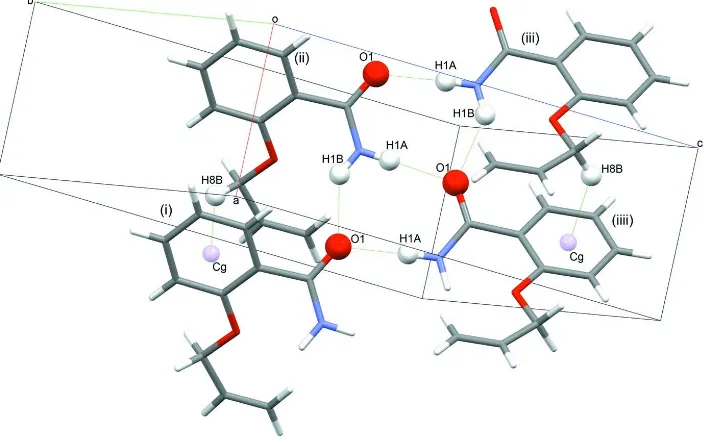
antiperiplanar, respectively. In the crystal, N—H O hydrogen bonds involving the amide groups generate C(4) andR23(7) motifs that organize the molecules into tapes along
thea-axis direction. There are C—H interactions between the propenoxy –CH2 group and the aromatic system of
neighboring molecules within the tape. The mean planes of the aromatic ring and the propenoxy group belonging to mol-ecules located on opposite sites of the tape form an angle of 83.16 (2).
Related literature
For crystal structures of similar compounds, see: Al Jasemet al.
(2012); Pagola & Stephens (2009); Johnstone et al. (2010); Pertlik (1990); Sasada et al. (1964). For uses of 2-alkoxy-benzamides, see: van de Waterbeemd & Testa (1983); Kusu-noki & Harada (1984). For the preparation of a related 2-alkoxybenzamide, see: Al Jasemet al.(2012).
Experimental
Crystal data
C10H11NO2 Mr= 177.20
Orthorhombic,P212121
a= 5.08891 (17) A˚
b= 11.2542 (4) A˚
c= 15.8802 (6) A˚
V= 909.48 (5) A˚3 Z= 4
CuKradiation = 0.74 mm1
T= 100 K
0.300.090.08 mm
Data collection
Agilent SuperNova Atlas diffractometer
Absorption correction: Gaussian (CrysAlis PRO; Agilent, 2012)
Tmin= 0.862,Tmax= 0.951
4718 measured reflections 1079 independent reflections 1016 reflections withI> 2(I)
Rint= 0.025
Refinement
R[F2> 2(F2)] = 0.033
wR(F2) = 0.087 S= 1.03 1079 reflections 126 parameters
H atoms treated by a mixture of independent and constrained refinement
max= 0.17 e A˚
3
min=0.18 e A˚
3
Table 1
Hydrogen-bond geometry (A˚ ,).
Cgis the centroid of the C1–C6 ring.
D—H A D—H H A D A D—H A
N1—H1A O1i
0.90 (2) 2.01 (2) 2.905 (2) 178 (17) N1—H1B O1ii 0.89 (3) 2.12 (3) 2.863 (2) 140 (2) N1—H1B O2 0.89 (3) 2.31 (2) 2.754 (2) 110.8 (18) C8—H8B Cgii
0.99 2.68 3.461 (2) 137
Symmetry codes: (i)xþ1 2;yþ
1
2;zþ1; (ii)xþ1;y;z.
Data collection: CrysAlis PRO(Agilent, 2012); cell refinement: CrysAlis PRO; data reduction: CrysAlis PRO; program(s) used to solve structure: SHELXS97(Sheldrick, 2008); program(s) used to refine structure: SHELXL97 (Sheldrick, 2008) within OLEX2 (Dolomanovet al., 2009); molecular graphics:PLATON(Spek, 2009); Mercury(Macraeet al., 2008); software used to prepare material for publication:SHELXL97,PLATON.
Supplementary data and figures for this paper are available from the IUCr electronic archives (Reference: GK2521).
References
Agilent (2012).CrysAlis PRO.Agilent Technologies, Yarnton, England. Al Jasem, Y., Hindawi, B. al, Thiemann, T. & White, F. (2012).Acta Cryst.E68,
o2639–o2640.
Dolomanov, O. V., Bourhis, L. J., Gildea, R. J., Howard, J. A. K. & Puschmann, H. (2009).J. Appl. Cryst.42, 339–341.
organic compounds
Acta Cryst.(2012). E68, o3169–o3170 doi:10.1107/S1600536812042250 Bugenhagenet al.
o3169
Acta Crystallographica Section EStructure Reports
Online
Richardson, P. R., Warren, J. E. & Wood, P. A. (2010).CrystEngComm,12, 1065–1078.
Kusunoki, T. & Harada, S. (1984).J. Dermatol.11, 277–281.
Macrae, C. F., Bruno, I. J., Chisholm, J. A., Edgington, P. R., McCabe, P., Pidcock, E., Rodriguez-Monge, L., Taylor, R., van de Streek, J. & Wood, P. A. (2008).J. Appl. Cryst.41, 466–470.
Pertlik, F. (1990).Monatsh. Chem.121, 129–139.
Sasada, Y., Takano, T. & Kakudo, M. (1964).Bull. Chem. Soc. Jpn,37, 940–946. Sheldrick, G. M. (2008).Acta Cryst.A64, 112–122.
Spek, A. L. (2009).Acta Cryst.D65, 148–155.
supporting information
sup-1
Acta Cryst. (2012). E68, o3169–o3170
supporting information
Acta Cryst. (2012). E68, o3169–o3170 [doi:10.1107/S1600536812042250]
2-(Prop-2-enyloxy)benzamide
Bernhard Bugenhagen, Yosef Al Jasem, Farah Barkhad, Bassam al Hindawi and Thies Thiemann
S1. Comment
In 2-propenoxybenzamide (2-allyloxybenzamide) (Figure 1), the O1—C7—C1—C6 torsion angle characterizing the twist
of the benzene ring relative to the amide group is -30.3 (2)° and the corresponding C8—O2—C2—C3 torsion angle for
the propoxy group is 5.9 (2)°. There is an intramolecular N1—H1B···O2 bond within each molecule (Table 1). When
compared to the structurally comparable 2-propoxybenzamide (Al Jasem et al., 2012), the torsion angle O1—C7—C1—
C6 is much larger in the title compound. The amide groups generate C(4) and R23(7) hydrogen-bond motifs that organize
the molecules into tapes along the a axis. The title compound exhibits a C10—H10A···O2 and a C8—H8··· π (Table 1)
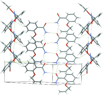
close contact, absent in propoxybenzamide (Figure 2). The C4—H4···O1 intermolecular interaction in
2-propenoxybenzamide links the neighboring tapes of molecules along the a axis with each other (Figure 3). However, in
2-propoxybenzamide, where also a C–H···O intermolecular interaction is found, the interaction proceeds from the carbon
ortho to the propoxy group, while in the present case, it proceeds from the carbon meta to the propenoxy group. As a
result of more close intermolecular contacts in 2-propenoxybenzamide as compared to 2-propoxybenzamide, the
difference in the packing between the two compounds is large. The main difference is that while in the
2-propoxybenzamide molecules are arranged into pairs by close contacts, where the pairs in one layer are not associated
through close contacts, in the title compound all neighboring molecules form close contacts to each other. Nevertheless,
both compounds exhibit particular molecular tapes, each compound with two different directions of tape propagation. In
the title compound, the average plane (0 1 - 1) of a tape propagation has an angle of 68.78 (2)° with the corresponding
plane (0 1 1) of the neighboring tape propagation. Due to the large dihedral angle between the benzene ring and the amide
group in 2-propenoxybenzamide, the average plane (-1 2 2) of the benzene ring and the propenoxy group of a molecule in
one stack makes an angle of 83.16 (2)° with the corresponding plane (1 2 2) of a molecule in the opposing motif within
one tape.
S2. Experimental
To powdered KOH (1.12 g, 20.0 mmol) in DMSO (18 ml) was added salicylamide (2.74 g, 20.0 mmol), and the resulting
mixture was stirred for 10 min. at rt. Thereafter, n-propenyl bromide (4.2 g, mmol, 34.7 mmol) was added dropwise. The
solution was stirred for 12 h at rt. Then, it was poured into water (200 ml) and extracted with chloroform (3 x 75 ml). The
organic phase was dried over anhydrous MgSO4, concentrated in vacuo, and the residue was subjected to column
chromatography on silica gel (CHCl3/MtBE/hexane v/v/v 1:1:1) to give 2-propenoxybenzamide (2.76 g, 78%) as colorless
crystals (m.p. 377 K). The crystal was grown from CHCl3/ MtBE/hexane (v/v/v 1:1:1).IR (KBr) νmax 3406, 3190, 1631,
1600, 1399, 1243, 996, 921, 757, 643, 627 cm-1; δH (400 MHz, CDCl3) 4.67 (2H, d, 3J = 5.6 Hz), 5.36 (1H, dd, 3J = 10.4
Hz, 2J = 1.2 Hz), 5.44 (1H, dd, 3J = 17.2 Hz, 2J = 1.2 Hz), 6.03 – 6.13 (1H, dt, 3J = 17.2 Hz, 3J = 10.4 Hz, 3J = 5.6 Hz),
6.25 (1H, bs, NH), 6.96 (1H, d, 3J = 8.0 Hz), 7.07 (1H, dd, 3J = 8.0 Hz, 3J = 8.0 Hz), 7.80 (1H, bs, NH), 8.20 (1H, dd, 3J =
All carbon-bound hydrogen atoms were placed in calculated positions with C—H
distances of 0.95 - 0.99 Å and refined as riding with Uiso(H)
=xUeq(C), where x = 1.5 for methyl and x = 1.2 for all other H-atoms.
The N-bound H atom positions were determined from difference electron
density map and refined freely. In the absence of significant anomalous
[image:4.610.115.486.171.463.2]scattering effects Friedel pairs have been merged.
Figure 1
A view of the title compound molecule with the atom-numbering scheme and the intramolecular interaction within the
supporting information
sup-3
[image:5.610.130.482.74.296.2]Acta Cryst. (2012). E68, o3169–o3170 Figure 2
Intermolecular attractions between molecules of the title compound. [Symmetry codes: i: 1+x,y,z; ii: x,y,z; iii: -1/2 + x,1/2
Figure 3
The crystal packing diagram showing the C—H···O intermolecular interactions between tapes formed via amide group
interactions.
2-(Prop-2-enyloxy)benzamide
Crystal data
C10H11NO2
Mr = 177.20
Orthorhombic, P212121
a = 5.08891 (17) Å
b = 11.2542 (4) Å
c = 15.8802 (6) Å
V = 909.48 (5) Å3
Z = 4
F(000) = 376
Dx = 1.294 Mg m−3 Melting point: 377 K
Cu Kα radiation, λ = 1.5418 Å
Cell parameters from 2824 reflections
θ = 3.9–72.6°
µ = 0.74 mm−1
T = 100 K
Needle, colourless 0.30 × 0.09 × 0.08 mm
Data collection
Agilent SuperNova Atlas diffractometer
Radiation source: SuperNova (Cu) X-ray Source
Mirror monochromator
Detector resolution: 10.4127 pixels mm-1
ω scans
Absorption correction: gaussian (CrysAlis PRO; Agilent, 2012)
Tmin = 0.862, Tmax = 0.951 4718 measured reflections 1079 independent reflections 1016 reflections with I > 2σ(I)
supporting information
sup-5
Acta Cryst. (2012). E68, o3169–o3170 θmax = 72.7°, θmin = 4.8°
h = −6→3
k = −12→13
l = −19→19
Refinement
Refinement on F2
Least-squares matrix: full
R[F2 > 2σ(F2)] = 0.033
wR(F2) = 0.087
S = 1.03
1079 reflections 126 parameters 0 restraints
Primary atom site location: structure-invariant direct methods
Secondary atom site location: difference Fourier map
Hydrogen site location: inferred from neighbouring sites
H atoms treated by a mixture of independent and constrained refinement
w = 1/[σ2(F
o2) + (0.0609P)2 + 0.1267P]
where P = (Fo2 + 2Fc2)/3 (Δ/σ)max < 0.001
Δρmax = 0.17 e Å−3 Δρmin = −0.18 e Å−3
Special details
Experimental. Numerical absorption correction based on gaussian integration over a multifaceted crystal model
Geometry. All e.s.d.'s (except the e.s.d. in the dihedral angle between two l.s. planes) are estimated using the full covariance matrix. The cell e.s.d.'s are taken into account individually in the estimation of e.s.d.'s in distances, angles and torsion angles; correlations between e.s.d.'s in cell parameters are only used when they are defined by crystal symmetry. An approximate (isotropic) treatment of cell e.s.d.'s is used for estimating e.s.d.'s involving l.s. planes.
Refinement. Refinement of F2 against ALL reflections. The weighted R-factor wR and goodness of fit S are based on F2,
conventional R-factors R are based on F, with F set to zero for negative F2. The threshold expression of F2 > σ(F2) is used
only for calculating R-factors(gt) etc. and is not relevant to the choice of reflections for refinement. R-factors based on F2
are statistically about twice as large as those based on F, and R- factors based on ALL data will be even larger.
Fractional atomic coordinates and isotropic or equivalent isotropic displacement parameters (Å2)
x y z Uiso*/Ueq
C1 0.2397 (4) 0.43317 (15) 0.31664 (10) 0.0181 (4)
C10 0.9389 (4) 0.59453 (19) 0.49447 (12) 0.0289 (4)
C2 0.4129 (3) 0.52840 (15) 0.30222 (11) 0.0188 (4)
C3 0.3949 (4) 0.59415 (17) 0.22777 (12) 0.0238 (4)
C4 0.2055 (4) 0.56545 (18) 0.16826 (11) 0.0262 (4)
C5 0.0314 (4) 0.47271 (17) 0.18190 (11) 0.0244 (4)
C6 0.0478 (4) 0.40808 (16) 0.25658 (11) 0.0208 (4)
C7 0.2401 (3) 0.35748 (15) 0.39466 (11) 0.0181 (4)
C8 0.7520 (4) 0.65550 (15) 0.35506 (12) 0.0231 (4)
C9 0.9255 (4) 0.66806 (17) 0.43007 (12) 0.0268 (4)
H10A 0.8310 0.5257 0.4958 0.035*
H10B 1.0565 0.6104 0.5396 0.035*
H1A 0.484 (5) 0.288 (2) 0.4772 (13) 0.028 (6)*
H1B 0.623 (5) 0.358 (2) 0.4113 (16) 0.040 (7)*
H3 0.5120 0.6584 0.2179 0.029*
H4 0.1950 0.6099 0.1175 0.031*
H5 −0.0975 0.4534 0.1408 0.029*
H6 −0.0741 0.3458 0.2667 0.025*
H8A 0.6389 0.7267 0.3496 0.028*
H8B 0.8604 0.6488 0.3035 0.028*
O1 0.0291 (2) 0.31561 (12) 0.42082 (8) 0.0224 (3)
O2 0.5922 (2) 0.55184 (11) 0.36411 (7) 0.0217 (3)
Atomic displacement parameters (Å2)
U11 U22 U33 U12 U13 U23
C1 0.0162 (8) 0.0185 (8) 0.0197 (8) 0.0023 (7) 0.0017 (7) −0.0006 (6)
C2 0.0149 (8) 0.0194 (8) 0.0221 (8) 0.0015 (7) 0.0019 (7) 0.0001 (7)
C3 0.0226 (9) 0.0230 (8) 0.0259 (9) 0.0029 (8) 0.0040 (7) 0.0037 (7)
C4 0.0309 (10) 0.0275 (10) 0.0202 (8) 0.0076 (9) 0.0018 (8) 0.0044 (7)
C5 0.0245 (9) 0.0280 (9) 0.0207 (8) 0.0049 (8) −0.0038 (7) −0.0032 (7)
C6 0.0180 (8) 0.0205 (8) 0.0239 (8) 0.0014 (7) −0.0004 (8) −0.0028 (7)
C7 0.0159 (8) 0.0173 (8) 0.0210 (8) 0.0005 (7) 0.0006 (7) −0.0019 (6)
C8 0.0213 (9) 0.0177 (8) 0.0304 (9) −0.0038 (8) 0.0011 (8) 0.0008 (7)
C9 0.0211 (9) 0.0240 (9) 0.0353 (10) −0.0040 (8) 0.0013 (8) −0.0066 (8)
C10 0.0270 (10) 0.0315 (9) 0.0283 (9) 0.0002 (9) −0.0022 (9) −0.0070 (8)
N1 0.0153 (7) 0.0260 (8) 0.0240 (7) −0.0007 (6) 0.0000 (6) 0.0071 (6)
O1 0.0153 (6) 0.0243 (6) 0.0276 (6) −0.0017 (5) 0.0006 (5) 0.0053 (5)
O2 0.0195 (6) 0.0210 (6) 0.0246 (6) −0.0045 (5) −0.0015 (5) 0.0033 (5)
Geometric parameters (Å, º)
C1—C2 1.406 (2) C7—N1 1.326 (2)
C1—C6 1.394 (2) C7—O1 1.244 (2)
C1—C7 1.504 (2) C8—H8A 0.9900
C2—C3 1.398 (2) C8—H8B 0.9900
C2—O2 1.367 (2) C8—C9 1.489 (3)
C3—H3 0.9500 C8—O2 1.429 (2)
C3—C4 1.388 (3) C9—H9 0.9500
C4—H4 0.9500 C9—C10 1.317 (3)
C4—C5 1.386 (3) C10—H10A 0.9500
C5—H5 0.9500 C10—H10B 0.9500
C5—C6 1.394 (2) N1—H1A 0.90 (2)
C6—H6 0.9500 N1—H1B 0.89 (3)
C1—C6—H6 119.4 C7—N1—H1A 123.2 (16)
C10—C9—C8 126.30 (18) C7—N1—H1B 124.0 (16)
C10—C9—H9 116.9 C8—C9—H9 116.9
C2—C1—C7 124.39 (15) C9—C10—H10A 120.0
C2—C3—H3 120.1 C9—C10—H10B 120.0
C2—O2—C8 117.72 (13) C9—C8—H8A 109.8
C3—C2—C1 120.00 (16) C9—C8—H8B 109.8
C3—C4—H4 119.6 H10A—C10—H10B 120.0
C4—C3—C2 119.89 (18) H1A—N1—H1B 113 (2)
C4—C3—H3 120.1 H8A—C8—H8B 108.2
C4—C5—H5 120.4 N1—C7—C1 118.44 (16)
supporting information
sup-7
Acta Cryst. (2012). E68, o3169–o3170
C5—C4—C3 120.84 (17) O1—C7—N1 122.25 (16)
C5—C4—H4 119.6 O2—C2—C1 116.64 (14)
C5—C6—C1 121.22 (17) O2—C2—C3 123.36 (16)
C5—C6—H6 119.4 O2—C8—H8A 109.8
C6—C1—C2 118.81 (15) O2—C8—H8B 109.8
C6—C1—C7 116.76 (16) O2—C8—C9 109.54 (15)
C6—C5—H5 120.4
Hydrogen-bond geometry (Å, º)
Cg is the centroid of the C1–C6 ring.
D—H···A D—H H···A D···A D—H···A
N1—H1A···O1i 0.90 (2) 2.01 (2) 2.905 (2) 178 (17)
N1—H1B···O1ii 0.89 (3) 2.12 (3) 2.863 (2) 140 (2)
N1—H1B···O2 0.89 (3) 2.31 (2) 2.754 (2) 110.8 (18)
C8—H8B···Cgii 0.99 2.68 3.461 (2) 137