1218
https://doi.org/10.1107/S2056989018010873 Acta Cryst.(2018). E74, 1218–1221research communications
Received 13 June 2018 Accepted 28 July 2018
Edited by K. Fejfarova, Institute of Biotechnology CAS, Czech Republic
Keywords:crystal structure; decaahydro-acridine; hydrogen bonds; halogen bonds; C— Br (ring) contacts.
CCDC reference:1859007
Supporting information:this article has supporting information at journals.iucr.org/e
The structure of
9-(3-bromo-6-chloro-2-hydroxy-
phenyl)-10-(2-hydroxyethyl)-3,6-diphenyl-3,4,5,6,7,9-hexahydro-2
H
-acridine-1,8-dione
Antar A. Abdelhamid,aFarouq E. Hawaiz,b* Alaa F. Mohamed,cShaaban K. Mohameddand Jim Simpsone
aChemistry Department, Faculty of Science, Sohag University, Sohag, Egypt,bChemistry Department, College of
Education, Salahaddin University-Hawler, Erbil, Kurdistan Region, Iraq,cNational Organization for Drug Control and
Research (NODCAR), Giza, Egypt,dChemistry and Environmental Division, Manchester Metropolitan University,
Manchester M1 5GD, England, Chemistry Department, Faculty of Science, Minia University, 61519 El-Minia, Egypt, and
eDepartment of Chemistry, University of Otago, PO Box 56, Dunedin, New Zealand. *Correspondence e-mail:
shaabankamel@yahoo.com
In the structure of the title compound C33H29BrClNO4, (I), the hexahydro-2H
-acridine ring system has a hydroxyethyl substituent on the N atom and a 3-bromo-6-chloro-2-hydroxyphenyl substituent on the central C atom at the 9-position. An unusual feature of the molecule is that the substituents at the 3-and 5-positions of the outer cyclohexenone rings are phenyl rings rather than the more common dimethyl substituents. C atoms on both of the cyclohexenone rings are disordered over two sites. In the crystal structure, O—H O, C— H O and C—H (ring) hydrogen bonds combine with an Br—O and unusual C—Br (ring) halogen bonds to generate a three dimensional network with molecules stacked along thea-axis direction.
1. Chemical context
Acridine derivatives form an important class of heterocycles containing nitrogen with a broad range of pharmaceutical properties. These include compounds that are used as anti-infammatory (Chen et al., 2002), anti-cancer (Gamega et al., 1999), anti-microbial (Kayaet al., 2011), anti-tubercular (Aly & Abadi 2004; Tripathiet al., 2006), anti-parasitic (Di Giorgio,
et al., 2005), anti-malarial (Kumar et al., 2009; Tomaret al., 2010), anti-viral (Gupta & Jaiswal, 2010; Tonelli et al., 2011) and fungicidal agents (Srivastava & Nizamuddin, 2004). Furthermore, acridines are used as dyes, fluorescent materials for the visualization of biomolecules and in laser technologies (Niknam & Damya, 2009). In this context we report here the synthesis and crystal structure of the title acridine derivative.
2. Structural commentary
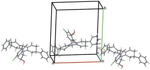
The title compound (I), consists of a hexahydro-2H-acridine ring system made up of a central dihydropyridine ring with an N-bound 2-hydroxyethyl substituent flanked by two cyclo-hexenone rings that carry phenyl substituents in the 3- and 5-positions, respectively (Fig. 1). The central C9 atom bears a 3-bromo-6-chloro-2-hydroxyphenyl substituent and the O20
hydroxy group forms an intramolecular hydrogen bond to the adjacent O8 carbonyl oxygen enclosing anS(8) ring. The C2 and C3 atoms of one cyclohexenone are disordered over two sites as is the C6 atom of the corresponding cyclohexenone. Their occupancy ratios refine to 0.521 (10):0.479 (10) for C2,C3 and 0.746 (9):0.254 (9) for C6. Only details of the major disorder components will be considered here. The central C9,N10,C11–C14 ring adopts a half-chair conformation and is inclined to the adjacent C1–C4,C11,C12 and C5–C8,C13,C14 rings at angles of 7.11 (18) and 21.64 (10), respectively, so the hexahydro-2H-acridine unit is far from planar. The 3-bromo-6-chloro-2-hydroxyphenyl ring subtends an angle of 84.39 (6)
to this central ring. The C1–C4,C11,C12 ring is best described as a severely flattened boat while the C5–C8,C13,C14 system is in a distorted half-chair conformation. The phenyl substituents on these outer cyclohexenone rings are inclined to their parent rings at angles of 76.87 (12) for C31–C36 and 86.27 (8)for
C61–C66. The N-bound 2-hydroxyethyl substituent points away from the convex face of the hexahydro-2H-acridine system as does the 3-bromo-6-chloro-2-hydroxyphenyl substituent.
3. Supramolecular features
The crystal structure of (I) is supported by a full range of classical and non-classical hydrogen bonds and C— H (ring) contacts, together with an intermolecular O Br halogen bond and an unusual C—Br (ring) contact. Classical O16—H16O O8 hydrogen bonds, Table 1, form
C(9) chains along theb-axis direction, linking the molecules in a head-to-tail fashion, Fig. 2. Chains also form along thea-axis direction through C65—H65 Cg7 contacts, Fig. 3, Table 1. C15—H15A O16 hydrogen bonds form inversion dimers that enclose R2
2(8) rings and are strengthened by C16—
H16 Cg8 interactions. Adjacent dimers are linked by C34— H34 Cl50 hydrogen bonds, forming double chains of
mol-ecules along the abdiagonal, Fig. 4. The extensive series of contacts is completed with inversion dimers that also form
research communications
Acta Cryst.(2018). E74, 1218–1221 Abdelhamidet al. C
[image:2.610.316.568.71.201.2]33H29BrClNO4
1219
Table 1Hydrogen-bond geometry (A˚ ,).
Cg7 andCg8 are the centroids of the C31–C36 and C61–C66 phenyl rings, respectively.
D—H A D—H H A D A D—H A
O20
—H20
O O8 0.85 (3) 1.79 (3) 2.626 (2) 170 (3) O16—H16O O8i 0.84 (4) 1.97 (4) 2.782 (2) 163 (4) C15—H15A O16ii 0.99 2.68 3.622 (3) 159
C5—H5A O8iii 0.99 2.69 3.669 (3) 172 C5—H5A O20iii 0.99 2.70 3.336 (3) 122
C15—H15B O1iii 0.99 2.47 3.451 (3) 172
C34—H34 Cl50iv 0.95 2.87 3.560 (3) 131
C16—H16B Cg8ii 0.99 2.66 3.529 (3) 147
C65—H65 Cg7v 0.95 2.78 3.648 (4) 152
Symmetry codes: (i)xþ1;yþ1 2;zþ
1
2; (ii)xþ1;yþ1;z; (iii)x;yþ 1 2;z
1 2;
(iv)xþ2;y1 2;zþ
1
2; (v)x1;y 1 2;z
[image:2.610.43.298.118.221.2]3 2.
Figure 2
C(9) chains of molecules of (I) alongb. In this and subsequent figures, hydrogen bonds are drawn as dashed lines.
Figure 3
Chains of molecules of (I) alonga. C—H contacts are shown as
dotted green lines with ring centroids shown as coloured spheres.
Figure 1
[image:2.610.47.294.519.700.2] [image:2.610.313.565.591.709.2]through O16 Br30v
halogen bonds [O Br = 3.0308 (18) A˚ ; symmetry code: (v) 1x, 1y, 1z] (Cavalloet al., 2016; Chifotides & Dunbar, 2013) and are supported by unusual C30—Br30 Cg4v
contacts [Br30 Cg4 = 3.6991 (10) A˚ , C30—
Br30 Cg4 = 83.89 (7); Cg4 is the centroid of the C10–C60
benzene ring] (Matteret al., 2009; Shuklaet al., 2017; Andleeb
et al., 2018). Both of these contacts are significantly shorter than the sum of the Br and O radii, 3.42 A˚ (Bondi, 1964) and that of the Br radius and an estimated half thickness of the benzene ring, 3.75 A˚ . The dimers are linked into chains running along the ac diagonal by a series of C—H O hydrogen bonds generating R2
1(8) and R 2
2(13) rings, with C5
acting as a bifurcated donor, Table 1, Fig. 5. Overall this plethora of intermolecular contacts combine to generate a complex three-dimensional network with molecules stacked along thea-axis direction, Fig. 6.
4. Database survey
A search of the Cambridge Structural Database (Version 5.39 Nov 2017 with three updates; Groomet al.2016) for an
acri-dine ring system with a phenyl or substituted benzene ring on the central C9 atom gave 94 hits, 76 of which represented unique occurrences. The majority of these, 58, have two methyl substituents at the 3- and 5-positions of the ring system. However, three instances reveal a pair of methyl substituents on the 3-position only, with the remaining 15 structures having no additional substitution on either of the cyclohexenone rings. Interestingly, no structures were observed with phenyl substituents at the 3- or the 3- and 5-positions of the hexahydro-2H-acridine ring system, empha-sizing the uniqueness of the structure reported here. Refining the search to structures with CH2CH substitution on the
acridine N atom reduced the hits to seven, four of which have hydroxyethyl substituents on N10 (Mohamed et al., 2013; Abdelhamidet al., 2016, 2014, 2011). Only one of the entries has a 2-hydroxypropyl N10 substituent (Khalilovet al., 2011), with pairs of methyl substituents on the 3- and 5-positions.
5. Synthesis and crystallization
The title compound was synthesized according to our previously reported method (Mohamedet al., 2013). Crystals suitable for X-ray diffraction were obtained by the slow evaporation method using ethanol/acetone (5:1) as the solvent mixture. Yield, 79%; m.p. 451 K.
6. Refinement
Crystal data, data collection and structure refinement details are summarized in Table 2. All H atoms were refined using a riding model withd(C—H) = 0.95 A˚ for aromatic, 0.99 A˚ for methylene and 1.00 A˚ for methine H atoms, all with Uiso = 1.2Ueq(C). The C2 and C3 atoms in the C1–C4,C11,C12 cyclohexenone ring and atom, C6, in the corresponding C5– C8,C13,C14 ring are disordered over two positions. Their
1220
Abdelhamidet al. C33H29BrClNO4 Acta Cryst.(2018). E74, 1218–1221
[image:3.610.46.309.70.253.2]research communications
Figure 5
Chains of molecule of (I) formed by C—H O hydrogen bonds, C—
[image:3.610.311.562.73.300.2]Br and O Br contacts, dotted green lines.
Figure 6
Overall packing of (I) viewed along thea-axis direction.
Figure 4
[image:3.610.48.295.564.720.2]occupancies were refined to sum to unity with the disordered atoms of the different rings allowed to refine separately. The occupancies converged to ratios of 0.521 (10): 0.479 (10) for C2 and C3 and 0.746 (9): 0.254 (9) for C6. Positions of the hydrogen atoms on adjacent methylene groups and phenyl rings were assigned taking this disorder into account but a somewhat close H15A H5Ccontact was still observed. One reflection with Fo>>>Fc, was omitted from the final refine-ment cycles.
Funding information
The authors thank the University of Otago for purchase of the diffractometer. JS thanks the Chemistry Department, University of Otago for support of his work.
References
Abdelhamid, A. A., Mohamed, S. K., Khalilov, A. N., Gurbanov, A. V. & Ng, S. W. (2011).Acta Cryst.E67, o744.
Abdelhamid, A. A., Mohamed, S. K. & Simpson, J. (2014).Acta Cryst.
E70, 44–47.
Abdelhamid, A. A., Mohamed, S. K. & Simpson, J. (2016).IUCrData,
1, x152425.
Agilent (2014). CrysAlis PRO. Agilent Technologies, Yarnton, Oxfordshire, England.
Allen, F. H., Johnson, O., Shields, G. P., Smith, B. R. & Towler, M. (2004).J. Appl. Cryst.37, 335–338.
Aly, E. I. & Abadi, A. H. (2004).Arch. Pharm. Res.27, 713–719. Andleeb, H., Khan, I., Bauza´, A., Tahir, M. N., Simpson, J., Hameed,
S. & Frontera, A. (2018).Acta Cryst.C74, 816–829. Bondi, A. (1964).J. Phys. Chem.68, 441–451.
Cavallo, G., Metrangolo, P., Milani, R., Pilati, T., Priimagi, A., Resnati, G. & Terraneo, G. (2016).Chem. Rev.116, 2478–2601. Chen, Y. L., Lu, C. M., Chen, I. L., Tsao, L. T. & Wang, J. P. (2002).J.
Med. Chem.45, 4689–4694.
Chifotides, H. T. & Dunbar, K. R. (2013).Acc. Chem. Res.46, 894– 906.
Di Giorgio, C., De Meo, M., Chiron, J., Delmas, F., Nikoyan, A., Jean, S., Dumenil, G., Timon-David, P. & Galy, J. P. (2005).Bioorg. Med. Chem.13, 5560–5568.
Farrugia, L. J. (2012).J. Appl. Cryst.45, 849–854.
Gamage, S. A., Spicer, J. A., Atwell, G. J., Finlay, G. J., Baguley, B. C. & Denny, W. A. (1999).J. Med. Chem.42, 2383–2393.
Groom, C. R., Bruno, I. J., Lightfoot, M. P. & Ward, S. C. (2016).Acta Cryst.B72, 171–179.
Gupta, H. C. & Jaiswal, V. (2010).Indian J. Heterocycl. Chem.19, 409–410.
Hunter, K. A. & Simpson, J. (1999). TITAN2000. University of Otago, New Zealand.
Kaya, M., Yıldırır, Y. & C¸ elik, G. Y. (2011).Med. Chem. Res.20, 293– 299.
Khalilov, A. N., Abdelhamid, A. A., Gurbanov, A. V. & Ng, S. W. (2011).Acta Cryst.E67, o1146.
Kumar, A., Srivastava, K., Kumar, S. R., Puri, S. K. & Chauhan, M. S. (2009).Bioorg. Med. Chem. Lett.19, 6996–6999.
Macrae, C. F., Bruno, I. J., Chisholm, J. A., Edgington, P. R., McCabe, P., Pidcock, E., Rodriguez-Monge, L., Taylor, R., van de Streek, J. & Wood, P. A. (2008).J. Appl. Cryst.41, 466–470.
Matter, H., Nazare´, M., Gu¨ssregen, S., Will, D. W., Schreuder, H., Bauer, A., Urmann, M., Ritter, K., Wagner, M. & Wehner, V. (2009).Angew. Chem. Int. Ed.48, 2911–2916.
Mohamed, S. K., Akkurt, M., Horton, P. N., Abdelhamid, A. A. & Remaily, M. A. A. E. (2013).Acta Cryst.E69, o85–o86.
Niknam, K. & Damya, M. (2009).Jnl Chin. Chem. Soc.56, 659–665. Sheldrick, G. M. (2015a).Acta Cryst.A71, 3–8.
Sheldrick, G. M. (2015b).Acta Cryst.C71, 3–8.
Shukla, R., Khan, I., Ibrar, A., Simpson, J. & Chopra, D. (2017).
CrystEngComm,19, 3485–3498.
Spek, A. L. (2009).Acta Cryst.D65, 148–155.
Srivastava, A. & Nizamuddin, A. (2004).Indian J. Heterocycl. Chem. 13, 261–264.
Tomar, V., Bhattacharjee, G., Kamaluddin, S. R., Rajakumar, S., Srivastava, K. & Puri, S. K. (2010).Eur. J. Med. Chem.45, 745– 751.
Tonelli, M., Vettoretti, G., Tasso, B., Novelli, F., Boido, V., Sparatore, F., Busonera, B., Ouhtit, A., Farci, P., Blois, S., Giliberti, G. & La Colla, P. (2011).Antiviral Res.91, 133–141.
Tripathi, R. P., Verma, S. S., Pandey, J., Agarwal, K. C., Chaturvedi, V., Manju, Y. K., Srivastva, A. K., Gaikwad, A. & Sinha, S. (2006).
Bioorg. Med. Chem. Lett.16, 5144–5147. Westrip, S. P. (2010).J. Appl. Cryst.43, 920–925.
research communications
Acta Cryst.(2018). E74, 1218–1221 Abdelhamidet al. C
[image:4.610.43.288.89.395.2]33H29BrClNO4
1221
Table 2Experimental details.
Crystal data
Chemical formula C33H29BrClNO4
Mr 618.93
Crystal system, space group Monoclinic,P21/c
Temperature (K) 100
a,b,c(A˚ ) 14.5669 (2), 15.4643 (2), 13.4979 (2)
(
) 107.280 (1)
V(A˚3) 2903.39 (7)
Z 4
Radiation type CuK
(mm1) 3.09
Crystal size (mm) 0.370.140.12
Data collection
Diffractometer Agilent SuperNova, Dual, Cu at zero, Atlas
Absorption correction Multi-scan (CrysAlis PRO; Agilent, 2014)
Tmin,Tmax 0.618, 1.000
No. of measured, independent and observed [I> 2(I)] reflections
23967, 6076, 5714
Rint 0.045
(sin/)max(A˚
1) 0.631
Refinement
R[F2> 2(F2)],wR(F2),S 0.040, 0.101, 1.07
No. of reflections 6076 No. of parameters 402
H-atom treatment H atoms treated by a mixture of independent and constrained refinement
max, min(e A˚
3) 0.66,0.57
Computer programs: CrysAlis PRO (Agilent, 2014), SHELXT(Sheldrick, 2015a),
SHELXL2014(Sheldrick, 2015b),TITAN(Hunter & Simpson, 1999),Mercury(Macrae
supporting information
sup-1 Acta Cryst. (2018). E74, 1218-1221
supporting information
Acta Cryst. (2018). E74, 1218-1221 [https://doi.org/10.1107/S2056989018010873]
The structure of
9-(3-bromo-6-chloro-2-hydroxyphenyl)-10-(2-hydroxy-ethyl)-3,6-diphenyl-3,4,5,6,7,9-hexahydro-2
H
-acridine-1,8-dione
Antar A. Abdelhamid, Farouq E. Hawaiz, Alaa F. Mohamed, Shaaban K. Mohamed and Jim
Simpson
Computing details
Data collection: CrysAlis PRO (Agilent, 2014); cell refinement: CrysAlis PRO (Agilent, 2014); data reduction: CrysAlis
PRO (Agilent, 2014); program(s) used to solve structure: SHELXT (Sheldrick, 2015a); program(s) used to refine
structure: SHELXL2014 (Sheldrick, 2015b) and TITAN (Hunter & Simpson, 1999); molecular graphics: Mercury (Macrae
et al., 2008); software used to prepare material for publication: SHELXL2014 (Sheldrick, 2015b), enCIFer (Allen et al.,
2004), PLATON (Spek, 2009), publCIF (Westrip 2010) and WinGX (Farrugia 2012).
9-(3-Bromo-6-chloro-2-hydroxyphenyl)-10-(2-hydroxyethyl)-3,6-diphenyl-3,4,5,6,7,9-hexahydro-2H
-acridine-1,8-dione
Crystal data
C33H29BrClNO4
Mr = 618.93
Monoclinic, P21/c
a = 14.5669 (2) Å b = 15.4643 (2) Å c = 13.4979 (2) Å β = 107.280 (1)° V = 2903.39 (7) Å3
Z = 4
F(000) = 1272 Dx = 1.416 Mg m−3
Cu Kα radiation, λ = 1.54184 Å Cell parameters from 16242 reflections θ = 4.2–76.6°
µ = 3.09 mm−1
T = 100 K
Rectangular plate, pale yellow 0.37 × 0.14 × 0.12 mm
Data collection
Agilent SuperNova, Dual, Cu at zero, Atlas diffractometer
Radiation source: SuperNova (Cu) X-ray Source
Detector resolution: 5.1725 pixels mm-1
ω scans
Absorption correction: multi-scan (CrysAlis PRO; Agilent, 2014) Tmin = 0.618, Tmax = 1.000
23967 measured reflections 6076 independent reflections 5714 reflections with I > 2σ(I) Rint = 0.045
θmax = 76.7°, θmin = 4.3°
h = −18→18 k = −19→14 l = −16→16
Refinement
Refinement on F2 Least-squares matrix: full R[F2 > 2σ(F2)] = 0.040
wR(F2) = 0.101
supporting information
sup-2 Acta Cryst. (2018). E74, 1218-1221
Hydrogen site location: mixed
H atoms treated by a mixture of independent and constrained refinement
w = 1/[σ2(F
o2) + (0.0434P)2 + 3.2091P] where P = (Fo2 + 2Fc2)/3
(Δ/σ)max = 0.001 Δρmax = 0.66 e Å−3 Δρmin = −0.57 e Å−3
Special details
Geometry. All esds (except the esd in the dihedral angle between two l.s. planes) are estimated using the full covariance
matrix. The cell esds are taken into account individually in the estimation of esds in distances, angles and torsion angles; correlations between esds in cell parameters are only used when they are defined by crystal symmetry. An approximate (isotropic) treatment of cell esds is used for estimating esds involving l.s. planes.
Refinement. One reflection with Fo >>> Fc was omitted from the final refinement cycles.
Fractional atomic coordinates and isotropic or equivalent isotropic displacement parameters (Å2)
x y z Uiso*/Ueq Occ. (<1)
O1 0.75525 (13) 0.16790 (13) 0.44418 (13) 0.0361 (4) C1 0.75604 (17) 0.20511 (17) 0.36498 (18) 0.0309 (5)
C2A 0.8507 (4) 0.2118 (5) 0.3297 (4) 0.0302 (15) 0.521 (10) H2A1 0.9018 0.2366 0.3886 0.036* 0.521 (10) H2A2 0.8706 0.1521 0.3196 0.036* 0.521 (10) C3A 0.8503 (3) 0.2523 (4) 0.2536 (4) 0.0197 (12) 0.521 (10) H3A 0.8850 0.3059 0.2857 0.024* 0.521 (10) C2B 0.8203 (4) 0.1718 (4) 0.3031 (5) 0.0198 (12) 0.479 (10) H2B1 0.8869 0.1711 0.3503 0.024* 0.479 (10) H2B2 0.8021 0.1109 0.2847 0.024* 0.479 (10) C3B 0.8224 (4) 0.2130 (4) 0.2140 (5) 0.0240 (15) 0.479 (10) H3B 0.7874 0.1693 0.1618 0.029* 0.479 (10) C31 0.9137 (2) 0.2237 (2) 0.1795 (3) 0.0519 (9)
C32 0.9205 (2) 0.1493 (2) 0.1268 (3) 0.0525 (9) H32 0.8802 0.1016 0.1290 0.063* C33 0.9860 (3) 0.1432 (2) 0.0700 (3) 0.0552 (8) H33 0.9902 0.0916 0.0334 0.066* C34 1.0447 (2) 0.2126 (2) 0.0674 (3) 0.0573 (9) H34 1.0896 0.2086 0.0289 0.069* C35 1.0389 (2) 0.2863 (2) 0.1194 (3) 0.0546 (9) H35 1.0792 0.3341 0.1170 0.065* C36 0.9734 (2) 0.2917 (2) 0.1762 (3) 0.0499 (8)
H36A 0.9700 0.3432 0.2132 0.060* 0.521 (10) H36B 0.9700 0.3432 0.2132 0.060* 0.479 (10) C4 0.75926 (14) 0.29215 (14) 0.17397 (16) 0.0201 (4)
H4A 0.7760 0.3489 0.1500 0.024* 0.521 (10) H4B 0.7374 0.2537 0.1128 0.024* 0.521 (10) H4C 0.8002 0.3445 0.1879 0.024* 0.479 (10) H4D 0.7311 0.2870 0.0978 0.024* 0.479 (10) C5 0.42495 (14) 0.37594 (13) 0.10511 (15) 0.0178 (4)
supporting information
sup-3 Acta Cryst. (2018). E74, 1218-1221
H5D 0.422 (8) 0.437 (7) 0.117 (8) 0.021* 0.254 (9) C6A 0.33431 (18) 0.3641 (2) 0.1396 (2) 0.0195 (9) 0.746 (9) H6A 0.3356 0.4086 0.1938 0.023* 0.746 (9) C6B 0.3349 (5) 0.3168 (5) 0.0936 (6) 0.015 (2) 0.254 (9) H6B 0.3341 0.2706 0.0415 0.019* 0.254 (9) C61 0.24605 (17) 0.3823 (2) 0.0451 (2) 0.0420 (7)
C62 0.20200 (18) 0.4592 (2) 0.0484 (2) 0.0396 (6) H62 0.2246 0.4951 0.1078 0.047* C63 0.12511 (18) 0.48598 (19) −0.0327 (2) 0.0359 (5) H63 0.0953 0.5399 −0.0281 0.043* C64 0.09078 (17) 0.43614 (17) −0.1202 (2) 0.0347 (6) H64 0.0377 0.4554 −0.1757 0.042* C65 0.13441 (19) 0.35737 (19) −0.1267 (2) 0.0406 (6) H65 0.1118 0.3221 −0.1866 0.049* C66 0.21263 (19) 0.33072 (19) −0.0430 (3) 0.0483 (8)
H66A 0.2430 0.2770 −0.0466 0.058* 0.746 (9) H66 0.2430 0.2770 −0.0466 0.058* 0.254 (9) C7 0.33159 (18) 0.2777 (2) 0.1859 (2) 0.0424 (7)
H7A 0.3299 0.2323 0.1337 0.051* 0.746 (9) H7B 0.2729 0.2722 0.2081 0.051* 0.746 (9) H7C 0.3036 0.2195 0.1671 0.051* 0.254 (9) H7D 0.2846 0.3109 0.2107 0.051* 0.254 (9) C8 0.41893 (15) 0.26609 (14) 0.27755 (16) 0.0217 (4)
supporting information
sup-4 Acta Cryst. (2018). E74, 1218-1221
Cl5′ 0.78601 (5) 0.56959 (4) 0.48926 (6) 0.04541 (18) C6′ 0.68789 (15) 0.42062 (15) 0.43232 (17) 0.0227 (4) H6′ 0.7206 0.4172 0.3811 0.027*
Atomic displacement parameters (Å2)
U11 U22 U33 U12 U13 U23
supporting information
sup-5 Acta Cryst. (2018). E74, 1218-1221
Cl5′ 0.0475 (4) 0.0350 (3) 0.0624 (4) −0.0218 (3) 0.0296 (3) −0.0141 (3) C6′ 0.0211 (9) 0.0255 (10) 0.0245 (10) −0.0003 (8) 0.0110 (8) −0.0004 (8)
Geometric parameters (Å, º)
supporting information
sup-6 Acta Cryst. (2018). E74, 1218-1221
C6B—C7 1.398 (8) C4′—H4′ 0.9500 C6B—C61 1.620 (8) C5′—C6′ 1.379 (3) C6B—H6B 1.0000 C5′—Cl5′ 1.745 (2) C61—C62 1.358 (5) C6′—H6′ 0.9500
supporting information
sup-7 Acta Cryst. (2018). E74, 1218-1221
supporting information
sup-8 Acta Cryst. (2018). E74, 1218-1221
supporting information
sup-9 Acta Cryst. (2018). E74, 1218-1221
C62—C61—C66—C65 −0.4 (4) C9—C1′—C2′—O2′ 2.7 (3) C6A—C61—C66—C65 −176.1 (2) C6′—C1′—C2′—C3′ −0.9 (3) C6B—C61—C66—C65 −174.8 (3) C9—C1′—C2′—C3′ −178.40 (18) C64—C65—C66—C61 0.0 (4) O2′—C2′—C3′—C4′ −179.7 (2) C5—C6B—C7—C8 −20.7 (8) C1′—C2′—C3′—C4′ 1.3 (3) C61—C6B—C7—C8 −138.1 (4) O2′—C2′—C3′—Br3′ −0.8 (3) C5—C6A—C7—C8 58.8 (3) C1′—C2′—C3′—Br3′ −179.80 (15) C61—C6A—C7—C8 −179.2 (2) C2′—C3′—C4′—C5′ −0.6 (3) C6B—C7—C8—O8 −175.2 (5) Br3′—C3′—C4′—C5′ −179.42 (18) C6A—C7—C8—O8 143.9 (2) C3′—C4′—C5′—C6′ −0.7 (4) C6B—C7—C8—C14 2.2 (6) C3′—C4′—C5′—Cl5′ 179.59 (18) C6A—C7—C8—C14 −38.7 (3) C4′—C5′—C6′—C1′ 1.2 (4) O1—C1—C11—C12 −177.9 (2) Cl5′—C5′—C6′—C1′ −179.13 (17) C2B—C1—C11—C12 −14.9 (5) C2′—C1′—C6′—C5′ −0.4 (3) C2A—C1—C11—C12 17.3 (5) C9—C1′—C6′—C5′ 177.2 (2)
Symmetry code: (i) −x+1, −y+1, −z+1.
Hydrogen-bond geometry (Å, º)
Cg7 and Cg8 are the centroids of the C31–C36 and C61–C66 phenyl rings, respectively.
D—H···A D—H H···A D···A D—H···A
O2′—H2′O···O8 0.85 (3) 1.79 (3) 2.626 (2) 170 (3) O16—H16O···O8ii 0.84 (4) 1.97 (4) 2.782 (2) 163 (4) C15—H15A···O16iii 0.99 2.68 3.622 (3) 159 C5—H5A···O8iv 0.99 2.69 3.669 (3) 172 C5—H5A···O2′iv 0.99 2.70 3.336 (3) 122 C15—H15B···O1iv 0.99 2.47 3.451 (3) 172 C34—H34···Cl5′v 0.95 2.87 3.560 (3) 131 C16—H16B···Cg8iii 0.99 2.66 3.529 (3) 147 C65—H65···Cg7vi 0.95 2.78 3.648 (4) 152