Crystal structure of 3-acetyl-4
H
-chromen-4-one
Yoshinobu Ishikawa
School of Pharmaceutical Sciences, University of Shizuoka, 52-1 Yada, Suruga-ku, Shizuoka 422-8526, Japan. *Correspondence e-mail: [email protected]
Received 22 June 2015; accepted 24 June 2015
Edited by W. T. A. Harrison, University of Aberdeen, Scotland
In the title compound, C11H8O3, the fused-ring system is
almost planar (r.m.s. deviation = 0.020 A˚ ), with the largest deviation from the least-squares plane [0.0462 (17) A˚ ] being for a pyran C atom. The dihedral angle between the plane of the fused-ring system and acetyl plane is 5.149 (16). In the
crystal, the fused rings are linked by aromatic – stacking interactions [centroid–centroid distance between the benzene and pyran rings = 3.643 (6) A˚ ] and C—H O hydrogen bonds, generating a three-dimensional network.
Keywords:crystal structure; chromone; hydrogen bond;–stacking.
CCDC reference:1408496
1. Related literature
For a related structure, see: Chandaet al.(2014). For further synthetic details, see: Yokoeet al.(1994); Liet al.(2012).
2. Experimental
2.1. Crystal data
C11H8O3
Mr= 188.18
Monoclinic,P21=n
a= 8.016 (13) A˚
b= 25.93 (6) A˚
c= 4.091 (8) A˚
Z= 4
MoKradiation
0.420.250.20 mm
2.2. Data collection
Rigaku AFC-7R diffractometer 2377 measured reflections 1962 independent reflections 1510 reflections withF2> 2.0(F2)
Rint= 0.018
3 standard reflections every 150 reflections
intensity decay:0.5%
2.3. Refinement
R[F2> 2(F2)] = 0.041
wR(F2) = 0.112
S= 1.03 1959 reflections
128 parameters
H-atom parameters constrained max= 0.31 e A˚
3
min=0.20 e A˚
3
Table 1
Hydrogen-bond geometry (A˚ ,).
D—H A D—H H A D A D—H A
C7—H5 O2i
0.95 2.40 3.292 (6) 155
C1—H1 O3ii
0.95 2.31 3.264 (5) 148
Symmetry codes: (i)x1;y;z; (ii)x;y;zþ3.
Data collection:WinAFC Diffractometer Control Software(Rigaku, 1999); cell refinement: WinAFC Diffractometer Control Software; data reduction: WinAFC Diffractometer Control Software; program(s) used to solve structure: SIR2008 (Burla, et al., 2007); program(s) used to refine structure:SHELXL97(Sheldrick, 2008); molecular graphics:CrystalStructure(Rigaku, 2010); software used to prepare material for publication:CrystalStructure.
Acknowledgements
The University of Shizuoka is acknowledged for instrumental support.
Supporting information for this paper is available from the IUCr electronic archives (Reference: HB7454).
References
Burla, M. C., Caliandro, R., Camalli, M., Carrozzini, B., Cascarano, G. L., De Caro, L., Giacovazzo, C., Polidori, G., Siliqi, D. & Spagna, R. (2007).J. Appl. Cryst.40, 609–613.
Chanda, T., Chowdhury, S., Koley, S., Anand, N. & Singh, M. S. (2014).Org. Biomol. Chem.12, 9216–9222.
Li, G., Zhang, Z. T., Dai, L. Y., Du, Y. L. & Xue, D. (2012).Helv. Chim. Acta,
95, 989–997.
Rigaku (1999).WinAFC Diffractometer Control Software. Rigaku Corpora-tion, Tokyo, Japan.
Rigaku (2010).CrystalStructure. Rigaku Corporation, Tokyo, Japan. Sheldrick, G. M. (2008).Acta Cryst.A64, 112–122.
Yokoe, I., Maruyama, K., Sugita, Y., Harashida, T. & Shirataki, Y. (1994).
Chem. Pharm. Bull.42, 1697–1699.
supporting information
Acta Cryst. (2015). E71, o527 [doi:10.1107/S2056989015012098]
Crystal structure of 3-acetyl-4
H
-chromen-4-one
Yoshinobu Ishikawa
S1. Comment
Many derivatives of the title compound are reported because of their chemical, biological and medicinal significance
(Yokoe et al. 1994, Chanda et al. 2014).
The mean deviation of the least-square plane for the non-hydrogen atoms of the fused-ring is 0.0201 Å, and the largest
deviation from the plane is 0.0462 (17) Å for C2. These mean that these atoms are essentially coplanar (Fig.1). The
dihedral angle between the fused-ring and acetyl plane is 5.149 (16) Å.
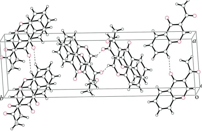
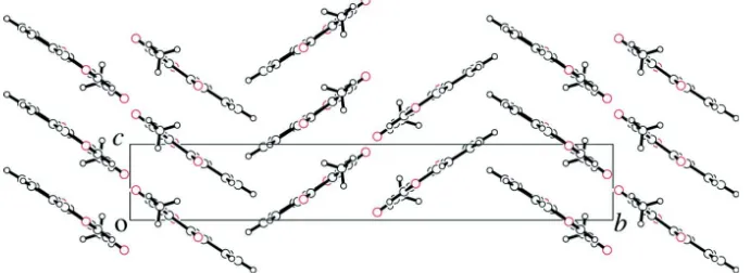
In the crystal, the molecules are linked by π–π stacking [centroid–centroid distance between the benzene and pyran
rings = 3.643 (6) Å], and C–H···O hydrogen bonds form sheets along [0 4 1] and [0 4 1], as shown in Fig.2 and Fig.3.
The crystal structure of a 2,5,6,7-substituted 3-acetylchromone derivative is reported (Chanda et al. 2014).
S2. Experimental
The title compound was synthesized from 3-(dimethylamino)-1-(2-hydroxyphenyl)prop-2-enone (Li et al. 2012)
according to the literature method (Yokoe et al. 1994). Single crystals suitable for X-ray diffraction were obtained by
slow evaporation of an ethyl acetate solution of the title compound at room temperature.
S3. Refinement
All hydrogen atoms were placed in geometrical positions [C–H 0.95 and 0.98 Å], and refined using a riding model with
Uiso(H) = 1.2Ueq of the parent atoms. The s.u.s for the cell parameters are rather large, possibly due to frost damage to the
Figure 1
The molecular structure of the title compound, with displacement ellipsoids drawn at the 50% probability level.
Hydrogen atoms are shown as small spheres of arbitrary radius.
Figure 2
[image:3.610.137.472.352.572.2]Figure 3
A view of the title compound down to the a-axis.
3-Acetyl-4H-chromen-4-one
Crystal data
C11H8O3 Mr = 188.18
Monoclinic, P21/n
Hall symbol: -P 2yn
a = 8.016 (13) Å
b = 25.93 (6) Å
c = 4.091 (8) Å
β = 94.79 (14)°
V = 847 (3) Å3
Z = 4
F(000) = 392.00
Dx = 1.475 Mg m−3
Mo Kα radiation, λ = 0.71069 Å Cell parameters from 25 reflections
θ = 15.2–17.5°
µ = 0.11 mm−1 T = 100 K
Prismatic, colorless 0.42 × 0.25 × 0.20 mm
Data collection
Rigaku AFC-7R diffractometer
ω scans
2377 measured reflections 1962 independent reflections 1510 reflections with F2 > 2.0σ(F2) Rint = 0.018
θmax = 27.6°
h = −5→10
k = 0→33
l = −5→5
3 standard reflections every 150 reflections intensity decay: −0.5%
Refinement
Refinement on F2 R[F2 > 2σ(F2)] = 0.041 wR(F2) = 0.112 S = 1.03 1959 reflections 128 parameters 0 restraints
Primary atom site location: structure-invariant direct methods
Secondary atom site location: difference Fourier map
Hydrogen site location: inferred from neighbouring sites
H-atom parameters constrained
w = 1/[σ2(Fo2) + (0.0509P)2 + 0.3687P]
where P = (Fo2 + 2Fc2)/3
(Δ/σ)max < 0.001
Δρmax = 0.31 e Å−3
Δρmin = −0.20 e Å−3
Special details
Refinement. Refinement was performed using all reflections. The weighted R-factor (wR) and goodness of fit (S) are based on F2. R-factor (gt) are based on F. The threshold expression of F2 > 2.0 σ(F2) is used only for calculating R-factor
x y z Uiso*/Ueq
O1 −0.18103 (13) 0.09136 (4) 1.0184 (3) 0.0256 (3) O2 0.27801 (13) 0.14078 (4) 0.7749 (3) 0.0296 (3) O3 0.22177 (14) 0.01208 (4) 1.3956 (4) 0.0352 (3) C1 −0.03986 (18) 0.06711 (6) 1.1256 (4) 0.0239 (4) C2 0.11774 (18) 0.08077 (5) 1.0636 (4) 0.0212 (3) C3 0.14176 (18) 0.12556 (6) 0.8560 (4) 0.0210 (3) C4 −0.01216 (18) 0.19777 (6) 0.5570 (4) 0.0233 (4) C5 −0.1584 (2) 0.22372 (6) 0.4646 (4) 0.0264 (4) C6 −0.31159 (19) 0.20407 (6) 0.5507 (4) 0.0268 (4) C7 −0.31830 (18) 0.15967 (6) 0.7318 (4) 0.0251 (4) C8 −0.01438 (17) 0.15278 (5) 0.7452 (4) 0.0202 (3) C9 −0.16842 (18) 0.13482 (5) 0.8296 (4) 0.0212 (3) C10 0.25597 (19) 0.04850 (6) 1.2256 (4) 0.0232 (4) C11 0.43401 (19) 0.06180 (6) 1.1822 (5) 0.0270 (4) H1 −0.0510 0.0374 1.2581 0.0286* H2 0.0912 0.2105 0.4924 0.0280* H3 −0.1554 0.2549 0.3429 0.0316* H4 −0.4124 0.2217 0.4826 0.0322* H5 −0.4223 0.1463 0.7889 0.0301* H6A 0.5082 0.0387 1.3165 0.0324* H7B 0.4557 0.0976 1.2506 0.0324* H8C 0.4550 0.0579 0.9509 0.0324*
Atomic displacement parameters (Å2)
U11 U22 U33 U12 U13 U23
O1 0.0172 (5) 0.0250 (6) 0.0351 (6) −0.0015 (4) 0.0041 (5) 0.0048 (5) O2 0.0169 (6) 0.0302 (6) 0.0419 (7) −0.0011 (5) 0.0043 (5) 0.0109 (5) O3 0.0269 (6) 0.0319 (7) 0.0468 (8) 0.0004 (5) 0.0032 (6) 0.0164 (6) C1 0.0211 (7) 0.0222 (7) 0.0282 (8) −0.0004 (6) 0.0018 (6) 0.0013 (6) C2 0.0183 (7) 0.0202 (7) 0.0250 (8) −0.0003 (6) 0.0018 (6) −0.0014 (6) C3 0.0175 (7) 0.0213 (7) 0.0243 (8) −0.0013 (6) 0.0026 (6) −0.0023 (6) C4 0.0207 (7) 0.0231 (7) 0.0261 (8) −0.0014 (6) 0.0015 (6) −0.0012 (6) C5 0.0273 (8) 0.0234 (8) 0.0280 (8) 0.0021 (6) −0.0001 (7) 0.0017 (7) C6 0.0205 (8) 0.0310 (9) 0.0285 (8) 0.0056 (6) 0.0000 (6) −0.0010 (7) C7 0.0178 (7) 0.0293 (8) 0.0283 (8) 0.0001 (6) 0.0028 (6) −0.0031 (7) C8 0.0175 (7) 0.0204 (7) 0.0227 (8) −0.0003 (6) 0.0017 (6) −0.0036 (6) C9 0.0192 (7) 0.0206 (7) 0.0239 (8) −0.0007 (6) 0.0023 (6) −0.0020 (6) C10 0.0217 (8) 0.0224 (7) 0.0253 (8) 0.0008 (6) 0.0017 (6) 0.0001 (6) C11 0.0193 (8) 0.0288 (8) 0.0327 (9) 0.0018 (6) 0.0011 (6) 0.0060 (7)
Geometric parameters (Å, º)
O2—C3 1.233 (3) C10—C11 1.493 (3) O3—C10 1.218 (3) C1—H1 0.950 C1—C2 1.356 (3) C4—H2 0.950 C2—C3 1.461 (3) C5—H3 0.950 C2—C10 1.498 (3) C6—H4 0.950 C3—C8 1.475 (3) C7—H5 0.950 C4—C5 1.377 (3) C11—H6A 0.980 C4—C8 1.399 (3) C11—H7B 0.980 C5—C6 1.401 (3) C11—H8C 0.980 C6—C7 1.373 (4)
C1—O1—C9 118.05 (15) O3—C10—C11 120.67 (16) O1—C1—C2 126.32 (18) C2—C10—C11 119.82 (17) C1—C2—C3 119.10 (15) O1—C1—H1 116.840 C1—C2—C10 115.94 (17) C2—C1—H1 116.843 C3—C2—C10 124.94 (16) C5—C4—H2 119.697 O2—C3—C2 125.01 (15) C8—C4—H2 119.697 O2—C3—C8 120.81 (18) C4—C5—H3 120.092 C2—C3—C8 114.17 (16) C6—C5—H3 120.097 C5—C4—C8 120.61 (17) C5—C6—H4 119.500 C4—C5—C6 119.81 (19) C7—C6—H4 119.487 C5—C6—C7 121.01 (16) C6—C7—H5 120.923 C6—C7—C9 118.14 (17) C9—C7—H5 120.937 C3—C8—C4 121.26 (16) C10—C11—H6A 109.476 C3—C8—C9 120.79 (17) C10—C11—H7B 109.464 C4—C8—C9 117.95 (15) C10—C11—H8C 109.473 O1—C9—C7 116.03 (16) H6A—C11—H7B 109.469 O1—C9—C8 121.52 (15) H6A—C11—H8C 109.475 C7—C9—C8 122.45 (17) H7B—C11—H8C 109.471 O3—C10—C2 119.50 (17)
C1—O1—C9—C7 −178.13 (12) H2—C4—C5—H3 −2.1 C1—O1—C9—C8 1.35 (19) H2—C4—C8—C3 2.2 C9—O1—C1—C2 −0.8 (3) H2—C4—C8—C9 −178.8 C9—O1—C1—H1 179.2 C4—C5—C6—C7 1.3 (3) O1—C1—C2—C3 −1.2 (3) C4—C5—C6—H4 −178.7 O1—C1—C2—C10 177.31 (13) H3—C5—C6—C7 −178.7 H1—C1—C2—C3 178.9 H3—C5—C6—H4 1.3 H1—C1—C2—C10 −2.7 C5—C6—C7—C9 0.4 (3) C1—C2—C3—O2 −177.82 (14) C5—C6—C7—H5 −179.6 C1—C2—C3—C8 2.4 (2) H4—C6—C7—C9 −179.6 C1—C2—C10—O3 1.2 (2) H4—C6—C7—H5 0.4
C2—C3—C8—C4 177.09 (12) C4—C8—C9—C7 0.5 (2) C2—C3—C8—C9 −1.87 (19) O3—C10—C11—H6A −3.2 C5—C4—C8—C3 −177.81 (13) O3—C10—C11—H7B −123.2 C5—C4—C8—C9 1.2 (2) O3—C10—C11—H8C 116.8 C8—C4—C5—C6 −2.0 (3) C2—C10—C11—H6A 175.7 C8—C4—C5—H3 177.9 C2—C10—C11—H7B 55.7 H2—C4—C5—C6 178.0 C2—C10—C11—H8C −64.3
Hydrogen-bond geometry (Å, º)
D—H···A D—H H···A D···A D—H···A
C7—H5···O2i 0.95 2.40 3.292 (6) 155
C1—H1···O3ii 0.95 2.31 3.264 (5) 148