• No results found

Illustrated Review of the Embryology and Development of the Facial Region, Part 2: Late Development of the Fetal Face and Changes in the Face from the Newborn to Adulthood

N/A
N/A
Protected

Academic year: 2020

Share "Illustrated Review of the Embryology and Development of the Facial Region, Part 2: Late Development of the Fetal Face and Changes in the Face from the Newborn to Adulthood"

Copied!
9
0
0

Loading.... (view fulltext now)

Full text

Figure

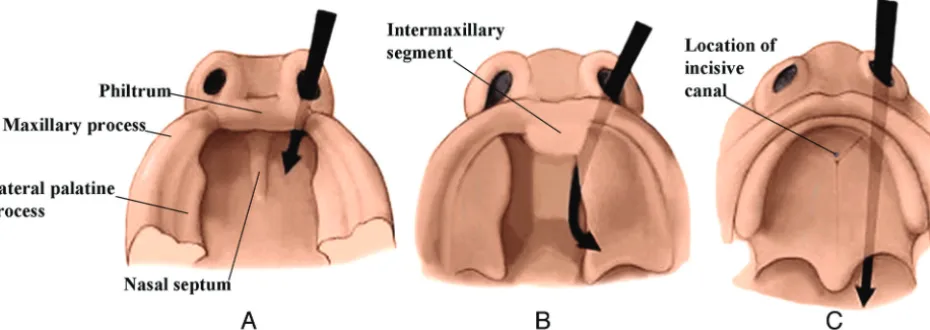
FIG 2. Serial frontal diagrams (A–D) from approximately 6–10 fetal weeks shown just posterior to the intermaxillary segment illustrate theprogressive development of the secondary palate and its fusion with the nasal septum.
FIG 3. Drawing from above and in front (A) of the developing lips and gums. The labiogingival lamina develops in the common mesenchymaltissue of this region
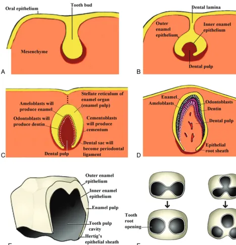
FIG 5. Drawings of the progressive development of the teeth from the tooth bud stage that comes from the dental lamina to the adult tooth.(Modified from http://embryology.ch/anglais/sdigestive/gesicht05.htm and www.embryo.chronolab.com/teeth.html).
FIG 7. Lateral drawings of a 7- to 8-week embryo (A) and an 8- to 10-week fetus (B) show that the opening of the external auditory canal remainsstationary but appears to rise because the progressive elongation of the jaw creates this impression
+5

References

Related documents