RESEARCH PERSPECTIVES
Acquisition Guidelines and Quality Assessment Tools for
Analyzing Neonatal Diffusion Tensor MRI Data
A.M. Heemskerk, A. Leemans, A. Plaisier, K. Pieterman, M.H. Lequin, and J. Dudink
ABSTRACT
SUMMARY: Diffusion tensor imaging is a valuable measure in clinical settings to assess diagnosis and prognosis of neonatal brain development. However, obtaining reliable images is not straightforward because of the tissue characteristics of the neonatal brain and the high likelihood of motion artifacts. In this review, we present guidelines on how to acquire DTI data of the neonatal brain and recommend high-quality data acquisition and processing as an essential means to obtain accurate and robust parametric maps. Sudden head move-ments are problematic for DTI in neonates, and these may lead to incorrect values. We describe strategies to minimize the corrupting effects both in terms of acquisition (eg, more gradient directions) and postprocessing (eg, tensor estimation methods). In addition, tools are described that can help assess whether a dataset is of sufficient quality for further assessment.
ABBREVIATIONS:MD⫽mean diffusivity; FA⫽fractional anisotropy; RESTORE⫽robust estimation of tensors by outlier rejection; TBSS⫽tract-based spatial statistics
B
oth premature birth and complications around term birth are risk factors for brain injury and subsequent neurodevelop-mental impairment. However, this injury often remains “silent” long after the threshold of irreversible injury has been crossed. The most important challenge is to define early proxy measure-ments of long-term neurodevelopmental outcome that will en-able intervention in the early stages of the still adaptive developing human brain. MR imaging is widely used to monitor develop-ment and injury of the neonate brain and to predict neurodevel-opmental outcome1-6(Fig 1). Advanced quantitative MR imagingtechniques such as DTI7-11can detect subtle differences between
normal and abnormal tissue. DTI has become invaluable for the assessment of brain development in preterm infants because it enables detection of white matter maturation during premyelination.1,3,12-14Several DTI studies have revealed that
ab-normal white matter maturation is correlated to neuromotor and neurocognitive performances to childhood.15-18 DTI
measure-ments can therefore provide early biomarkers to be targeted in neuroprotective intervention trials.
Although diffusion MR imaging of the neonatal brain is gain-ing popularity, time constraints often impede acquisition of high quality data. The total time in the scanner should be kept to a minimum, not only for ethical reasons but also to prevent hemodynamic instability. Moreover, a longer acquisition time increases the chance of motion. The feasibility of an MR imag-ing study and the optimal settimag-ings for the MR imagimag-ing scan are determined by the available time in combination with MR hardware and the requirements for obtaining reliable and meaningful data.
From the information above, it may be clear that acquisition, processing, and interpretation of the images with this ad-vanced MR imaging technique are not as straightforward as with the conventional MR imaging sequences. Recent reviews have covered the general concerns within the DTI pipeline that can severely influence the measured results and therefore the study outcome.11,19,20Many of the specifics of diffusion MR
imaging acquisition in adult brains are also applicable to im-aging the neonatal brain. However, the latter presents chal-lenges related to size, tissue composition, and motion and therefore requires specific acquisition settings. Targeted acqui-sition schemes and postprocessing methods are necessary to account for motion and to obtain reliable DTI parameter maps.
In this review, we present guidelines on how to acquire diffusion MR imaging data of the neonatal brain. Furthermore, we recommend high-quality data acquisition and processing as essential means to obtain accurate and robust parametric Received October 3, 2012; accepted after revision November 15.
From the Division of Neonatology, Department of Pediatrics (A.M.H., A.P., K.P., J.D.), and Division of Pediatric Radiology, Department of Radiology (A.M.H., A.P., M.H.L., J.D.), Erasmus Medical Center, Rotterdam, The Netherlands; and Image Sciences Institute (A.L.), University Medical Center Utrecht, Utrecht, The Netherlands.
Please address correspondence to Jeroen Dudink, MD, PhD, Division of Neonatol-ogy, Department of Pediatrics, Erasmus Medical Center, Dr Molewaterplein 60, 3015 GJ Rotterdam, Netherlands; e-mail: [email protected].
maps. We will first describe some general topics related to DTI ac-quisition. Next, we broadly describe the specific concerns and acqui-sition settings related to DTI in the neonate. Finally, we discuss the main steps related to DTI acquisition and processing and the ways they influence the neonatal DTI data quality.
DTI OF THE NEONATE
Imaging the neonatal brain is more difficult than imaging the adult brain. The neonate head is much smaller, and the brain is developing at a fast rate, has a high water content, and is unmy-elinated.21MR imaging acquisition settings therefore must be
ad-justed to neonatal MR imaging relaxation times and diffusion coefficients. Additionally, there is a greater likelihood of mo-tion artifacts in the images because neonates have higher heart and breathing rates and will not lie motionless because seda-tion is usually not given in this situaseda-tion. Fig 2 shows examples of motion artifacts. Thus, the optimal acquisition strategies established for the adult brain22are not necessarily applicable
to the neonatal brain because diffusivities, anisotropy, T2, and SNR will differ. Additionally, optimal acquisition depends on multiple, interrelated parameters, including but not limited to, TR, TE, acquisition matrix, field of view, section thickness, acquisition time, number of averages, maximum b-value, number of b-values, number of non-DWIs, and number of gradient directions.
SETUP FOR IMAGING NEONATES
Neonatal MR imaging is challenging be-cause many neonates are receiving respi-ratory support and are vulnerable to he-modynamic instability.23-25 A recentstudy showed that despite of the use of a dedicated guideline, adverse events dur-ing neonatal MR imagdur-ing scanndur-ing were common.26Because of the increased
ap-plication of neonatal MR imaging, the provision of a safe environment during the MR imaging procedures is of great concern. An MR-compatible incubator with a specialized neonatal head coil al-lows the infant to be kept stable and safe during transport and MR imaging scan. A small-size neonatal head coil typically offers better SNR than the larger adult head coils.27With improved SNR, the
scan time can be reduced and/or the spa-tial resolution for the conventional scans can be improved.
Two common strategies are used to limit motion. One is sedation, which in neonates can compromise breathing and therefore must be performed with great caution. The second strategy is promoting sleep by placing the infant in the incubator in a comfortable and se-cure way.28This will generally help, but
it takes some practice to comfort the child and prevent it from waking up when imaging starts.29The infant’s
per-ception of acquisition noise can be reduced with the use of mold-able earplugs, neonatal earmuffs, and/or an acoustic hood.30
RESOLUTION, TR, TE
A striking difference between the neonatal and adult brain is the inverse contrast between white and gray matter on both the T1-and T2-weighted images.31This is caused by the high water
con-tent and the unmyelinated white matter of the neonate. T1 and T2 relaxation times quickly decrease over the first year of life, and the exact pattern is different for the different brain structures. Typical T2 relaxation times are approximately 300 ms at⬍30 weeks’ ges-tational age, approximately 200 ms at term age,32and
approxi-mately 100 ms at adulthood.33T1 relaxation times for white
mat-ter are approximately 1600 ms at mat-term age and approximately 500 ms at 2 years of age.31For DTI measurements, a longer TR can be
needed to reduce the effect of the high T1 relaxation time (ie, saturation and therefore reduced SNR and/or bias to other proton pools). Fortunately, the long T2 relaxation time does favor the DTI measurement because there is more signal left at a similar TE. However, the DTI estimates for different ages or brain structures must be interpreted carefully because they can be influenced by the varying T1 and T2 relaxation times, possibly caused by altered relative contributions of the different water compartments (eg, intracellular versus extracellular).
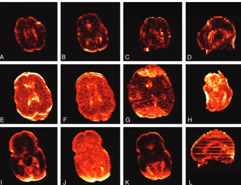
FIG 1. Anatomic, MD, and FA map of a neonate.A,At 30 weeks’ gestational age;B,at term age; and
[image:2.594.57.372.49.368.2]All structures in the neonatal brain are smaller than in the adult brain, thereby requiring a higher resolution. Typical in-plane resolutions range from 0.6⫻0.6 mm2to 2⫻2 mm2, with a
section thickness of 1.9 –3.0 mm (Table 1), which is still relatively large; therefore images should be interpreted cautiously.34
Addi-tionally, the in-plane resolution is often obtained with a low ac-quisition matrix that is zero-filled to a higher matrix; the images therefore are smoother and boundaries are less sharp. The imag-ing resolution and the partial volume effects determine what structures can be resolved. For fiber-tracking purposes, the reso-lution is ideally isotropic and contiguous sections are needed without section gap. The use of isotropic voxels prevents prefer-ential averaging of fiber orientation along a certain axis.11
In addition, the resolution and SNR are related because higher resolution means lower SNR. Therefore, SNR constraints can limit the resolution. Higher resolutions are obtained by decreas-ing the field of view or increasdecreas-ing the matrix size. However, both options have their limitations because the field of view must fit the whole image volume, and higher matrix size introduces artifacts and increases scan time.
GENERAL DTI ACQUISITION CONSIDERATIONS
Generally, a single-shot EPI sequence allows fast acquisition but, unfortunately, is prone to artifacts that affect the data acquisition and the processing.11,19,20,35The most critical artifacts are imagedistortions caused by magnetic susceptibility effects and eddy cur-rents. Among the solutions proposed to reduce the susceptibility artifacts are parallel imaging and B0 field correction. Eddy cur-rents can be reduced by applying bipolar gradients and dual-echo or twice-refocused sequences.36For a more comprehensive and
detailed description of these artifacts and other problems, the au-thors recommend the recent review by Jones and Cercignani19or
other reviews regarding DTI.11,20,35
Diffusion MR imaging is intrinsically a low SNR technique, and the SNR can significantly influence the reliability of each ten-sor estimate. For example, in adult brains, low SNR levels reduce
the accuracy of the diffusion estimates (eg, increased FA) and decrease preci-sion (eg, larger standard deviations).37
The SNR dependence is influenced by the underlying diffusivities and FA val-ues and by the acquisition settings such as b-value.37,38
The magnetic field strength affects the quality of the images both positively and negatively. At 3T, the SNR is gener-ally higher than at 1.5T. However, the images acquired at 3T are more sensitive to susceptibility artifacts, which are prone around air-tissue boundaries. Therefore, the field strength must be considered carefully when setting up a protocol.
The performance of the gradient sys-tems is an important determinant of DTI image quality. Stronger gradients allow for higher diffusion weighting within a shorter time, thereby reducing TE and improving SNR and reducing artifacts. Gradient systems with advanced eddy-current compensation are preferred because these limit image distortions.
Hardware and software of the MR imaging systems differ be-tween manufacturers and are constantly being upgraded. There-fore, imaging protocols cannot always be exchanged between sites, and what is achievable at one scanner might not be possible at another. Additionally, even equally equipped MR imaging sys-tems can introduce a bias.39
The coil should adequately cover the volume of interest and exhibit sufficient SNR. Preferably, a multichannel coil should be used with the option to use parallel imaging. Parallel imaging techniques can shorten the EPI readout, thereby reducing imag-ing artifacts and TE.40
DIFFUSION WEIGHTING
The b-value indicates the amount of diffusion weighting that is applied. The optimal b-value depends on the tensor information of interest but is generally approximately 1.09/ADC.22For clinical
adult brain DTI, a b-value of 1000 s/mm2is common practice,
which is a concession between a longer optimal b-value and the need for shorter echo times to ensure enough SNR. As mentioned, the neonatal brain contains more water and is less myelineated than the adult brain. Therefore, the neonatal MD is higher than the adult MD (2.0 ⫻10⫺3mm2/s versus 0.7⫻ 10⫺3mm2/s)
and the optimal b-value should be lower to accurately estimate the diffusion tensor. Currently, there is no consensus on the b-value in neonates, and b-values range between 600 –1100 s/mm2(Table
1). Simulation studies are needed to determine the optimal b-value, just as has been performed for adult brains.
At least 6 diffusion gradient directions and 1 non-DWI are needed to estimate the diffusion tensor. However, more direc-tions increase the accuracy and precision of the diffusion esti-mates. Tensor estimates with 30 directions are statistically rota-tionally invariant.41 Increasing the number of directions also
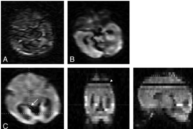
FIG 2. Effects of motion and pulsation.A,Almost complete signal loss caused by head motion;B,
signal drop-out caused by pulsation; andC,3-plane view in which the effects of pulsation(arrow)
[image:3.594.55.374.47.260.2]entails increasing the number ofb⫽0 acquisitions because 1b⫽ 0 image is needed for every 8 –10 DWIs.22The optimal
distribu-tion of the gradients is a uniform distribudistribu-tion along the surface of a sphere,22which minimizes the orientation dependency of the
tensor estimates. Additionally, the order of the gradients ideally should be uniformly distributed in case the acquisition is cor-rupted by motion or intercor-rupted acquisition.42,43Dubois et al43
have proposed optimal diffusion gradient orientation schemes that allow calculation of the diffusion tensor estimates with the use of partial datasets.
The higher likelihood of motion in the neonates requires a higher number of gradient directions. The relative contribution of a cor-rupted image is less if more gradient directions are acquired. There-fore, it may be preferable to increase the number of gradient direc-tions at the cost of reducing TR (and thereby SNR) to keep the total scan time approximately equal. In addition, we recommend the use of extra b⫽0 images because these images also can be affected by intersection and intrasection motion, and good-qualityb⫽0 images are needed to correctly calculate the tensor estimates.
MOTION
Subject-related motion can affect the resulting parameter maps. The motion can be evident between images or within a single image. During the long acquisition, adult subjects tend to slowly move their heads even if they are instructed to lay still and the head is secured by padding.44A slow displacement and/or
rota-tion causes a misalignment between images. This can be adjusted for by image registration. The motion within a single image is caused by cardiac pulsation or sudden large amplitude subject-related motion. The latter occurs rarely in adults (1.4%)45but is a
severe problem in neonates. Fig 2 shows the effects of severe mo-tion, cardiac pulsamo-tion, and the large differences in signal intensi-ties throughout the image volume.
Cardiac pulsation causes both a local deformation of the tissue
and additional signal loss. The tensor estimates are then less reli-able. Because neonates have much faster heart rates than adults, the likelihood that the diffusion sensitization is obtained during a pulsation is larger, leading to a higher likelihood of artifacts. The effects of cardiac pulsation can be reduced by triggering the ac-quisition on the cardiac cycle; however, scan times become longer because the effective TR should be at least 5 times the T1 of the tissue. For neonates, cardiac gating seems possible only by moni-toring the ECG because the delay time for the pulse wave on the pulse oximeter is too long (249 ms for adults46) compared with
the heart beat (approximately every 350 ms).
Subject motion is a major concern for neonatal imaging because neonates tend to move about even when sedated. Intrasection mo-tion from a sudden shake of the head may corrupt the image and result in miscalculation of the DTI estimates. A typical motion-cor-rupted image is characterized by severe signal loss; this is caused by tissue displacement during the diffusion encoding, which also causes signal loss next to the dephasing caused by diffusion. This severe signal loss cannot be recovered, and an apparent high displacement would be calculated. Improper dealing with the motion artifacts may result in biased MD and high FA values.
Only a few studies have reported data rejection on the grounds of severe artifacts. Rejection was necessary in 15– 60% of the sub-jects,15,47-49and the incidence of less noticeable artifacts will be
larger. In a pilot study of 27 preterm neonates, we investigated the statistical outliers of the tensor regression (see “DTI Quality and Pathology” section) and showed that data of 60% of the subjects were corrupted by motion (defined as⬎10 sections with⬎30% outliers) (Table 2).50This high occurrence of motion can have
[image:4.594.55.531.56.272.2]devastating results. Unfortunately, the typical tensor regression method used by MR imaging vendors and commercial processing tools is insufficient with corrupted data, and it appears that many DTI users are unaware of the motion-related problems. Table 1: Published DTI acquisition settings for neonates from different groups
PMA FS
Neo
Coil TR TE FOV Thk
Image
Resolution b-Value No. b= 0
No.
Dir Sedation Reference
40–45 3 ⫺ 7745 48 180 2 1.41 800 1 32 ⫹ van Pul et al67
Term 3 ⫺ 8400 84 220 2.2 2.20 750 ? ? ⫾ Wintermark et al68
31–41 1.5 ⫹ 4000 60 210 3.0 0.82 1000 1 ? ⫺ Arrigoni et al69
38–41 3 ⫺ ⬎3000 71 150 1.9 1.88 700 5 30 ⫺ Oishi et al70
Term 3 7680 82 2.0 2.00 1000 7 42 ⫺ Wang et al71
37–43 1.5 6000 88 200 2.5 1.56 1000 1 15 ⫹ Hasegawa et al72
40–44 3 7465 54 2.0 1.40 1000 1 32 ⫹ de Bruine et al73
Term 1.5 ⫹ 4047 59 210 3.0 0.82 1000 1 6 ⫺ Righini et al74
24–33/TEA 1.5 ⫹ 7000 100 3.0 1.40 600 1 6 ⫺ Bonifacio et al75
24–33/TEA 1.5 ⫹ 4900 104 160 3.0 1.30 600/700 1 12 ⫺ Bonifacio et al75
Term 3 5200 73 2.0 2.00 1000 1 6 ⫺ Gilmore et al76
25–32 1.5 ⫹ 9150 98 200 3.0 0.78 1000 1 25 ⫺ Dudink et al77
35–42 1.5 ⫺ 5888 92 220 2.3 2.00 600 1 32 ⫺ Liu et al78
39–41 1.5 ⫺ 7000 74 180 2.2 1.40 700 1 15 ⫹ Skiold et al5
Term 1.5 ⫺ 8000 100 240 3.0 1.88 700 1 10 ⫺ Malik et al79
Term 1.5 ⫺ 6000 106 230 2.5 0.90 1100 16 44 ⫺ Rose et al80
38–45 3 ⫺ 8000 79 224 2.0 1.75 750 1 15 ⫾ Anjari et al13
30 1.5 ⫹ 11725 90.5 220 3.0 0.86 750 3 25 ⫺ Erasmus MC – Sophia
Term 1.5 ⫹ 11725 85.6 220 3.0 0.66 1000 3 25 ⫺ Erasmus MC – Sophia
Note:—Image resolution is all squared (mm); b-values are s/mm2
.
The solutions to prevent corrupted images are limited. Obvi-ously, the movement should be minimized a priori by placing the child comfortably and supporting the head with pads. The acqui-sition strategies to reduce the signal loss are limited because the
sudden gross motion occurs during the time the diffusion encoding is performed. Therefore, navigator echoes or altered readout sequences appear not to be useful. However, shortening the diffusion time by stronger gradients or lower b-value might reduce the effects. Fortunately, there are strategies to decrease the detrimental effects of corrupted images. These in-clude the oversampling of gradient di-rections, the removal of corrupted im-ages, and/or the use of more advanced tensor regression methods.
TEMPERATURE
Another issue one must be aware of is that diffusion is temperature-depen-dent. A 1° difference in temperature leads to a 2–3% difference in ADC.51,52
It will therefore be necessary to maintain the child at a constant temperature dur-ing scanndur-ing. Also, consistent core tem-peratures are necessary in studying group differences or performing a longi-tudinal study because the differences in MD or FA between ages, groups, and therapies are small.
PROCESSING
The basic steps in data postprocessing are motion/distortion correction, esti-mation of the diffusion tensor, and com-putation of the parametric maps. The data should be inspected before and af-ter each step to ensure good image qual-ity and absence of artifacts. Postprocess-ing results in data reduction, and it can be difficult to spot erroneous results on the parametric maps.
MOTION/DISTORTION
CORRECTION
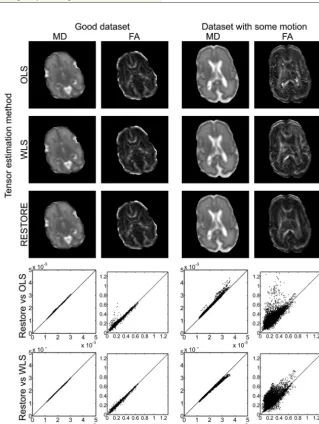
Registration and correction of the im-ages is necessary to adjust for slight vari-ation in brain position and to correct for the eddy current. A quick check for mis-registration between images is to exam-ine the FA maps for rims of high anisot-ropy at the edges of the brain or at the interface between CSF and white matter. Because most available registration tools are optimized for the contrast in adult brain, one should check whether the registration software is adequate for the neonatal brain, in view of its different contrast. In addition, most tools perform their registration on the whole image volume and it is therefore logical that section drop-outs caused by motion severely alter the registration, especially in the case of affine reg-FIG 3. Effects of different tensor estimation methods. Data of 2 datasets are displayed: on the
[image:5.594.54.374.139.564.2]left, data without gross motion artifacts, and on the right, data with gross motion artifacts for one of the gradient directions. Both MD and FA maps are depicted, showing no visible differences for the good dataset and clearly visible differences for the dataset with motion. Especially the FA maps show that the ordinary least-squares (OLS) estimation results in very high FA values that are not related to the known anatomy. In the graphs, the pixel value of the OLS or weighted least-squares (WLS) tensor estimation is plotted against the pixel value of the RESTORE tensor estimation (x-axis), and the line of identity is included. The spread around the line of identity is broader for the dataset, with motion indicating an effect of the tensor estimation method on the resulting FA or MD value. For the graphs, the images were eroded to exclude the outer rim, which contains poor-quality data caused by partial volume effects. Scaling: MD, 0 –2⫻10⫺3mm2/s; FA, 0 –1.
Table 2: Outliers in a small pilot study (27 preterm infants scanned at 30 weeks’ gestational age)
>10% Outliers >30% Outliers >50% Outliers
No. of subjects⬎10 sections 27 15 10
No. of subjects⬎20 sections 26 10 1
Mean No. of sections with outliers 50 15 9
Range No. of sections with outliers 17–79 1–38 0–24
Mean percentage of sections with outliers 9.5 2.9 1.7
istration. A final step is to also correct the diffusion encoding gradients with the same rotational correction as the images.44
Neglecting to do so will induce a mismatch between the actual diffusion weighting and the expected diffusion weighting and therefore result in errors in the estimates of the diffusion tensor and the fiber orientation.
It is of paramount importance to ensure that motion correc-tion, which is often necessary in neonatal scanning, does not neg-atively affect the DTI dataset. Intersection movement is problem-atic for volume registration, and the b⫽0 volume should be carefully checked on misalignment between sections. Large signal drop-outs are problematic with through-plane motion correction because thereafter, the incorrect signal intensities affect 2 sections, resulting in possible incorrect tensor estimations. Zhou et al47
investigated the use of local texture features to identify and reject outlier images automatically before estimating the diffusion ten-sor. Their method is fast and removes sections that are corrupted and cannot be used for tensor estimation, resulting in more accu-rate data. Implementation of fast detection techniques that are based on image characteristics will improve data quality and might offer possibilities to repeat corrupted gradient directions while scanning.53
ESTIMATION OF THE TENSOR
Several tensor estimation methods have been developed to es-timate the diffusion tensor. Each of the approaches is based on different principles, and generally speed and accuracy are in-versely proportional: methods range from fast but less accu-rate, to slow but more accurate.9,49,54The main problem in
estimating the tensor is the presence of data outliers caused by motion, pulsation, artifacts, noise, and so on. Depending on the method used, these outliers can have a significant impact on the resulting eigenvalues and eigenvectors. The linear least-squares method is commonly used by vendors and is the de-fault setting for commonly used DTI postprocessing software such as FSL (http://www.fmrib.ox.ac.uk/fsl)55; however, this
method proved to be the least appropriate to estimate the ten-sor because the method assumptions are restrictive and phys-ically implausible.56Fig 3 shows the effect of the tensor
[image:6.594.57.534.45.410.2]Therefore, for neonatal DTI data, with its high likelihood of corrupted data, we must use proper tensor estimation procedures to obtain reliable diagnostic images or research data. RESTORE54
is a method that detects and removes outlier data in the tensor estimation. This method requires additional gradient encoding directions to eliminate erroneous data. For neonatal DTI mea-surements, Morris et al49investigated the use of removing outliers
based on the RESTORE algorithm before motion correction because the latter can result in averaging outliers with uncor-rupted voxels. With this new method, they found an increased sensitivity to outliers, which is important as outliers correlated strongly with subject movement.
QUALITY ASSURANCE
It is of paramount importance to ensure that all the steps from data acquisition to data analysis are correct and that the parame-ter maps are of sufficient quality before analyzing the data and drawing conclusions. Good-quality data are of paramount im-portance for the neonatal population because the chances of
arti-facts and corrupted data are larger than for the average adult population, and ar-tifacts will bias the quantification and increase the variation. Data of sufficient quality will provide quantitative mea-sures with low variation.
Quality assurance begins by care-fully checking the b⫽0 images for ac-curacy and absence of artifacts and intersection movement. Intersection movement causes severe problems with the image registration, resulting in erro-neous results; therefore, those image volumes must be excluded from further analysis and hence our previous sugges-tion to increase the number of b ⫽ 0 volumes. Thereafter, the DWIs are in-spected for large signal dropouts, which are easily observed by through-plane projections (Fig 2C). Image misalign-ments caused by motion or eddy cur-rents can be visualized by a high FA around the rim of the brain or by calcu-lating the standard deviations across the DW images, in which large SD at the rims indicates misregistration. This is also a good method to check whether images were registered accurately or if registration issues persist or even are in-troduced. Artifacts in the data can also be spotted by locating the areas where FA is⬎1. By definition, this should not be plausible but occurs when the DW signal intensity is larger than the b⫽0 signal intensity.
The tensor estimation model also en-ables possibilities to spot artifacts by means of the residuals of the tensor fit and the calculation of data outliers. Re-siduals represent the difference between the fit and the data signal, with high residuals indicating either a poor estimation model or underlying data artifacts. The average residual maps as depicted in Fig 4 are helpful to detect and locate artifacts, assuming that high residuals are caused by artifacts and that good image data result in a homogeneous residuals map.20 High residuals are generally
found in areas with partial volume effects (rims) and in regions with low signal intensities,20and therefore one should extra
care-fully interpret results from those regions.
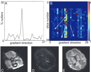
In addition, the residuals are used to detect signal outliers. The location and extent of outliers is a functional tool to investigate the DTI quality. The example in Fig 5 shows that 2 DTI volumes have a high percentage of outliers, which corresponds to cor-rupted images. We have found that determining the volumes and sections with high percentage outliers is an excellent method to assess data corruption caused by gross motion.50However,
label-ing a point as an outlier also is dependent on the presence or absence of other (larger) outliers. Indeed, one expects a more or less even FIG 5. Detection of outliers.A,Percentage of outliers per DWI is a tool to indicate potential
problems with the DTI dataset. In this case, DWI 11 and 12 have a high percentage of outliers.B,
Percentage outliers per section facilitates easy detection of the problematic sections. During the acquisition of gradient direction 12, several sections are affected by movement of the infant.C
andD,Examples of the resulting images;E,nonaffected image. Because of the interleaved section acquisition, there is an alternating pattern for the odd and even sections. Scaling is similar for DWI.
[image:7.594.55.373.44.295.2] [image:7.594.55.377.377.455.2]distribution of outliers with high-quality data, whereas corrupted sections cause a significant increase in outliers (Fig 6). Further re-search is needed to determine the relation between the distribution of outliers and the resulting parameter maps, the needed processing steps, and suitability of a dataset for further analysis.
DATA ANALYSIS
After the computation of the tensor estimates, the parametric maps can be used for further data analysis to assess the white matter integrity or structure. Commonly used techniques are re-gion of interest selection, voxel-based analysis, tract-based spatial statistics, and fiber tractography. Region of interest selection is used to compare white matter properties within a specific region of the brain. The lower number of comparisons between subjects increases the statistical power, and small differences between sub-jects may be found. The technique is also independent of individ-ual variations in shape and size of the brain. Major limitations include reliance on the accuracy of the region of interest place-ments, the likelihood of missing differences in regions that are not included, the time-consuming nature when the regions of interest are manually drawn, and the observer dependency.
Whole-brain analysis can be performed with voxel-based analyses, in which all voxels are analyzed in terms of a tensor estimate. Because of individual variation in size and shape of the brain, all individual brains must first be aligned to a template (normalization) before they can be compared.57,58Normalization
is the main challenge in voxel-based analysis because inaccurate nor-malization results in incorrect comparison of individuals.59This is
particularly problematic in patients with cerebral injuries where nor-malization results in distortion of the brains. TBSS overcomes this limitation by aligning the FA images from multiple subjects to a “mean FA skeleton.” With this technique, only voxels that are at the center of tracts common to all the individuals in the population are
included. Because voxels with poor alignment are excluded, one of the limitations of this strategy is that it may miss variations in the periphery of white matter tracts. Other limitations and concerns re-lated to TBSS have been addressed by others.60,61
Fiber tractography enables delineation and comparison of the orientation and direction of a specific white matter tract between individuals. This method is independent of variation in brain or-ganization. In the deterministic approach,62the algorithm moves
in the direction of the principal eigenvector (1); this is assumed
to be parallel to the dominant fiber orientation in each voxel. The main limitation of deterministic tractography is uncertainty about the reliability of a reconstructed trajectory. Probabilistic tractography takes the uncertainty in1into account by
propa-gating a distribution of possible orientations from the seed point.63Although probabilistic tractography seems more reliable,
it remains dependent on the placement of the seed points, which is manually done.3,12,35,64-66
Although these analytic measurements provide in vivo infor-mation about the orientation, organization, and microstructural properties of the cerebral white matter, DTI analysis remains a proxy technique and not a direct visualization of the cerebral white matter. Moreover, because tensor estimations are subject to error, we emphasize the importance of appropriate acquisition settings with regard to gestational age, a quality check before post-processing, reliable postprocessing techniques, and good knowl-edge of neuro anatomy.35
DTI QUALITY AND PATHOLOGY
[image:8.594.58.535.46.288.2]injury. Large portions of the brain are affected, and these conse-quently have lower MD values as the result of edema. These data-sets appear to be of lesser quality in terms of outliers and residuals than the nonpathologic datasets without motion corruption. However, a closer look reveals that the outliers are randomly dis-tributed in time and that the residuals are high in the areas with low MD values. This is expected because the diffusivities in the pathologic areas are lower and therefore the b-value might not be optimal to accurately determine MD and FA values in these areas. Although data quality assurance is not directly a vital issue for clinical practice in which the main goal is to visualize the areas with altered MD and FA values, it is imperative for the analysis of the DTI values to exclude any bias in DTI indices.
CONCLUSIONS
High-quality, quantitative data are essential to ensure reliable and meaningful imaging findings. Both data quality and analysis will improve with the use of protocols and image-processing tools specifically designed for neonatal imaging, seeing that tissue com-position and occurrence of artifacts in neonates are significantly different from those in the adult population. Optimized acquisi-tion and targeted processing will increase the diagnostic and prog-nostic value of the scans but will also considerably improve inter-vention or outcome studies because a reduced data spread will lead to stronger correlations, will require fewer subjects, and will allow earlier determination of valuable intervention methods.
REFERENCES
1. Counsell SJ, Rutherford MA, Cowan FM, et al.Magnetic resonance imaging of preterm brain injury.Arch Dis Child Fetal Neonatal Ed
2003;88:F269 –74
2. Huppi PS, Barnes PD.Magnetic resonance techniques in the evalu-ation of the newborn brain.Clin Perinatol1997;24:693–723 3. Ment LR, Hirtz D, Huppi PS.Imaging biomarkers of outcome in the
developing preterm brain.Lancet Neurol2009;8:1042–55
4. Lequin MH, Dudink J, Tong KA, et al.Magnetic resonance imaging in neonatal stroke.Semin Fetal Neonatal Med2009;14:299 –310 5. Skiold B, Horsch S, Hallberg B, et al.White matter changes in
ex-tremely preterm infants, a population-based diffusion tensor im-aging study.Acta Paediatr2010;99:842– 49
6. Gimenez M, Miranda MJ, Born AP, et al.Accelerated cerebral white matter development in preterm infants: a voxel-based morphome-try study with diffusion tensor MR imaging. Neuroimage
2008;41:728 –34
7. Basser PJ, Mattiello J, LeBihan D.Estimation of the effective
self-diffusion tensor from the NMR spin echo. J Magn Reson
1994;103:247–54
8. Pierpaoli C, Jezzard P, Basser PJ, et al.Diffusion tensor MR imaging of the human brain.Radiology1996;201:637– 48
9. Le Bihan D, van Zijl P.From the diffusion coefficient to the diffu-sion tensor.NMR Biomed2002;15:431–34
10. Mori S, van Zijl PC.Fiber tracking: principles and strategies: a tech-nical review.NMR Biomed2002;15:468 – 80
11. Jones DK, Leemans A.Diffusion tensor imaging.Methods Mol Biol
2011;711:127– 44
12. Dudink J, Kerr JL, Paterson K, et al.Connecting the developing pre-term brain.Early Hum Dev2008;84:777– 82
13. Anjari M, Srinivasan L, Allsop JM, et al.Diffusion tensor imaging with tract-based spatial statistics reveals local white matter abnor-malities in preterm infants.Neuroimage2007;35:1021–27 14. Bassi L, Chew A, Merchant N, et al.Diffusion tensor imaging in
preterm infants with punctate white matter lesions.Pediatr Res
2011;69:561– 66
15. van Kooij BJ, van Pul C, Benders MJ, et al.Fiber tracking at term displays gender differences regarding cognitive and motor out-come at 2 years of age in preterm infants.Pediatr Res2011;70:626 –32 16. Bassi L, Ricci D, Volzone A, et al.Probabilistic diffusion tractogra-phy of the optic radiations and visual function in preterm infants at term equivalent age.Brain2008;131:573– 82
17. Drobyshevsky A, Bregman J, Storey P, et al.Serial diffusion tensor imaging detects white matter changes that correlate with motor outcome in premature infants.Dev Neurosci2007;29:289 –301 18. van Kooij BJ, de Vries LS, Ball G, et al.Neonatal tract-based spatial
statistics findings and outcome in preterm infants.AJNR Am J Neu-roradiol2012;33:188 –94
19. Jones DK, Cercignani M.Twenty-five pitfalls in the analysis of dif-fusion MRI data.NMR Biomed2010;23:803–20
20. Tournier JD, Mori S, Leemans A.Diffusion tensor imaging and be-yond.Magn Reson Med2011;65:1532–56
21. Johansen-Berg H, Behrens TE, eds.Diffusion MRI: from quantita-tive measurement to in-vivo neuroanatomy. 2009. Waltham, Massachusetts: Academic Press, Elsevier.
22. Jones DK, Horsfield MA, Simmons A.Optimal strategies for mea-suring diffusion in anisotropic systems by magnetic resonance im-aging.Magn Reson Med1999;42:515–25
23. Battin M, Maalouf EF, Counsell S, et al.Physiological stability of preterm infants during magnetic resonance imaging.Early Hum Dev1998;52:101–10
24. Benavente-Fernandez I, Lubian-Lopez PS, Zuazo-Ojeda MA, et al.
Safety of magnetic resonance imaging in preterm infants.Acta Pae-diatr2010;99:850 –53
25. Merchant N, Groves A, Larkman DJ, et al.A patient care system for early 3.0 Tesla magnetic resonance imaging of very low birth weight infants.Early Hum Dev2009;85:779 – 83
26. Plaisier A, Raets MM, van der Starre C, et al.Safety of routine early MRI in preterm infants.Pediatr Radiol2012;42:1205–11
27. Keil B, Alagappan V, Mareyam A, et al.Size-optimized 32-channel brain arrays for 3 T pediatric imaging. Magn Reson Med
2011;66:1777– 87
28. Legendre V, Burtner PA, Martinez KL, et al.The evolving practice of developmental care in the neonatal unit: a systematic review.Phys Occup Ther Pediatr2011;31:315–38
29. Neubauer V, Griesmaier E, Baumgartner K, et al.Feasibility of cere-bral MRI in non-sedated preterm-born infants at term-equivalent age: report of a single centre.Acta Paediatr2011;100:1544 – 47 30. Nordell A, Lundh M, Horsch S, et al.The acoustic hood: a
patient-independent device improving acoustic noise protection during neonatal magnetic resonance imaging. Acta Paediatr 2009;98: 1278 – 83
31. Holland BA, Haas DK, Norman D, et al.MRI of normal brain mat-uration.AJNR Am J Neuroradiol1986;7:201– 08
32. Counsell SJ, Kennea NL, Herlihy AH, et al.T2 relaxation values in
the developing preterm brain. AJNR Am J Neuroradiol
2003;24:1654 – 60
33. Kumar R, Delshad S, Macey PM, et al.Development of T2-relaxation values in regional brain sites during adolescence.Magn Reson Im-aging2011;29:185–93
34. Vos SB, Jones DK, Viergever MA, et al.Partial volume effect as a hidden covariate in DTI analyses.Neuroimage2011;55:1566 –76 35. Mukherjee P, Chung SW, Berman JI, et al.Diffusion tensor MR
im-aging and fiber tractography: technical considerations.AJNR Am J Neuroradiol2008;29:843–52
36. Reese TG, Heid O, Weisskoff RM, et al.Reduction of eddy-current-induced distortion in diffusion MRI using a twice-refocused spin echo.Magn Reson Med2003;49:177– 82
37. Pierpaoli C, Basser PJ.Toward a quantitative assessment of diffu-sion anisotropy.Magn Reson Med1996;36:893–906
38. Damon BM.Effects of image noise in muscle diffusion tensor (DT)-MRI assessed using numerical simulations. Magn Reson Med
2008;60:934 – 44
measurement using a temperature-controlled fluid for quality con-trol in multicenter studies.J Magn Reson Imaging2011;34:983– 87 40. Bammer R, Schoenberg SO.Current concepts and advances in
clin-ical parallel magnetic resonance imaging.Top Magn Reson Imaging
2004;15:129 –58
41. Jones DK.The effect of gradient sampling schemes on measures derived from diffusion tensor MRI: a Monte Carlo study.Magn Reson Med2004;51:807–15
42. Cook PA, Symms M, Boulby PA, et al.Optimal acquisition orders of diffusion-weighted MRI measurements. J Magn Reson Imaging
2007;25:1051–58
43. Dubois J, Poupon C, Lethimonnier F, et al.Optimized diffusion gra-dient orientation schemes for corrupted clinical DTI data sets.
MAGMA2006;19:134 – 43
44. Leemans A, Jones DK.The B-matrix must be rotated when correct-ing for subject motion in DTI data.Magn Reson Med2009;61: 1336 – 49
45. Ling J, Merideth F, Caprihan A, et al.Head injury or head motion? Assessment and quantification of motion artifacts in diffusion ten-sor imaging studies.Hum Brain Mapp2012;33:50 – 62
46. Pierpaoli C, Marenco S, Rohde G, et al.Analyzing the contribution of cardiac pulsation to the variability of quantities derived from the diffusion tensor.ISMRMToronto, Canada, July 10 –16, 2003:70 47. Zhou Z, Liu W, Cui J, et al.Automated artifact detection and
re-moval for improved tensor estimation in motion-corrupted DTI data sets using the combination of local binary patterns and 2D partial least squares.Magn Reson Imaging2011;29:230 – 42 48. Dudink J, Lequin M, van Pul C, et al.Fractional anisotropy in white
matter tracts of very-low-birth-weight infants. Pediatr Radiol
2007;37:1216 –23
49. Morris D, Nossin-Manor R, Taylor MJ, et al.Preterm neonatal dif-fusion processing using detection and replacement of outliers prior to resampling.Magn Reson Med2011;66:92–101
50. Heemskerk AM, Plaisier A, Reiss I, et al.DTI in neonates: data cor-ruption due to motion.ISMRMMelbourne, Australia, May 5–11, 2012
51. Tofts PS, Lloyd D, Clark CA, et al.Test liquids for quantitative MRI measurements of self-diffusion coefficient in vivo.Magn Reson Med
2000;43:368 –74
52. Kozak LR, Bango M, Szabo M, et al.Using diffusion MRI for mea-suring the temperature of cerebrospinal fluid within the lateral ventricles.Acta Paediatr2010;99:237– 43
53. Aksoy M, Forman C, Straka M, et al.Real-time optical motion cor-rection for diffusion tensor imaging. Magn Reson Med 2011; 66:366 –78
54. Chang LC, Jones DK, Pierpaoli C.RESTORE: robust estimation of tensors by outlier rejection.Magn Reson Med2005;53:1088 –95 55. Smith SM, Jenkinson M, Woolrich MW, et al.Advances in functional
and structural MR image analysis and implementation as FSL. Neu-roimage2004;23(Suppl 1):S208 –19
56. Koay CG, Chang LC, Carew JD, et al.A unifying theoretical and algorithmic framework for least squares methods of estimation in diffusion tensor imaging.J Magn Reson2006;182:115–25
57. Van Hecke W, Leemans A, D’Agostino E, et al.Nonrigid coregistra-tion of diffusion tensor images using a viscous fluid model and mu-tual information.IEEE Trans Med Imaging2007;26:1598 – 612 58. Van Hecke W, Sijbers J, D’Agostino E, et al.On the construction of
an inter-subject diffusion tensor magnetic resonance atlas of the healthy human brain.Neuroimage2008;43:69 – 80
59. Van Hecke W, Leemans A, Sage CA, et al.The effect of template selection on diffusion tensor voxel-based analysis results. Neuroim-age2011;55:566 –73
60. Edden RA, Jones DK.Spatial and orientational heterogeneity in the statistical sensitivity of skeleton-based analyses of diffusion tensor MR imaging data.J Neurosci Methods2011;201:213–19
61. Van Hecke W, Leemans A, De Backer S, et al.Comparing isotropic and anisotropic smoothing for voxel-based DTI analyses: a simula-tion study.Hum Brain Mapp2010;31:98 –114
62. Basser PJ, Pajevic S, Pierpaoli C, et al.In vivo fiber tractography using DT-MRI data.Magn Reson Med2000;44:625–32
63. Jones DK.Tractography gone wild: probabilistic fibre tracking us-ing the wild bootstrap with diffusion tensor MRI.IEEE Trans Med Imaging2008;27:1268 –74
64. Feldman HM, Yeatman JD, Lee ES, et al.Diffusion tensor imaging: a review for pediatric researchers and clinicians.J Dev Behav Pediatr
2010;31:346 –56
65. Jones DK.Studying connections in the living human brain with diffusion MRI.Cortex2008;44:936 –52
66. Huppi PS, Dubois J.Diffusion tensor imaging of brain develop-ment.Semin Fetal Neonatal Med2006;11:489 –97
67. van Pul C, van Kooij BJ, de Vries LS, et al.Quantitative fiber tracking in the corpus callosum and internal capsule reveals microstructural abnormalities in preterm infants at term-equivalent age. AJNR Am J Neuroradiol2012;33:678 – 84
68. Wintermark P, Hansen A, Gregas MC, et al.Brain perfusion in as-phyxiated newborns treated with therapeutic hypothermia.AJNR Am J Neuroradiol2011;32:2023–29
69. Arrigoni F, Parazzini C, Righini A, et al.Deep medullary vein in-volvement in neonates with brain damage: an MR imaging study.
AJNR Am J Neuroradiol2011;32:2030 –36
70. Oishi K, Mori S, Donohue PK, et al.Multi-contrast human neonatal brain atlas: application to normal neonate development analysis.
Neuroimage2011;56:8 –20
71. Wang Y, Gupta A, Liu Z, et al.DTI registration in atlas based fiber analysis of infantile Krabbe disease.Neuroimage2011;55:1577– 86 72. Hasegawa T, Yamada K, Morimoto M, et al.Development of corpus
callosum in preterm infants is affected by the prematurity: in vivo assessment of diffusion tensor imaging at term-equivalent age. Pe-diatr Res2011;69:249 –54
73. de Bruine FT, van Wezel-Meijler G, Leijser LM, et al.Tractography of developing white matter of the internal capsule and corpus callo-sum in very preterm infants.Eur Radiol2011;21:538 – 47
74. Righini A, Doneda C, Parazzini C, et al.Diffusion tensor imaging of early changes in corpus callosum after acute cerebral hemisphere lesions in newborns.Neuroradiology2010;52:1025–35
75. Bonifacio SL, Glass HC, Chau V, et al.Extreme premature birth is not associated with impaired development of brain microstructure.
J Pediatr2010;157:726 –32 e1
76. Gilmore JH, Kang C, Evans DD, et al.Prenatal and neonatal brain structure and white matter maturation in children at high risk for schizophrenia.Am J Psychiatry2010;167:1083–91
77. Dudink J, Buijs J, Govaert P, et al.Diffusion tensor imaging of the cortical plate and subplate in very-low-birth-weight infants.Pediatr Radiol2010;40:1397– 404
78. Liu Y, Baleriaux D, Kavec M, et al.Structural asymmetries in motor and language networks in a population of healthy preterm neonates at term equivalent age: a diffusion tensor imaging and probabilistic tractography study.Neuroimage2010;51:783– 88
79. Malik GK, Trivedi R, Gupta A, et al.Quantitative DTI assessment of periventricular white matter changes in neonatal meningitis.Brain Dev2008;30:334 – 41