ORIGINAL RESEARCH
HEAD & NECK
Performance of Iterative Image Reconstruction in CT of the
Paranasal Sinuses: A Phantom Study
B. Schulz, M. Beeres, B. Bodelle, R. Bauer, F. Al-Butmeh, A. Thalhammer, T.J. Vogl, and J.M. Kerl
ABSTRACT
BACKGROUND AND PURPOSE: CT in low dose technique is the criterion standard imaging modality for evaluation of the paranasal sinus. Our aim was to evaluate the dose-reduction potential of a recently available sinogram-affirmed iterative reconstruction technique, regarding noise, image quality, and time duration when evaluating this region.
MATERIALS AND METHODS:CT was performed on a phantom head at different tube voltages (120 kV, 100 kV) and currents (100 mAs, 50 mAs, 25 mAs). Each protocol was reconstructed (in soft tissue and bony kernel) by using standard filtered back-projection and 5 different SAFIRE strengths, and image noise was evaluated. Subjective image quality was evaluated on noise-aligned image triplets acquired at tube currents of 100% (FBP), 50% (SAFIRE), and 25% (SAFIRE) by using a 5-point scale (1⫽worst, 5⫽best). The time duration for image reconstruction was noted for calculations with FBP and SAFIRE.
RESULTS:SAFIRE reduced image noise by 15%– 85%, depending on the iterative strength, rendering kernel, and dose parameters. Noise reduction was stronger at a bone kernel algorithm both in 1- and 3-mm images (P⬍.05). Subjective quality evaluation of the noise-adapted images showed preference for those acquired at 100% tube current with FBP (4.7–5.0) versus 50% dose with SAFIRE (3.4 – 4.4) versus 25% dose with SAFIRE (2.0 –3.1). The time duration for FBP image sets was 2.9 – 6.6 images per second versus SAFIRE with 0.9 –1.6 images per second.
CONCLUSIONS: For CT of the paranasal sinus, SAFIRE algorithms are suitable for image-noise reduction. Because image quality decreases with dosage, careful choice of the appropriate iterative method is necessary to achieve an optimal balance between image noise and quality.
ABBREVIATIONS:CTDIvol⫽volume CT dose index; FBP⫽filtered back-projection; IRIS⫽iterative reconstruction in image space; SAFIRE⫽sinogram-affirmed iterative reconstruction
C
T is the imaging technique of choice for the evaluation of inflammatory disorders of the paranasal sinuses.1-3CTimag-ing adds valuable information to the clinical diagnosis of rhinosi-nusitis regarding the extent and severity of inflammation and ex-quisitely demonstrates anatomy and surgically relevant anatomic variants. Due to a typically younger patient population and the proximity of radiosensitive organs such as the eye lenses and thy-roid gland, increased concern is focused on radiation dosage.4,5
Given the relatively small diameter of the head in comparison with the trunk and the high intrinsic contrast of the evaluated
structures, CT can be performed with adapted dose parameters— depending on the indication.2,6 Reducing the tube current is
eventually limited by increased noise leading to a decrease in im-age quality, however. Recently, iterative reconstruction tech-niques for CT have been introduced to decrease image noise as an alternative to the standard filtered back-projection method. By decreasing graininess, this technique will be able to reduce the necessary radiation dose 35%–76%, while maintaining equivalent image quality.7-9
A second generation of iterative reconstruction processes, si-nogram-affirmed iterative reconstruction, is now commercially available. In brief, SAFIRE estimates the noise content in raw data caused by fluctuations in neighboring voxels and subtracts the noise stepwise in several validation loops. The result of the first correction loop is compared with the “master data,” and an up-dated image is generated for the next iteration, leading to further noise reduction. In contrast to its predecessor, iterative recon-struction in image space, which performs a single correction loop,
Received May 10, 2012; accepted after revision July 20.
From the Department of Diagnostic and Interventional Radiology, Clinic of the Goethe University, Frankfurt, Germany.
Please address correspondence to Boris Schulz, MD, Department of Diagnostic and Interventional Radiology, Clinic of the Goethe University, Haus 23 C UG, The-odor-Stern-Kai 7, 60590 Frankfurt, Germany; e-mail: [email protected]
SAFIRE uses up to 5 repetitive correction loops aimed at further decreasing image noise.
The purpose of this phantom study was to evaluate the noise and image quality of FBP and iterative reconstruction techniques when performing facial CT at different dose levels.
MATERIALS AND METHODS
Data Acquisition and Image Reconstruction
Examinations were performed on a phantom head consisting of a human skull cast in gelatin. For this study, a 128-section CT de-vice (Somatom Definition Flash; Siemens, Erlangen, Germany) was used to examine the phantom head. The scan was performed with a single-source spiral technique with parameters recom-mended by the manufacturer (collimation, 20⫻0.6 mm; rotation time, 1 second; pitch value, 1.0; tube current modulation deacti-vated). Six examinations were conducted with different settings of the tube voltage and current: 120 kV/100 mAs, 120 kV/50 mAs, 120 kV/25 mAs, 100 kV/100 mAs, 100 kV/50 mAs, and 100 kV/25 mAs. Dose measurements were derived from the study protocol as volume CT dose index and dose-length product. The effective dose was measured according to the European guidelines by using a conversion factor of k⫽0.0023.10Image sets were reconstructed
in a transverse direction by using bone and soft-tissue reconstruc-tion kernels (“hard kernel” versus “soft kernel”) with each secreconstruc-tion having a thickness of 3 and 1 mm, respectively. SAFIRE software (syngo CT 2011A, VA40; Siemens) allows 5 different reconstruc-tion strengths (I⫽weakest to V ⫽strongest) controlling the amount of noise suppression. Each dataset was reconstructed in FBP, IRIS, and the 5 different SAFIRE levels, and the time dura-tion for each reconstrucdura-tion technique was measured.
Assessment of Image-Quality Parameters
Image quality was measured by placing a region of interest with a diameter of 10 mm in the left maxillary sinus of every image set. A mean SD in Hounsfield units of 3 repeated region-of-interest measurements was defined as image noise.
Subjective image quality was evaluated on a diagnostic moni-tor for 2 triplets of image sets (120 kV and 100 kV) acquired at 100 mAs, 50 mAs, and 25 mAs. The images selected for the triplets were equal in terms of image noise using SAFIRE. Seven radiolo-gists, with experience ranging from 2 to 29 years (average, 13 years) who were blinded to the tube current and reconstruction technique evaluated the sharpness of relevant structures (frontal and maxillary sinus, ethmoidal sinus, sphenoidal roof, bony or-bital boundaries, mastoid cells) within the images on a 5-point scale (1⫽poor image quality, 2⫽fair image quality, 3⫽ mod-erate image quality, 4⫽good image quality, 5⫽excellent image quality).
Statistical Analysis
Image noise was transformed to percentile values, with FBP set at 100% image noise. The Studentttest on paired samples was used to test for significances among the reconstruction techniques. In-terobserver agreement between the raters was evaluated by using the weightedstatistic to estimate consistency. APvalue of␣⬍ .05 was considered significant.
RESULTS
Dosage
The average dose-length product of the different examination protocols ranged from 228 mGy⫻cm (120 kV/100 mAs) to 37 mGy⫻cm (100 kV/25 mAs), resulting in estimated effective doses from 0.52 to 0.09 mSv (Table 1).
Image Noise
The image noise of the FBP images was always greater compared with the iterative reconstruction algorithms IRIS and SAFIRE (Table 2). The greatest image-noise reduction, up to 85%, was achieved in the 1-mm image set (hard kernel) acquired at 120 kV/50 mAs by using the strongest iteration mode SAFIRE V
(23.6-Table 1: Examination parameters and the corresponding calculated radiation doses
Tube Voltage (kV) Tube Current (mAs) CTDIvol (mGy) Dose-Length Product (mGy × cm)
Effective Dose (mSv)
120 100 9.45 228 0.52
120 50 4.79 115 0.26
120 25 2.44 59 0.14
100 100 5.93 144 0.33
100 50 3.02 73 0.17
[image:2.594.301.534.65.158.2]100 25 1.54 37 0.09
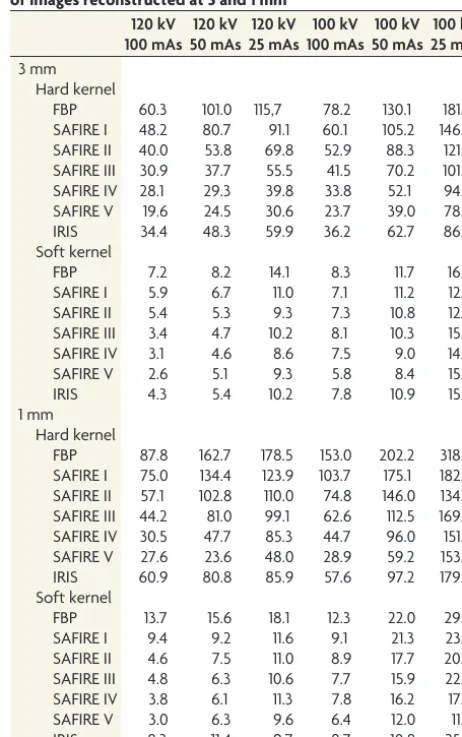
Table 2: Mean image noise measured as an SD in Hounsfield units of images reconstructed at 3 and 1 mm
120 kV 100 mAs 120 kV 50 mAs 120 kV 25 mAs 100 kV 100 mAs 100 kV 50 mAs 100 kV 25 mAs 3 mm Hard kernel
FBP 60.3 101.0 115,7 78.2 130.1 181.3 SAFIRE I 48.2 80.7 91.1 60.1 105.2 146.4 SAFIRE II 40.0 53.8 69.8 52.9 88.3 121.0 SAFIRE III 30.9 37.7 55.5 41.5 70.2 101.7 SAFIRE IV 28.1 29.3 39.8 33.8 52.1 94.0 SAFIRE V 19.6 24.5 30.6 23.7 39.0 78.1 IRIS 34.4 48.3 59.9 36.2 62.7 86.5 Soft kernel
FBP 7.2 8.2 14.1 8.3 11.7 16.7
SAFIRE I 5.9 6.7 11.0 7.1 11.2 12.8 SAFIRE II 5.4 5.3 9.3 7.3 10.8 12.1 SAFIRE III 3.4 4.7 10.2 8.1 10.3 15.4 SAFIRE IV 3.1 4.6 8.6 7.5 9.0 14.9 SAFIRE V 2.6 5.1 9.3 5.8 8.4 15.6
IRIS 4.3 5.4 10.2 7.8 10.9 15.6
1 mm Hard kernel
FBP 87.8 162.7 178.5 153.0 202.2 318.5 SAFIRE I 75.0 134.4 123.9 103.7 175.1 182.9 SAFIRE II 57.1 102.8 110.0 74.8 146.0 134.7 SAFIRE III 44.2 81.0 99.1 62.6 112.5 169.7 SAFIRE IV 30.5 47.7 85.3 44.7 96.0 151.7 SAFIRE V 27.6 23.6 48.0 28.9 59.2 153.2 IRIS 60.9 80.8 85.9 57.6 97.2 179.2 Soft kernel
FBP 13.7 15.6 18.1 12.3 22.0 29.8 SAFIRE I 9.4 9.2 11.6 9.1 21.3 23.6 SAFIRE II 4.6 7.5 11.0 8.9 17.7 20.6 SAFIRE III 4.8 6.3 10.6 7.7 15.9 22.6 SAFIRE IV 3.8 6.1 11.3 7.8 16.2 17.5 SAFIRE V 3.0 6.3 9.6 6.4 12.0 11.7
[image:2.594.300.531.190.558.2]versus 167.7-HU FBP). For the examination protocol with the smallest radiation dosage (100 kV/25 mAs), the average image-noise reductions of the iterative techniques were 42.3% (3 mm, hard kernel), 49.2% (1 mm, hard kernel), 13.8% (3 mm, soft kernel), and 32.2% (1 mm, soft kernel).
The mean image-noise reductions of all examination proto-cols were 47.5% for 3-mm images and 49.4% for 1-mm images rendered with hard kernel (Table 3). With the soft kernel, mean overall image-noise reduction was less, only 24.8% (3-mm im-ages) and 40.9% (1-mm imim-ages). The differences in image-noise reduction between the hard versus soft kernel were statistically significant (hard versus soft kernel on 3-mm images,P⬍.01; hard versus soft kernel on 1-mm images,P⫽.006).
Apart from a few exceptions, SAFIRE V performed the best in image-noise reduction, while SAFIRE I diminished noise the least. On average, IRIS performed noise reduction at an equal noise level either to SAFIRE III (1 mm and 3 mm, hard kernel) or to SAFIRE I (3 mm, soft kernel) and SAFIRE II (1 mm, soft kernel).
Time Performance Calculations
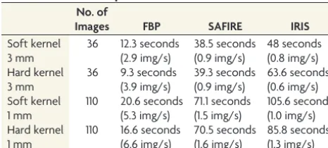
As shown in Table 4, the FBP method required 12.3 seconds for reconstruction of 36 images with the soft kernel at a thickness of 3 mm (IRIS, 48 seconds; SAFIRE, 38.5 seconds). The new SAFIRE reconstruction method was 20%– 67% faster than IRIS. When we took into account all image thicknesses and kernels in this study, the average time needed for reconstruction of a SAFIRE image set was higher by an average factor of 3.7 compared with standard FBP.
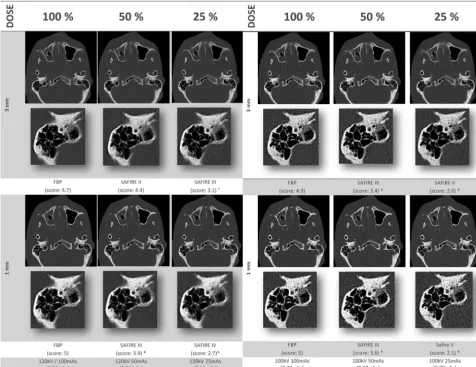
Subjective Image Quality of Noise-Adapted Image Sets
Regarding the 4 image triplet series (120 kV/100 kV in 3- and 1-mm thicknesses), the 7 readers voted every time in favor of the datasets with the highest tube current, which were calculated in the FBP technique (5-point scale, 4.71–5.0). Figure 1 illustrates the image sets that were graded by the readers. The 50% tube current images were evaluated worse than the 100% ones, from
3.4 to 4.4, while the images using a quarter of the maximum tube current scored worst, 2–3.1 points. Most of the differences reached statistical significance, as noted in Fig 1. Overall interob-server agreement among the 7 raters was good, with a mean weightedvalue of⫽0.711 (median,⫽0.729; SD, 0.130; minimum,⫽0.400; maximum,⫽0.933).
DISCUSSION
The main focus of our study was to investigate the performance and image quality of the latest generation of iterative reconstruc-tion methods for CT of the paranasal sinus region. Because exam-ination protocols vary between different hospitals, we analyzed image noise at various dose preferences.
In comparison with standard FBP, all iterative reconstruction techniques reduced image noise by 15%– 85%, depending on the algorithm strength and type of image. Bone kernel images were significantly more susceptible to iterative-based noise reduction than soft-tissue images, potentially due to the generally increased ambient noise level caused by the sharp edge-enhancing quality of the hard kernel algorithm. In our study, iterative reconstruction techniques with a soft-tissue kernel were more efficient at 120 kV than the 100-kV image series; at the lowest dosage settings, the soft-tissue kernel algorithm showed the least benefit by using IRIS or SAFIRE.
Regarding the subjective image quality, the readers, unaware of tube current and type of reconstruction, independently voted in favor of the image sets acquired at the highest tube current (FBP images), though the images were aligned in terms of image noise. This result held true for both 120 and 100 kV image triplets. The reason may be an overall increased softening of cortical structures and bone edges caused by the iterative process. Increased image blurring has been discussed in several publications that investi-gated iterative reconstruction techniques.11-13Silva et al12
sug-gested that the diminished noise manifests as an oversmoothing of the images. We have read with interest the results of Bulla et al,8
who examined IRIS and concluded that iterative reconstructed paranasal sinus CT images did not lose image quality even though the tube current was reduced by⬎50%. However, their work-group subjectively evaluated image noise and diagnostic conclu-siveness on a common 5-point scale, which might be too impre-cise for evaluating the image quality of a CT dataset.
Because the images rated in our study were aligned in terms of image noise, 1 conclusion would be that the lack of sufficient photons cannot be compensated for by simply increasing iterative reconstruction strength. Mitsumori et al,13who examined
[image:3.594.54.285.65.130.2]adap-tive statistical iteraadap-tive reconstruction on liver CT, recommended the use of balanced reconstruction preferences with a mild itera-tive influence to prevent a “waxy” image impression that may constrain diagnostic validity. Although we are aware that de-creased image noise can lead to inde-creased image quality, this field requires more and precise scientific evaluation. Especially when one designs new examination protocols with reduced dosage pa-rameters based on iterative reconstruction techniques, the ap-pearance of an increased blurriness or artificial image impression should be considered. Furthermore, regarding the heterogeneous reduction in image noise for the different datasets rendered with
Table 3: Average noise reduction of SAFIRE for soft tissue and bony kernel
Hard Kernel (3 mm)
Hard Kernel (1 mm)
Soft Kernel (3 mm)
Soft Kernel (1 mm) Mean noise reduction 47.5% 49.4% 24.8% 40.9% Median noise reduction 48.5% 50.7% 24.2% 39.3%
SD 16.8% 17.6% 15.9% 16.9%
Table 4: Time efficiency of FBP and iterative image reconstruction techniquesa
No. of
Images FBP SAFIRE IRIS
Soft kernel 36 12.3 seconds 38.5 seconds 48 seconds 3 mm (2.9 img/s) (0.9 img/s) (0.8 img/s) Hard kernel 36 9.3 seconds 39.3 seconds 63.6 seconds 3 mm (3.9 img/s) (0.9 img/s) (0.6 img/s) Soft kernel 110 20.6 seconds 71.1 seconds 105.6 seconds 1 mm (5.3 img/s) (1.5 img/s) (1.0 img/s) Hard kernel 110 16.6 seconds 70.5 seconds 85.8 seconds 1 mm (6.6 img/s) (1.6 img/s) (1.3 img/s)
Note:—img indicates images.
aCalculations were done with a section thickness of either 3 mm (3-mm increments)
[image:3.594.53.286.163.268.2]soft and hard kernel preferences, it seems mandatory to choose an individual reconstruction method for a particular image type.
Regarding the time frame for image calculation, the iterative techniques require a time increase by a factor of 3.7 (SAFIRE) to 5.2 (IRIS), compared with corresponding FBP reconstructions. This has to be considered, especially when generating large data-sets with thin images, small increments, or multiple multiplanar reconstructions, all of which can lead to a dataset of thousands of images. During a typical daily task, calculating complete datasets by using iterative reconstruction techniques would lead to a con-siderable queue of images and could cause delays in interpreting the CT studies. However, with the increased computing power of modern workstations, this disadvantage may be negligible in the near future.
As deduced from the results of our phantom study, we con-sider 2 interesting application areas that might be suitable for iterative reconstruction techniques: One may be the evaluation of bony structures with decreased dose parameters, because these structures are usually evaluated by using a sharp-edged rendered dataset (eg, hard kernel). However, the clinical value is still mixed in this research field: While Pontana et al9did not notice any loss
of image quality with iterative reconstructed CT, Yanagawa et al14
noticed an increase in false-positive pathologic findings when us-ing the adaptive statistical iterative reconstruction technique. Again, these results oppose the findings of the workgroup of Hu et al,15who concluded that using 40% dose-reduction IRIS images
produced a dataset with quality equal to or even superior to that of the 100% dose using FBP.
Another field for application could be standard-dose CT ex-aminations that are evaluated by using a soft-tissue kernel and undergo marginal iterative enhancement, because significant im-age-noise reduction can be achieved with even slight iterative al-terations at higher dose levels. This hypothesis is underlined by a recent phantom study that researched image-noise suppression of an adaptive iterative dose-reduction method and concluded that the 40% decrease in image noise has the potential to reduce radi-ation dosage in future clinical applicradi-ations.16
A limitation of our study is the artificial nature of the methods used to assess a phantom head for image quality. Because the gelatin material of the phantom head has an average attenuation of 130 HU, denser than that of human soft tissue, the results of iterative noise reduction and even the base level of image noise
FIG 1.Noise-adapted image triplets (120 kV versus 100 kV: 3- versus 1-mm thickness) at tube-current-adjusted dose levels of 100%, 50%, and 25%, which were evaluated by 7 readers regarding the subjective image quality. The right mastoid region of each image is amplified to emphasize the differences in detail. Beneath each reconstruction algorithm, the average rating score is noted (the asterisk indicates significant differences of
[image:4.594.55.532.50.417.2]itself might be exaggerated. Furthermore, the noise results regard-ing the soft-tissue kernel have to be interpreted critically because the absolute values are quite contiguous with a dispersion of only 4 –10 HU. Dedicated analyses of live CT studies are necessary for actual analysis of the clinical value of iterative reconstructions for images rendered with a soft-tissue kernel. In addition, the influ-ence of artifacts caused by patient movement on iterative tech-niques cannot be estimated by a phantom model. Another limi-tation of the study is the restriction of the analyzed iterative technique itself to 1 specific algorithm (SAFIRE, Siemens), though the overall conclusion is likely still generalizable to other vendors and methodologies.
CONCLUSIONS
Sinogram-affirmed iterative reconstruction (SAFIRE) algo-rithms are suitable for image-noise reduction for CT of the paranasal sinus region. Because image quality decreases with dosage, careful choice of the appropriate iterative method is necessary to achieve an optimal balance between image noise and quality.
Disclosures: Ralf Bauer—UNRELATED:Payment for Lectures (including service on Speakers Bureaus): Siemens AG. Josef Matthias Kerl—UNRELATED:Consultancy: Siemens Healthcare AG (no compensation paid),Payment for Lectures (including service on Speakers Bureaus): Siemens Healthcare AG.
REFERENCES
1. Vogl TJ, Mack MG, Balzer J.Chronic infections of the paranasal sinuses[in German].Radiologe2000;40:500 – 06
2. Dammann F.Imaging of paranasal sinuses today[in German]. Ra-diologe2007;47:576, 578 – 83
3. Aaløkken TM, Hagtvedt T, Dalen I, et al.Conventional sinus radi-ography compared with CT in the diagnosis of acute sinusitis. Den-tomaxillofac Radiol2003;32:60 – 62
4. Mazonakis M, Tzedakis A, Damilakis J, et al.Thyroid dose from common head and neck CT examinations in children: is there an excess risk for thyroid cancer induction? Eur Radiol 2007;17: 1352–57
5. National Council on Radiation Protection and Measure-ments.NCoRPaM.Induction of Thyroid Cancer by Ionizing Radiation.
NCRP report 80. Bethesda, Maryland: NCRP Publications; 1985 6. Brem MH, Zamani AA, Riva R, et al.Multidetector CT of the
para-nasal sinus: potential for radiation dose reduction.Radiology2007; 243:847–52
7. Funama Y, Taguchi K, Utsunomiya D, et al.Combination of a low-tube-voltage technique with hybrid iterative reconstruction (iDose) algorithm at coronary computed tomographic angiogra-phy.J Comput Assist Tomogr2011;35:480 – 85
8. Bulla S, Blanke P, Hassepass F, et al.Reducing the radiation dose for low-dose CT of the paranasal sinuses using iterative reconstruction: feasibility and image quality.Eur J Radiol2012;81:2246 –50
9. Pontana F, Duhamel A, Pagniez J, et al.Chest computed tomography using iterative reconstruction vs filtered back projection. Part 2. Image quality of low-dose CT examinations in 80 patients.Eur Ra-diol2011;21:636 – 43
10. Bongartz G, Golding SJ, Jurik AG, et al.European Guidelines on Quality Criteria for Computed Tomography:Report EUR 1999; 16262. http://www.drs.dk/guidelines/ct/quality/. Accessed Novem-ber 2012
11. Leipsic J, Labounty TM, Heilbron B, et al.Adaptive statistical itera-tive reconstruction: assessment of image noise and image quality in coronary CT angiography.AJR Am J Roentgenol2010;195:649 –54 12. Silva AC, Lawder HJ, Hara A, et al.Innovations in CT dose reduction
strategy: application of the adaptive statistical iterative reconstruc-tion algorithm.AJR Am J Roentgenol2010;194:191–99
13. Mitsumori LM, Shuman WP, Busey JM, et al.Adaptive statistical iterative reconstruction versus filtered back projection in the same patient: 64 channel liver CT image quality and patient radiation dose.Eur Radiol2012;22:138 – 43
14. Yanagawa M, Honda O, Kikuyama A, et al.Pulmonary nodules: ef-fect of adaptive statistical iterative reconstruction (ASIR) tech-nique on performance of a computer-aided detection (CAD) sys-tem— comparison of performance between different-dose CT scans.Eur J Radiol2012;81:2877– 86
15. Hu XH, Ding XF, Wu RZ, et al.Radiation dose of non-enhanced chest CT can be reduced 40% by using iterative reconstruction in image space.Clin Radiol2011;66:1023–29