Original Article
Sensitization to tamoxifen by tanshinone IIA
in tamoxifen-resistant breast cancer cells
in vitro
Cheng Yang1,2, Xu Zhang2, Bo Han2, Xingsong Tian1
1Department of Breast and Thyroid Surgery, Provincial Hospital Affiliated to Shandong University, 324 Jingwu
Weiqi Road, Jinan 250021, Shandong, P. R. China; 2Department of General Surgery, Qingdao City Center Hospital,
127 Siliu South Road, Qingdao 266042, Shandong, P. R. China
Received November 13, 2016; Accepted December 12, 2016; Epub February 15, 2017; Published February 28, 2017
Abstract: The treatment success of tamoxifen is mainly dependent on the expression of the estrogen receptor (ER) in the breast carcinoma. However, a large percent of responding patients ultimately develop tamoxifen resis-tance. Given that increasing evidence has shown that miRNAs are involved in modulating tamoxifen resistance and Tanshinone IIA (TSA) exhibits great anti-cancer effects on both ER-positive and -negative breast cancer cells, the present study examined the effects of TSA on tamoxifen resistance. To this end, we derived a tamoxifen-resistant breast cancer cell line (i.e., MCF-7-TamR) using MCF-7 cells. We evaluated the effects of tamoxifen and TSA treat-ment on the proliferation, clonogenic potential, and apoptosis of MCF-7 and MCF-7-TamR cells, and explored the expression of miRNAs after TSA treatment in MCF-7 and MCF-7-TamR cells. Our results showed that 0.1, 0.5, or 1 µM, but not 5 or 10 µM, tamoxifen failed to alter cell proliferation in tamoxifen-resistant MCF-7-TamR cells. However, a low dose of TSA (0.05 µM) treatment, which alone failed to alter the proliferation in either MCF-7 or MCF-7-TamR cells, was able to attenuate cell proliferation and clonogenic potential, and increase apoptosis in tamoxifen-resis-tant MCF-7-TamR cells when co-treated with 1 µM of tamoxifen. Furthermore, 0.05 µM of TSA treatment alone enhanced the expression of miRNA-22 in MCF-7 or MCF-7-TamR cells, which is correlated with attenuated expres-sion of c-Myc. Taken together, our results suggested that low dose of TSA promotes the sensitivity to tamoxifen in tamoxifen-resistant cells in vitro likely involving miRNA-22 and c-Myc mediated signaling pathways.
Keywords: Tashinone IIA, tamoxifen-resistance, breast cancer, sensitization, microRNA
Introduction
As the second most common type of cancer in women [1], breast cancer can be classified into three subtypes based on the expression of the estrogen receptor (ER), progesterone receptor (PR), and cell surface receptor of human epi-dermal growth receptor 2 (HER2), which have been the most commonly used predictive fac-tors in chemotherapy selections for breast can-cer patients [2, 3]. However, current chemo-therapies for breast cancer often lead to the development of drug resistance. For example, tamoxifen has long been used for the syste- mic treatment of patients with breast cancer. The treatment success of tamoxifen is main- ly dependent on the expression of the estro- gen receptor (ER) in breast carcinoma [4, 5]. However, a large percent of responding pa- tients ultimately develop tamoxifen resistance [6, 7]. Therefore, searching for effective
regi-mens that could prevent or reverse the tamo- xifen resistance may bring great benefits in breast cancer treatment and research.
epithelial-mesenchymal transition through HIF-1α down-regulation, reversing hypoxia-induced chemotherapy resistance in breast cancer cell lines [17]. Interestingly, TSA has been shown to alter the expression of various microRNAs in cardiac myocytes [18-20]. However, little is known about the effects of TSA on miRNA ex- pressions in the breast cancer cell lines. MiRNAs play an important role in the regu- lation of the expression of various genes. An increasing number of studies have reported that miRNAs are involved in modulating tamoxi-fen resistance [21, 22]. Specifically, estradiol treatment increases accumulation of miRNA-98 and miRNA-21 in MCF-7 cells [23]. Such a change in miRNA expressions may alter pa- tient response to tamoxifen treatment. Fur- thermore, estradiol can reduce the express- ion of miRNA-181a and miRNA-26a, which in turn attenuate cell proliferation [24]. Addition- ally, more and more miRNAs, including 375, miRNAs 221/222, 200, miRNA-342, and miRNA-519a, have been discovered as potential biomarkers for the tamoxifen re- sponse [25-29].
Therefore, the present study was undertaken to examine the effects of TSA on tamoxifen resis-tance. To this end, we derived a tamoxifen-resistant breast cancer cell line (i.e., MCF-7-TamR) using MCF-7 cells. We evaluated the ef- fects of tamoxifen and TSA treatment on the proliferation and apoptosis of 7 and MCF-7-TamR cells. Meanwhile, the putative mecha-nisms underlying the effects of TSA on tamoxi-fen resistance were explored. Specifically, we explored the effects of TSA treatment on the expression of several miRNAs.
Materials and methods Cell culture
The human mammary carcinoma cell line MCF-7 was obtained from the Cell Resource Center of Peking Union Medical College (Bei- jing, China), and was maintained in 10% fetal bovine serum-supplemented Dulbecco’s mo- dified Eagle’s medium (DMEM) (Gibco, Grand. Island, NY, U.S.A.). We derived Tamoxifen-re- sistant MCF-7 cell line (MCF-7-TamR) by contin-uously exposing it to tamoxifen (1 μM; Diluted in 0.1% ethanol) for more than 12 months us- ing the method described in previous studies [30, 31]. All cell cultures were maintained in a humidified incubator at 37°C and 5% CO2.
Consistent with previous studies, MCF-7-TamR cell line exhibited ER positive [32].
Cell proliferation assays
Cell counting kit-8 (CCK-8; Boppard, Shanghai, China) was used to measure the cell prolifera-tion. MCF-7 or MCF-7-TamR cells were seeded in full growth medium, which was replaced with RPMI-1640 24 h later, and the cells were cul-tured for additional 48 h. Next, approximate 3000 cells in 100 μl medium were seeded in a 96-well plate for 24 h and then the medium was replaced by 200 μl of full growth medium, which contained Tamoxifen or Tanshinone IIA. The cells were cultured for 5 days. Five repli-cates for each treatment were conducted on the plate. When the cell proliferation assay started, 10 μl of CCK-8 solution was added to the medium and then the plate was placed in incubator at 37°C. Two hours later, cell num-bers were evaluated by measuring the absor-bance at 450 nm using an ELx800 Univer- sal Microplate Reader (Biotek Instrument Inc., Highland Park, VT, USA).
Apoptosis test
MCF-7 or MCF-7-TamR cells (1.5×10⁵/well) were treated with either 0.1% ethanol (control) or Tanshinone IIA in full growth medium contain-ing 1 μM tamoxifen for 5 days. Cells were then stained with FITC-conjugated anti-Annexin V antibodies. Cell apoptosis was further analy- zed by using the Annexin V-FITC Apoptosis De- tection kit (Abcam, Shanghai, China) with flow cytometry (Biosciences, Franklin Lakes, NJ, USA).
Soft agar assay
micro-scope, and images were taken with digital camera.
RNA extraction and quantitative real-time-PCR (qPCR) for miRNA
Total miRNA-enriched RNAs from cell culture were extracted from MCF-7 and MCF-7-TamR cells following 0.05 µM of TSA treatment for 5 days using TRI Reagent (Sigma, St Louis, MO, USA). The purity and concentration of RNAs was evaluated using NanoDrop 1000 spectro-photometer (Thermo Scientific, Wilmington, DE, USA). RNAs were then converted into cDNA us- ing ImPro-II reverse transcriptase (Promega, Beijing, China) followed by qPCR using a SYBR PrimeScript miRNA RT-PCR kit (Takara Biotechnology Co. Ltd, Dalian, China) with the miRNA primer sets for 22, 221, miR-222, miR-200a, miR-200b, or miR-200c (San- gon Biotech, Shanghai, China) on the 7500 Real-Time PCR systems (Applied Biosystems, Carlsbad, CA, USA). U6RNA was used as an internal standard for normalization, and the 2-ΔCT (ΔC
T=CTmicroRNA-CTU6 RNA) method was used to quantify relative amount of miRNA. The change in miRNA expression was then convert-ed to fold-change, relative to control group. Real-time PCR primers are shown in Table 1.
Western blotting
The expression of c-myc (~65 kDa) was deter-mined by Western blot following 0.05 µM of TSA treatment for 5 days. MCF-7 or MCF-7-TamR cells were lysed in RIPA buffer (50 mM Tris-HCl, pH 7.4, 150 mM NaCl, 1% Triton X-100,
were boiled for 5 min before loading. 10% run-ning gel (25% of 40% acrylamide stock, Beyo- time; 0.375 M of 1.5 M Tris-HCl, pH 8.8; 1% of 10% sodium dodecyl sulfate; 1% of 10% ammonium persulfate; 0.1% Tetramethylethy- lenediamine) was utilized. The gel was trans-ferred to a same size Nitrocellulose transfer membrane (Thermo Scientific, Waltham, MA, USA) within transfer buffer (25 mM Tris base, 192 mM glycine, 0.037% sodium dodecyl sul-fate, and 20% methanol) under 45 V for 40 min, and probed with the first antibody against c-Myc (#9402; Santa Cruz Biotechnology, Santa Cruz, CA, USA) with a 1/1000 dilution in blocking buffer (50 mM Tris base; 100 mM NaCl; 0.02% Tween 20; and 3% BSA) overnight. The membrane was washed by TTBS (0.1% Tween 20, 10 mM Tris base, 100 mM NaCl, pH 7.5) for three times before adding secondary antibody (ab6721, Abcam, Shanghai, China) with 1/5000 dilution in blocking buffer for 2 hours. Background color was reduced careful- ly by washing with TTBS. The results were visu-alized using ECL kit (Abcam, Shanghai, China), and protein levels were normalized to GAPDH and quantified using Tanon Gel image system (Tanon, Shanghai, China).
Statistical analysis
[image:3.612.91.386.83.283.2]Results were collected as the average of at least five independent experiments. All the data were presented as means ± standard deviation. The data were analyzed using multi-factor ANOVA followed by Tukey’s post hoc test with GraphPad PRISM software package
Table 1. List of real-time PCR primers
MicroRNA Sequences of probes
Mir 22 F: 5’-ACACTCCAGCTGGGTTCGACGGTCAACTTC-3’
R: 5’-CTCAACTGGTGTCGTGGAGTCGGCAATTCAGTTGAGACAGTTCT-3’ Mir 221 F: 5’-AGCTACATTGTCTGCTGG-3’
R: 5’-GTATCCAGTGCAGGGTCC-3’ Mir 222 F: 5’-AGCTACATCTGGCTACTGG-3’
R: 5’-GTATCCAGTGCAGGGTCC-3’ Mir 200a F: 5’-CTTACCGGACAGTGCTG-3’
R: 5’-GAACATGTCTGCGTATCTC-3’ Mir 200b F: 5’-CTTACTGGGCAGCATTG-3’
R: 5’-GAACATGTCTGCGTATCTC-3’ Mir 200c F: 5’-GTCTTACCCAGCAGTGT-3’
R: 5’-GAACATGTCTGCGTATCTC-3’ U6 F: 5’-GCGCGTCGTGAAGCGTTC-3’ R: 5’-GTGCAGGGTCCGAGGT-3’
(GraphPad Software, Inc.). Data were deter-mined to be statistically different when P<0.05.
Results
Effects of tamoxifen or TSA treatment on the proliferation of MCF-7 and MCF-7-TamR cells
We established tamoxifen resistant breast can-cer cell lines MCF-7-TamR cells by continuously exposing MCF-7 cells to 1 μM of tamoxifen for about 12 months using a method described in previous studies. We first examined the effects of different concentrations of tamoxifen or TSA alone on the proliferation of MCF-7 and MCF- 7-TamR cells using CCK8. Overall, MCF-7-TamR cells exhibited higher proliferation than MCF-7 cells after tamoxifen treatment (cell line main effect, F(1, 48)=29.12, P=0.0001), and tamoxifen dose-dependently decreased the proliferation of MCF-7 and MCF-7-TamR cells (dose main and interaction effects, F(5, 48)=13.27-12.24, P= 0.001; Figure 1A). Furthermore, the lowest effective dose of tamoxifen was 0.5 µM in MCF-7 cells (Tukey test, P<0.01; Figure 1A), but the lowest effective dose of tamoxifen was 5 µM in MCF-7-TamR cells (Tukey test, P<0.01;
Figure 1A). Therefore, 1 µM of tamoxifen was employed as the optimal dosage in further in- vestigation of the effects of TSA on tamoxifen resistance.
Similar to the effects of tamoxifen treatment, TSA treatment dose-dependently decreased
potential, and apoptosis of MCF-7 and MCF-7-TamR cells
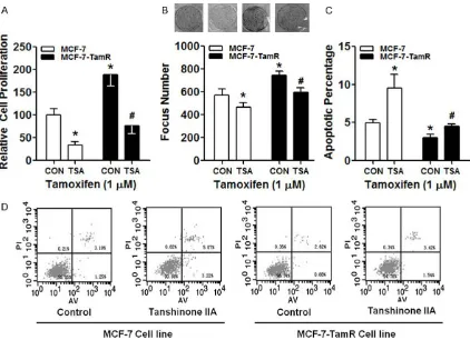
We then explored the effect of co-treatment of TSA and tamoxifen on the proliferation of MCF-7 and MCF-7-TamR cells. Cell proliferation was examined following the treatments of either 0.1% ethanol (control) or 0.05 µM of TSA in full growth medium containing 1 μM of tamoxifen for 5 days. Consistently, MCF-7-TamR cells exhibited higher proliferation than MCF-7 cells under the treatment of 0.1% ethanol and 1 μM of tamoxifen (cell line main effect, F(1,
16)=17.84, P=0.001; Figure 2A). Interestingly, 0.05 µM of TSA treatment reduced the pro- liferation in either MCF-7 or MCF-7-TamR cells (treatment main effect, F(1, 16)=19.38, P=0.001; interaction effect F(1, 16)=10.07, P=0.01; Figure 2A), as compared to control. This is surprising because 0.05 µM of TSA treatment alone fail- ed to alter the proliferation in either MCF-7 or MCF-7-TamR cells (Figure 1B). More specifi- cally, 0.05 µM of TSA and 1 μM of tamoxifen treatment decreased the proliferation of MCF-7-TamR cells to a similar level of MCF-7 cells after 0.1% ethanol treatment (Figure 2A). Furthermore, we conducted the soft agar colo-ny formation assay in order to assess the ef- fects of co-treatment of TSA and tamoxifen on the clonogenic potential of MCF-7 and MCF-7-TamR cells, which has been shown to correlate with tumor formation in vivo [33]. As shown in Figure 1. Effects of tamoxifen or TSA treatment on the proliferation of MCF-7
and MCF-7-TamR cells. Five days after (A) tamoxifen or (B) TSA treatment, the cell proliferation assay was conducted using CCK-8 kit. Cell numbers were evaluated by measuring the absorbance at 450 nm. Five replicates for each treatment were conducted on the plate. Asterisks (P<0.05) represent the ANOVA simple main effect, as compared to MCF-7 cell line. Ponds (P<0.05) represent the ANOVA simple main or main effects, as compared to no treat-ment control.
the proliferation of MCF-7 and MCF-7-TamR cells (dose main effect, F(5, 48)=25.11, P= 0.0001; Figure 1B). However, in contrast to the effects of tamoxifen, higher doses of TSA (i.e., 0.1 µM or more) treatment decreased the pro-liferation of MCF-7 and MCF- 7-TamR cells similarly regard-less of the cell lines (cell line main effect, F(1, 48)=1.54, P= 0.31; Figure 1B). Specifical- ly, the lowest effective dose of TSA was 0.1 µM, and cell proliferation was significantly accelerated (Tukey test, P< 0.01; Figure 1B).
Figure 2B, MCF-7-TamR cells treated with 0.1% ethanol and 1 μM of tamoxifen treatment ex- hibited higher focus number as compared with MCF-7 cells (cell line main effect, F(1, 16)=16.31,
P=0.01; Figure 2B). However, 0.05 µM of TSA and 1 μM of tamoxifen treatment reduced the focus number in either MCF-7 or MCF-7-TamR cells (treatment main effect, F(1, 16)=19.24, P= 0.001; interaction effect F(1, 16)=9.71, P=0.02;
Figure 2B).
Additionally, we examined the effect of co-treat-ment of TSA and tamoxifen on the apoptosis of MCF-7 and MCF-7-TamR cells. Cell apoptosis was examined with flow cytometry following the treatments of either 0.1% ethanol (control) or 0.05 µM of TSA in full growth medium con-taining 1 μM tamoxifen for 5 days. During
apop-tosis, phosphatidylserine (PS) translocation in the cell membrane is assumed to be an early feature of apoptosis, and Annexin V has the ability to bind to the translocated PS on the cell membrane during the early apoptotic stage. Propidium iodide (PI) can bind to DNA in the middle and late stage of apoptosis when cell membrane and nucleus membrane are perme-able. Therefore, FITC-conjugated Annexin V and PI were used to identify apoptotic cells in the present study. In general, MCF-7 cells exhibited higher apoptosis than MCF-7-TamR cells under the treatment of 0.1% ethanol and 1 μM of tamoxifen (cell line main effect, F(1, 16)=8.83,
[image:5.612.95.517.69.375.2]effect F(1, 16)=7.98, P=0.03; Figure 2C and 2D), as compared to control. More specifically, 0.05 µM of TSA treatment increased the apoptosis of MCF-7-TamR cells to a similar level in MCF-7 cells after 0.1% ethanol treatment (Figure 2C
and 2D).
Effects of TSA treatment on miRNA expression in MCF-7 and MCF-7-TamR cells
We next explored the putative mechanisms underlying the sensitizing effects of 0.05 µM of TSA treatment on tamoxifen resistance by examining the expression of various miRNAs, including miRNA-22, miRNA 221/222, and miRNA 200 families, following 0.05 µM of TSA treatment. As shown in Figure 3, the expres-sion of miRNA-22 and miRNA 200 families, including 200a, 200b, and 200c was
attenuat-ed in MCF-7-TamR cells, but the expression of miRNA 221/222 was enhanced in MCF-7-TamR cells (all cell line main effect, F(1, 16)= 13.96-27.84, P=0.0001-0.001), as compared to MCF-7 cells. However, 0.05 µM of TSA treat-ment only altered the expression of miRNA- 22 in either MCF-7 or MCF-7-TamR cells (treat-ment main effect, F(1, 16)=13.96, P=0.001; inter-action effect F(1, 16)=8.29, P=0.02; Figure 3A). Specifically, 0.05 µM of TSA treatment incre- ased the expression of miRNA-22 in MCF-7-TamR cells to a similar level in MCF-7 cells after 0.1% ethanol treatment.
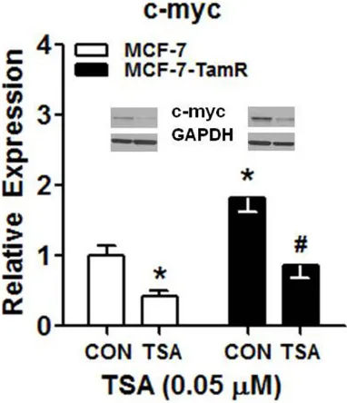
Effects of TSA treatment on c-Myc expression in MCF-7 and MCF-7-TamR cells
[image:6.612.92.520.75.400.2]c-Myc, we explored the effects of TSA treat-ment on the expression of c-Myc in MCF-7 and MCF-7-TamR cells. As shown in Figure 4, the expression of c-Myc was robustly increased in MCF-7-TamR cells, as compared to MCF-7 cells (cell line main effect, F(1, 16)=10.78, P=0.01). However, 0.05 µM of TSA treatment reduced the expression of c-Myc in either MCF-7 or MCF-7-TamR cells (treatment main effect, F(1,
16)=9.04, P=0.02; interaction effect F(1, 16)= 7.58, P=0.03). Specifically, 0.05 µM of TSA treatment attenuated the expression of c-Myc in MCF-7-TamR cells to a similar level in MCF- 7 cells after 0.1% ethanol treatment.
Discussion
The present study first examined the putative effects of TSA on the tamoxifen resistance. We have demonstrated that 0.1, 0.5, or 1 µM, but not 5 or 10 µM, tamoxifen failed to alter cell proliferation in tamoxifen-resistant MCF-7-TamR cells. However, a low dose of TSA (0.05 µM) treatment, which alone failed to alter the
[image:7.612.93.284.75.297.2]proliferation in either MCF-7 or MCF-7-TamR cells, was able to attenuate cell proliferation and clonogenic potential, and increase apopto-sis in tamoxifen-reapopto-sistant MCF-7-TamR cells when co-treated with 1 µM of tamoxifen. Fur- thermore, 0.05 µM of TSA treatment alone at- tenuated the expression of c-Myc in MCF-7 or MCF-7-TamR cells, which is correlated with enhanced expression of miRNA-22 following 0.05 µM of TSA treatment. Taken together, our results suggested that a low dose of TSA like- ly promotes the sensitivity to tamoxifen in ta- moxifen-resistant cells in vitro via miRNA-22 and c-Myc mediated signaling pathways. Our findings of the anti-tamoxifen resistance effects of TSA have shed new lights on the molecular mechanisms underlying the antican-cer effects of TSA. Traditional Chinese medi-cine practice has been using Danshen (Salvia miltiorrhiza Bunge) widely in the treatment of coronary artery disease and cerebrovascular diseases for centuries. It has shown minimal side effects for these treatments. Previous studies have shown that Danshen contains at least about 20 different phenolic acids and more than 30 diterpene compounds. Among these compounds, abundant tanshinones, in- cluding tanshinone I, tanshinone IIA, cryptotan-shinone, dihydrotanshinone and tanshinone II have been isolated [9]. Importantly, an incre- asing number of studies have demonstrated that tanshinones show some activities against human cancer cells. For instance, as one of the major diterpenes isolated from Danshen, tan-shinones show cytotoxic effects on various hu- man cancer cell lines, which are derived from various human carcinomas of the liver, neuro-glia, ovary, lung, mouth, and colon [15, 34-37]. The cytotoxic effects of TSA can induce apopto-sis in various human cancer cells, including leu-kemia, human hepatocellular carcinoma, and nasopharyngeal carcinoma cells [34, 38, 39], as well as both ER-positive and -negative bre- ast cancer cells [12-15]. Interestingly, recent studies have shown that TSA can inhibit the angiogenesis and growth of breast cancer in vivo [40], which is likely due to its suppress- ing effects on protein synthesis of hypoxia-inducible factor 1α (HIF-1α) and expression of vascular endothelial growth factor (VEGF) via the mTOR/p70S6K/4E-BP1 signaling pathway [40]. Such an effect on down-regulation of HIF-1α expression can ameliorate hypoxia-induced Figure 4. Effects of TSA treatment on c-Myc
doxorubicin resistance [41]. Adding to the liter-ature, the present study has shown that TSA at a relatively low concentration, which did not have apparent cytotoxic effects on breast can-cer cell lines, can promote the sensitivity of tamoxifen-resistant breast cancer cells to tamoxifen treatment. This effect is correlated with enhanced expression of miRNA-22 and decreased expression of c-Myc. Thus, future studies will be necessary to explore in depth the molecular mechanisms underlying this phenomenon.
Systemic treatment with tamoxifen for the patients with breast cancer has been routinely performed for over three decades [42]. The success rate of tamoxifen treatment is large- ly relying on the expression level of the estro-gen receptor (ER) in the breast carcinoma [42-44]. Clinical studies have revealed that more than half of patients with advanced ER-posi- tive breast cancer fail to respond to tamoxifen. Even in the initially responding patients, tamox-ifen-resistant phenotype will ultimately deve- lop following prolonged tamoxifen treatment [42]. Numerous studies have elucidated that multiple mechanisms can lead to intrinsic and acquired tamoxifen resistance, including the alterations in the structure and function of the ER, the tumor environment, genetic alterations in the breast cancer cells, or pharmacology of tamoxifen per se [4, 42]. For example, breast cancer antiestrogen resistance (BCAR) genes have been reported to play a critical role in the intrinsic and acquired tamoxifen resistance in human breast cancer cells [4, 45]. Primary breast tumors that are ER-positive and are associated with intrinsic resistance to tamoxi-fen treatment often exhibit high levels of BC- AR1/pl30C as protein expression [42]. Further- more, tamoxifen resistance is associated with increased expression of alternative G-protein coupled receptor GPR-30 (GPER) and estrogen receptor splice products (e.g., ERα36) on the plasma membrane [4], and may recruit the sig-naling pathways involving the epidermal growth factor EGF, the inflammation associated tran-scription factor NF-κB, and IGF-1 [4]. It is not clear whether the expressions of these genes are modulated by the miRNAs, it will be impor-tant to explore this question in the future. While miRNA analysis in a Danish Breast Can- cer Co-operative Group (DBCG) study
discov-ered that no single miRNA profile was able to predict tamoxifen treatment outcome [46], increasing evidence has revealed the impor-tant role of various miRNAs in modulation of tamoxifen resistance. Specifically, expression of miR-320a is up-regulated in tamoxifen-re- sistant ER-positive breast cancer cells, and it is negatively correlated with the expression of ARPP-19 and ERRγ, and the downstream gene expression of c-Myc and Cyclin D1, which may relate to tamoxifen resistance [47]. In addition, MiR-873 can reduce the transcriptional ac- tivity of ERα and tamoxifen resistance via tar-geting CDK3 in breast cancer cells [48]. Adding to this literature, results in the present study have suggested that TSA might increase ta- moxifen sensitivity by enhancing the expres-sion of miRNA-22. While TSA treatment fail- ed to alter the expression of miRNA-221/222 or the families of miRNA-200 in the present study, previous studies have shown that these miRNAs are important in modulation of tamo- xifen resistance [49-52]. Given the emerging numbers of miRNAs have been demonstrated in tamoxifen resistance, it will be necessary to systematically investigate the effects of TSA on miRNA expressions in breast cancer cells in order to provide in depth understandings of such a phenomenon.
profiles of miRNAs, laboratory studies on spe-cific miRNAs have shown the direct functional link between miRNA function and the breast tumor proliferation, invasion, and metastasis. Specifically, over-expression of miR-373 and miR-520c in lymph node metastases of breast tumor likely promote the tumor invasion and metastasis via suppression of the CD44 gene, which codes for a hyaluronan receptor, a met- astasis suppressor in breast cancer [59, 60]. Inhibition of HRAS and high mobility group AT-hook2 (HMGA2) genes, which are involved in self-renewal and differentiation, by let-7 miRNA family can increase the proliferation of breast tumor-initiating cells derived from cell lines and primary patient tumors [61]. Addi- tionally, the present study has confirmed that the expression of miRNA-22 is up-regulated in tamoxifen-resistant breast cancer cells, which is consistent with the results from previous studies [62, 63]. Therefore, it will be also im- portant to examining the effects of TSA on these miRNAs in order to explore the antican-cer effects of TSA in a broader range.
In summary, the present study was the first to show that TSA can promote the sensitivity to tamoxifen treatment in tamoxifen-resistant breast cancer cells in vitro, and this phenome-non may involve the miRNA-22 and c-Myc sig-naling pathways. Hence, it will be necessary to evaluate the in vivo effects of TSA on tamoxifen resistance in the future. The concept of combi-nation of tamoxifen and anti-miRNA treatment may help alleviate the issue of tamoxifen re- sistance in clinical therapy. In support of this, co-delivery of anti-miRNA-21 and 4-Hydroxy- tamoxifen has been shown to inhibit the pro- liferation of human breast cancer cells [64]. Therefore, the line of research on the effects of TSA on miRNA expression in breast cancer cells would help the development of this drug combination for use in the treatment of tamo- xifen-resistance breast cancer.
Acknowledgements
This research was funded by Science and Technology Plan Project of Shandong Pro- vince (Grant No. 2009GG10002060) & Medi- cal and Health Science and Technology Deve- lopment Plan Project of Shandong Province (Grant No. 2011HZ071) & National Natural
Science Foundation of Shandong Province (Grant No. ZR2014HM115).
Disclosure of conflict of interest
None.
Address correspondence to: Dr. Xingsong Tian, De- partment of Breast and Thyroid Surgery, Provincial
Hospital Affiliated to Shandong University, 324
Jingwu Weiqi Road, Jinan 250021, Shandong, P. R. China. Tel: 008615168887531; Fax: 053168776- 940; E-mail: Xingsongtian@gmail.com
References
[1] Ferlay J, Shin HR, Bray F, Forman D, Mathers C and Parkin DM. Estimates of worldwide bur-den of cancer in 2008: GLOBOCAN 2008. Int J Cancer 2010; 127: 2893-2917.
[2] Andersen J and Poulsen HS. Immunohisto- chemical estrogen-receptor determination in
paraffin-embedded tissue - prediction of re -sponse to hormonal treatment in advanced breast-cancer. Cancer 1989; 64: 1901-1908. [3] Slamon DJ, Godolphin W, Jones LA, Holt JA,
Wong SG, Keith DE, Levin WJ, Stuart SG, Udove J, Ullrich A and Press MF. Studies of the her-2/neu proto-oncogene in human-breast and ovarian-cancer. Science 1989; 244: 707-712.
[4] Nass N and Kalinski T. Tamoxifen resistance: from cell culture experiments towards novel biomarkers. Pathol Res Pract 2015; 211: 189-197.
[5] Huang B, Warner M and Gustafsson JA. Estrogen receptors in breast carcinogenesis and endocrine therapy. Mol Cell Endocrinol 2015; 3: 240-4.
[6] Milani A, Geuna E, Mittica G and Valabrega G. Overcoming endocrine resistance in meta-static breast cancer: current evidence and future directions. World J Clin Oncol 2014; 5: 990-1001.
[7] Viedma-Rodriguez R, Baiza-Gutman L, Sala- manca-Gomez F, Diaz-Zaragoza M, Martinez-Hernandez G, Ruiz Esparza-Garrido R, Velaz- quez-Flores MA and Arenas-Aranda D. Me- chanisms associated with resistance to tamox-ifen in estrogen receptor-positive breast can-cer (review). Oncol Rep 2014; 32: 3-15. [8] Xu S and Liu P. Tanshinone II-A: new
perspec-tives for old remedies. Expert Opin Ther Pat 2013; 23: 149-153.
[10] Yang RF, Liu AJ, Ma XJ, Li L, Su DF and Liu JG. Sodium tanshinone IIA sulfonate protects car-diomyocytes against oxidative stress-mediated apoptosis through inhibiting JNK activation. J Cardiovasc Pharmacol 2008; 51: 396-401. [11] Lam BY, Lo AC, Sun X, Luo HW, Chung SK and
Sucher NJ. Neuroprotective effects of tanshi-nones in transient focal cerebral ischemia in mice. Phytomedicine 2003; 10: 286-291. [12] Nicolin V, Fancellu G and Valentini R. Effect of
tanshinone II on cell growth of breast cancer cell line type MCF-7 and MD-MB-231. Ital J Anat Embryol 2014; 119: 38-43.
[13] Su CC, Chien SY, Kuo SJ, Chen YL, Cheng CY and Chen DR. Tanshinone IIA inhibits human breast cancer MDA-MB-231 cells by decreas-ing LC3-II, Erb-B2 and NF-kappaBp65. Mol Med Rep 2012; 5: 1019-1022.
[14] Lu Q, Zhang P, Zhang X and Chen J. Experi- mental study of the anti-cancer mechanism of tanshinone IIA against human breast can-cer. Int J Mol Med 2009; 24: 773-780.
[15] Nizamutdinova IT, Lee GW, Son KH, Jeon SJ, Kang SS, Kim YS, Lee JH, Seo HG, Chang KC and Kim HJ. Tanshinone I effectively in- duces apoptosis in estrogen receptor-positive (MCF-7) and estrogen receptor-negative (MDA-MB-231) breast cancer cells. Int J Oncol 2008; 33: 485-491.
[16] Gong Y, Li YL, Abdolmaleky HM, Li LL and Zhou JR. Tanshinones inhibit the growth of
breast cancer cells through epigenetic modifi -cation of Aurora a expression and function. PLoS One 2012; 7: e33656.
[17] Fu PF, Du FY, Chen W, Yao MY, Lv KZ and Liu Y. Tanshinone IIA blocks epithelial-mesenchy-mal transition through HIF-1 alpha downregu-lation, reversing hypoxia-induced chemothera-py resistance in breast cancer cell lines. Oncol Rep 2014; 31: 2561-2568.
[18] Zhang L, Wu YL, Li YM, Xu CQ, Li XL, Zhu DL, Zhang Y, Xing S, Wang HY, Zhang ZH and Shan HL. Tanshinone IIA improves miR-133 expression through MAPK ERK1/2 pathway in hypoxic cardiac myocytes. Cell Physiol Bio- chem 2012; 30: 843-852.
[19] Zhang Y, Zhang L, Chu WF, Wang B, Zhang JL, Zhao M, Li XL, Li BX, Lu YJ, Yang BF and Shan HL. Tanshinone IIA inhibits miR-1 expression through p38 MAPK signal path- way in post-infarction rat cardiomyocytes. Cell Physiol Biochem 2010; 26: 991-998.
[20] Shan HL, Li XL, Pan ZW, Zhang L, Cai BZ, Zhang Y, Xu CQ, Chu WF, Qiao GF, Li BX, Lu YJ and Yang BF. Tanshinone IIA protects against sud-den cardiac death induced by lethal arrhyth-mias via repression of microRNA-1. Br J Phar- macol 2009; 158: 1227-1235.
[21] Zhou J, Teng R, Wang Q, Xu C, Guo J, Yuan C, Shen J, Hu W, Wang L and Xie S. Endocrine
resistance in breast cancer: current status and a perspective on the roles of miRNAs (Review). Oncol Lett 2013; 6: 295-305. [22] Gupta A, Caffrey E, Callagy G and Gupta
S. Oestrogen-dependent regulation of miRNA biogenesis: many ways to skin the cat. Biochem Soc Trans 2012; 40: 752-758.
[23] Bhat-Nakshatri P, Wang G, Collins NR, Thom- son MJ, Geistlinger TR, Carroll JS, Brown M, Hammond S, Srour EF, Liu Y and Nakshatri H. Estradiol-regulated microRNAs control estra- diol response in breast cancer cells. Nucleic Acids Res 2009; 37: 4850-4861.
[24] Maillot G, Lacroix-Triki M, Pierredon S, Grata- dou L, Schmidt S, Benes V, Roche H, Dalenc F, Auboeuf D, Millevoi S and Vagner S. Wide- spread estrogen-dependent repression of mi-crornas involved in breast tumor cell growth. Cancer Res 2009; 69: 8332-8340.
[25] Ward A, Balwierz A, Zhang JD, Kublbeck M, Pawitan Y, Hielscher T, Wiemann S and Sahin O. Re-expression of microRNA-375 reverses both tamoxifen resistance and accompanying EMT-like properties in breast cancer. Oncogene 2013; 32: 1173-1182.
[26] Wei Y, Lai X, Yu S, Chen S, Ma Y, Zhang Y, Li H, Zhu X, Yao L and Zhang J. Exosomal miR-221/222 enhances tamoxifen resistance in recipient ER-positive breast cancer cells. Breast Cancer Res Treat 2014; 147: 423-431. [27] Bai JX, Yan B, Zhao ZN, Xiao X, Qin WW, Zhang
R, Jia LT, Meng YL, Jin BQ, Fan DM, Wang T and Yang AG. Tamoxifen represses miR-200 mi-croRNAs and promotes epithelial-to-mesenchy-mal transition by up-regulating c-Myc in endo-metrial carcinoma cell lines. Endocrinology 2013; 154: 635-645.
[28] He YJ, Wu JZ, Ji MH, Ma T, Qiao EQ, Ma R and Tang JH. miR-342 is associated with estro-gen receptor-alpha expression and response to tamoxifen in breast cancer. Exp Ther Med 2013; 5: 813-818.
[29] Ward A, Shukla K, Balwierz A, Soons Z, Konig R, Sahin O and Wiemann S. MicroRNA-519a is a novel oncomir conferring tamoxifen resis-tance by targeting a network of tumour-sup-pressor genes in ER+ breast cancer. J Pathol 2014; 233: 368-379.
[30] Briand P and Lykkesfeldt AE. Long-term cul- tivation of a human breast cancer cell line,
MCF-7, in a chemically defined medium. Effect
of estradiol. Anticancer Res 1986; 6: 85-90. [31] Lykkesfeldt AE, Madsen MW and Briand P.
Altered expression of estrogen-regulated ge- nes in a tamoxifen-resistant and ICI 164,384 and ICI 182, 780 sensitive human breast can-cer cell line, MCF-7/TAMR-1. Cancan-cer Res 1994; 54: 1587-1595.
mRNA splice variants in the tamoxifen resis-tant human breast cancer cell line, MCF-7/ TAMR-1 compared to the parental MCF-7 cell line. Mol Cell Endocrinol 1995; 109: 197-207. [33] Tiang JM, Butcher NJ and Minchin RF. Small
molecule inhibition of arylamine N-acetyltrans- ferase Type I inhibits proliferation and inva- siveness of MDA-MB-231 breast cancer cells. Biochem Biophys Res Commun 2010; 393: 95-100.
[34] Yuan SL, Wei YQ, Wang XJ, Xiao F, Li SF and Zhang J. Growth inhibition and apoptosis in-duction of tanshinone II-A on human hepato-cellular carcinoma cells. World J Gastroenterol 2004; 10: 2024-2028.
[35] Yuxian X, Feng T, Ren L and Zhengcai L. Tan- shinone II-A inhibits invasion and metastasis of human hepatocellular carcinoma cells in vitro and in vivo. Tumori 2009; 95: 789-795. [36] Lee WY, Cheung CC, Liu KW, Fung KP, Wong
J, Lai PB and Yeung JH. Cytotoxic effects of tanshinones from Salvia miltiorrhiza on doxo-rubicin-resistant human liver cancer cells. J Nat Prod 2010; 73: 854-859.
[37] Zhang X, Zhang PR, Chen J and Lu Q. [A study on the effect of tanshinone IIA against human breast cancer in vivo]. Sichuan Da Xue Xue Bao Yi Xue Ban 2010; 41: 62-67.
[38] Sung HJ, Choi SM, Yoon Y and An KS. Tanshinone IIA, an ingredient of Salvia miltior-rhiza BUNGE, induces apoptosis in human leukemia cell lines through the activation of caspase-3. Exp Mol Med 1999; 31: 174-178. [39] Tseng PY, Lu WC, Hsieh MJ, Chien SY and Chen
MK. Tanshinone IIA Induces apoptosis in hu-man oral cancer kb cells through a mitochon-dria-dependent pathway. Biomed Res Int 2014; 2014: 540516.
[40] Li G, Shan C, Liu L, Zhou T, Zhou J, Hu X, Chen Y, Cui H and Gao N. Tanshinone IIA inhib-its HIF-1alpha and VEGF expression in breast cancer cells via mTOR/p70S6K/RPS6/4E- BP1 signaling pathway. PLoS One 2015; 10: e0117440.
[41] Fu P, Du F, Chen W, Yao M, Lv K and Liu Y. Tanshinone IIA blocks epithelial-mesenchymal transition through HIF-1alpha downregulation, reversing hypoxia-induced chemotherapy re-sistance in breast cancer cell lines. Oncol Rep 2014; 31: 2561-2568.
[42] Dorssers LC, Van der Flier S, Brinkman A, van Agthoven T, Veldscholte J, Berns EM, Klijn JG, Beex LV and Foekens JA. Tamoxifen resistan- ce in breast cancer: elucidating mechanisms. Drugs 2001; 61: 1721-1733.
[43] van Agthoven T, Sieuwerts AM, Meijer D, Meijer-van Gelder ME, van Agthoven TL, Sar- wari R, Sleijfer S, Foekens JA and Dorssers LC. Selective recruitment of breast cancer anti-
estrogen resistance genes and relevance for breast cancer progression and tamoxifen ther-apy response. Endocr Relat Cancer 2010; 17: 215-230.
[44] Meijer D, van Agthoven T, Bosma PT, Nooter K and Dorssers LC. Functional screen for genes responsible for tamoxifen resistance in human breast cancer cells. Mol Cancer Res 2006; 4: 379-386.
[45] Wallez Y, Riedl SJ and Pasquale EB. Association of the breast cancer antiestrogen resistance protein 1 (BCAR1) and BCAR3 scaffolding pro-teins in cell signaling and antiestrogen resis-tance. J Biol Chem 2014; 289: 10431-10444. [46] Lyng MB, Laenkholm AV, Sokilde R, Gravgaard
KH, Litman T and Ditzel HJ. Global microRNA
expression profiling of high-risk ER+ breast
cancers from patients receiving adjuvant ta- moxifen mono-therapy: a DBCG study. PLoS One 2012; 7: e36170.
[47] Lu M, Ding K, Zhang G, Yin M, Yao G, Tian H, Lian J, Liu L, Liang M, Zhu T and Sun F. MicroRNA-320a sensitizes tamoxifen-resistant breast cancer cells to tamoxifen by targeting ARPP-19 and ERRgamma. Sci Rep 2015; 5: 8735.
[48] Cui J, Bi M, Overstreet AM, Yang Y, Li H, Leng Y, Qian K, Huang Q, Zhang C, Lu Z, Chen J, Sun T, Wu R, Sun Y, Song H, Wei X, Jing P, Meredith A and Yang X. MiR-873 regulates ERalpha tran-scriptional activity and tamoxifen resistance via targeting CDK3 in breast cancer cells. Oncogene 2014; 34: 3895-907.
[49] Gan R, Yang Y, Yang X, Zhao L, Lu J and Meng QH. Downregulation of miR-221/222 enhanc-es sensitivity of breast cancer cells to tamoxi-fen through upregulation of TIMP3. Cancer Gene Ther 2014; 21: 290-296.
[50] Zhao JJ, Lin J, Yang H, Kong W, He L, Ma X, Coppola D and Cheng JQ. MicroRNA-221/222 negatively regulates estrogen receptor alpha and is associated with tamoxifen resistance in breast cancer. J Biol Chem 2008; 283: 31079-31086.
[51] Miller TE, Ghoshal K, Ramaswamy B, Roy S, Datta J, Shapiro CL, Jacob S and Majumder S. MicroRNA-221/222 confers tamoxifen resis-tance in breast cancer by targeting p27Kip1. J Biol Chem 2008; 283: 29897-29903.
[52] Manavalan TT, Teng Y, Litchfield LM, Muluhngwi
P, Al-Rayyan N and Klinge CM. Reduced ex- pression of miR-200 family members contrib-utes to antiestrogen resistance in LY2 hu- man breast cancer cells. PLoS One 2013; 8: e62334.
ion profiling of human breast cancer identifies
new markers of tumor subtype. Genome Biol 2007; 8: R214.
[54] Iorio MV, Ferracin M, Liu CG, Veronese A, Spizzo R, Sabbioni S, Magri E, Pedriali M, Fabbri M, Campiglio M, Menard S, Palazzo JP, Rosenberg A, Musiani P, Volinia S, Nenci I, Calin GA, Querzoli P, Negrini M and Croce CM. MicroRNA gene expression deregulation in human breast cancer. Cancer Res 2005; 65: 7065-7070.
[55] Huang S, Chen Y, Wu W, Ouyang N, Chen J, Li H, Liu X, Su F, Lin L and Yao Y. MiR-150 pro-motes human breast cancer growth and malig-nant behavior by targeting the pro-apoptotic purinergic P2X7 receptor. PLoS One 2013; 8: e80707.
[56] Arigoni M, Barutello G, Riccardo F, Ercole E, Cantarella D, Orso F, Conti L, Lanzardo S, Taverna D, Merighi I, Calogero RA, Cavallo F and Quaglino E. MiR-135b coordinates pro-gression of erbb2-driven mammary carcino-mas through suppression of MIDI and MTCH2. Am J Pathol 2013; 182: 2058-2070.
[57] Hoppe R, Achinger-Kawecka J, Winter S, Fritz P, Lo WY, Schroth W and Brauch H. Increased expression of miR-126 and miR-10a predict prolonged relapse-free time of primary oestro-gen receptor-positive breast cancer following tamoxifen treatment. Eur J Cancer 2013; 49: 3598-3608.
[58] Qi L, Bart J, Tan LP, Platteel I, Sluis T, Huitema S, Harms G, Fu L, Hollema H and Berg A. Ex- pression of miR-21 and its targets (PTEN,
PDCD4, TM1) in flat epithelial atypia of the
breast in relation to ductal carcinoma in situ and invasive carcinoma. BMC Cancer 2009; 9: 163.
[59] Huang Q, Gumireddy K, Schrier M, le Sage C, Nagel R, Nair S, Egan DA, Li A, Huang G, Klein-Szanto AJ, Gimotty PA, Katsaros D, Coukos G, Zhang L, Pure E and Agami R. The microRNAs miR-373 and miR-520c promote tumour inva-sion and metastasis. Nat Cell Biol 2008; 10: 202-210.
[60] Lopez JI, Camenisch TD, Stevens MV, Sands BJ, McDonald J and Schroeder JA. CD44 at-tenuates metastatic invasion during breast cancer progression. Cancer Res 2005; 65: 6755-6763.
[61] Yu F, Yao H, Zhu P, Zhang X, Pan Q, Gong C, Huang Y, Hu X, Su F, Lieberman J and Song E. Let-7 regulates self renewal and tumorige-nicity of breast cancer cells. Cell 2007; 131: 1109-1123.
[62] Manavalan TT, Teng Y, Appana SN, Datta S,
Kalbfleisch TS, Li Y and Klinge CM. Differential
expression of microRNA expression in tamoxi-fen-sensitive MCF-7 versus tamoxifen-resis-tant LY2 human breast cancer cells. Cancer Lett 2011; 313: 26-43.
[63] Xiong J, Yu D, Wei N, Fu H, Cai T, Huang Y, Wu C, Zheng X, Du Q, Lin D and Liang Z. An estro-gen receptor alpha suppressor, microRNA-22, is downregulated in estrogen receptor alpha-positive human breast cancer cell lines and clinical samples. FEBS J 2010; 277: 1684-1694.