Original Article
Diffusion tensor imaging of patients with behavioral
variant frontotemporal dementia: a controlled study
Guanjun Li1*, Yingying Tang2*, Ling Yue1, Jinghua Wang1, Jianye Zhang3, Shifu Xiao1, Huafang Li4 1Department of Geriatric, Shanghai Mental Health Center, Shanghai Jiaotong University School of Medicine, Shanghai 200030, China; 2Shanghai Mental Health Center, Shanghai Jiaotong University School of Medicine, Shanghai 200030, China; 3Imaging Center, Shanghai Mental Health Center, Shanghai Jiaotong University School of Medicine, Shanghai 200030, China; 4Medical Institution Conducting Clinical Trial for Human Used Drug, Shang-hai Mental Health Center, ShangShang-hai 200030, China. *Equal contributors.
Received September 7, 2015; Accepted December 5, 2015; Epub February 15, 2016; Published February 29, 2016
Abstract: Background: This study aimed to investigate the clinically important early symptoms of behavioral variant frontotemporal dementia (bvFTD) and the characteristics of bvFTD in structural imaging and to explore the value of diffusion tensor magnetic resonance imaging (MRI-DTI) in the early diagnosis of bvFTD. Material and Methods: Siemens 3T Verio MRI with echoplanar imaging (EPI) was employed in 8 patients with suspected bvFTD diagnosed according to the FTDC diagnostic consensus, 8 matched Alzheimer’s disease (AD) patients and 8 healthy controls, and 3DT1 and DWI images were collected. Results: DTI showed the bilateral thalamic radiation, cingulate gyrus and hippocampus had significantly reduced FA (P<0.001). After FEW correction, FA in these regions was compa-rable between bvFTD patients and AD patients. The MD of the anterior frontal lobe and temporal lobe increased significantly in bvFTD patients, suggesting more severe white matter damage. Analysis of lateralization effect of FA showed lateralization distribution in the left thalamic radiation, inferior longitudinal fasciculus and cingulate gyrus in bvFTD patients as compared to healthy controls. Conclusion: MRI-DTI shows evident white matter lesions in bvFTD patients, and the injury to white matter at left thalamic radiation, left cingulate gyrus, forceps minor, left fasciculus occipitofrontalis inferior, right inferior longitudinal fasciculus and left uncinate fasciculus deteriorates. Moreover, the asymmetrical damage to the left thalamic radiation, left inferior longitudinal fasciculus and left cingulated is helpful for the early differential diagnosis.
Keywords: Diffusion tensor imaging, frontotemporal lobar degeneration, dementia
Introduction
Behavioural variant frontotemporal dementia (bvFTD) is a common type of frontotemporal lobar degeneration (FTLD). In USA, the preva-lence of FTLD is about 20 cases per 100000 people younger than 65 years [1], which is about half of the prevalence of Alzheimer’s dis-ease (AD) in the same age group [2-4]. FTLD is frequently found in patients aged 40-65 years and accounts for 12-22% of pre-senile demen-tia (<65 years) [2, 5]. The clinical manifesta-tions of bvFTD include loss of insight, affective blunting, impairment of interpersonal commu-nication, compromised social competence and insidious onset, which are five core symptoms [6]. Although bvFTD patients have behavioral features, the early diagnosis of bvFTD is still difficult.
Typical bvFTD is progressive. However, in some patients meeting the diagnostic criteria for bvFTD, progression was not observed during the follow up, suggesting a “benign” course [7]. MRI of these patients fails to show atrophy of the frontal and temporal lobes. Currently, imag-ing examination is still a major tool used for the early diagnosis and the determination of prog-nosis of bvFTD. The imaging examinations of bvFTD include MRI, functional MRI, PET and others [8-12]. Structural imaging techniques have been mature and can be used to directly observe the location and severity of brain atro-phy, for voxel-based morphometery (VBM) and for quantification [8, 13, 14].
grey matters closely related to the pathogene-sis of bvFTD. Seeley et al. found these regions were involved in patients with very early bvFTD [17]. Recent study also reveals that the white matters around these regions are also associ-ated with bvFTD, and of great importance, their changes occur earlier. Hornberger et al. found that the behavioral disinhibition was related to the orbitofrontal cortex, anterior temporal lobe, MPFC and white matter neural correlates [18]. Recent pathological study also confirms that white matter damage is a major feature of FTLD, frontal cortex is rich in connecting fibers, and a large amount of axons in the frontal cor-tex connect with other brain regions [9]. Di- ffusion tensor imaging (DTI) is a non-invasive imaging technique. Whitwell et al. investigated bvFTD with DTI and found that the mean diffu-sivity (MD) of grey matter (GM) increased, sug-gesting that cellular structure is damaged. They also noted that white matter tracts connecting with these GM structures were also injured [19]. Zhang et al. found the fractional anisotro-py (FA) of white matter in the anterior brain regions reduced as compared to AD patients and healthy controls, suggesting that the white matter injury is more severe in FTD patients although there is also white matter injury in both AD patients and FTD patients [20]. Several studies on DTI have indicated that abnormali-ties in white matter tracts connecting with the frontal lobe or crossing the temporal lobe are more frequently found in bvFTD patients, which is helpful for the early differential diagnosis of bvFTD. In this study, patients with early bvFTD, matched AD patients and healthy controls were recruited to investigate the roles of DTI in the diagnosis and differential diagnosis of bvFTD. Subjects and methods
Subjects
Patients: Patients (n=8) diagnosed with proba-ble bvFTD and receiving neuropsychological tests and MRI-DTI were recruited from the Shanghai Mental Health Center. Inclusion crite-ria: Informed consent was obtained and pa- tients were willing to receive information collec-tion, MRI and neuropsychological tests; pati- ents were diagnosed with probable bvFTD according to the diagnostic criteria for bvFTD of International behavioural variant FTD criteria consortium [21]; there were no major and un-
stable physical illnesses; there were no centr- al nervous system diseases including cerebro-vascular disease and Parkinson’s disease. Ex- clusion criteria: There were contradictions to MRI (such as presence of metal implants inclu- ding cardiac pacemaker); there were severe physical illnesses.
AD patients: Eight patients with AD receiving MRI-DTI were recruited from the Shanghai Mental Health Center. Patients were diagnosed with probable AD according to the diagnostic criteria of National Institute of Neurological and Communicative Diseases and Stroke/Al- zheimer’s Disease and Related Disorders As- sociation (NINCDS-ADRDA) [22]. These patients were matched with bvFTD patients in the age, gender, education level, score of mini mental state examination (MMSE) and score of clinical dementia rating (CDR), and also cooperated with MRI and neuropsychological tests.
Healthy controls (HC): Eight healthy controls also recruited from the Shanghai Mental Health Center and received MRI-DTI. They matched with bvFTD patients in age, gender and educa-tion level. Clinical examinaeduca-tions and scoring with MMSE failed to show cognition impairment and they cooperated with MRI.
Ethics
This was a parallel, controlled study which was approved by the Ethics Committee of Shanghai Mental Health Center.
Clinical information
MRI Acquisition and preprocessing
Sagittal 3DT1 sequences were acquired with the matrix of 256×256, 192 slices, Plane reso-lution of 1×1 mm2, slice thickness of 1 mm, TR of 2300 ms, TE of 2.96 ms, TI of 900 ms and flip angle of 9°. DWI (diffusion weighted imag-ing, DWI) images were obtained with Siemens 3T Verio (Siemens, Erlangen, Germany) using EPI sequence with time repetition (TR)=7600 ms, time echo (TE)=97 ms, voxel size=2.3 mm×2.3 mm×2.3 mm, FoV=230 mm, matrix size=122×122 and 55 contiguous slices in the axial orientation. The protocol lasted 9 minutes including 64 gradient directions with b=1000 s/mm2, and 1 gradient direction with b=0. Raw DWI images were preprocessed using FDT (FMRIB’s Diffusion Toolbox, http://fsl.fmrib.ox. ac.uk/fsl/fslwiki/FDT). DWI images were first
into Montreal Neurological Institute (MNI) 152 standard space and generated the mean FA image. The mean FA skeleton was obtained with an FA threshold of 0.15. Then all the indi-vidual’s FA images were projected onto the mean FA tract skeleton. MD images were simi-larly processed and also projected onto the mean FA tract skeleton using the tbss_non_FA module. Finally both skeletonized FA and MD images were obtained for each subject for the statistical analysis.
Statistical analysis
Permutation tests were used for group compar-isons for each voxel on FA and MD measure-ments with the randomize tool in FSL (steps of random permutation: 5000). Multiple compari-sons across space were corrected by family wise error (FWE) and the threshold for signifi-cance was set to P<0.01. Statistical analysis of demographic data was performed with SPSS version 11.5. Data for descriptive statistical analysis are expressed as mean ± standard deviation or rate (constituent ratio). Quantitative data were compared with one way analysis of variance for means among groups or indepen-dent sample t test between two groups when normal distribution was observed. Categorical data were compared with Chi square test or Fisher exact test.
Results
The demographics of bvFTD patients, AD pa- tients and healthy controls are shown in Table 1. Results showed there were no marked differ-ences in the gender, age and education level among three groups. In addition, significant dif-Table 1. Demographics of bvFTD patients, AD patients and
healthy controls
bvFTD AD HC Statistic P
n 8 8 8
Gender (M/F) 5/3 4/4 5/3 1.018* 0.601
Age 52.6±16.6 63.2±4.5 52.4±5.9 3.017** 0.069
Education 11.7±3.4 11±2.9 10.3±1.6 0.621** 0.546
MMSE 20.9±3.9 20.2±5.1 29.7±0.7 0.430# 0.522
ADL 27.1±8.9 24±4.5 15.6±1.9 0469# 0.504
CDR 2.0±0.8 1.3±0.6 0.0±0.16 2.131# 0.165
Notes: bvFTD: Behavioral variant frontotemporal dementia; AD: Alzheim-er’s disease; HC: healthy controls. *Chi square test, χ2; **One way analysis
of variance among three groups, F; #One way analysis of variance between
[image:3.612.90.344.98.202.2]bvFTD patients and AD patients, F.
Figure 1. Massive white matter with significantly re -duced FA in bvFTD patients as compared to HC. FA: Fractional Anisotropy; FTD: Behavioral variant fron-totemporal dementia; AD: Alzheimer’s disease; HC: healthy controls.
corrected for eddy currents using the Linear Image Registration Tool (FLIRT) in FSL 5.0.4. Then binary tensor masks for each individual were gener-ated using BET. With eddy-corrected DWI images and tensor masks, the diffusion tensor was estimated for each voxel using probability tractog-raphy algorithm by the dtifit module [23]. The scalar FA and MD images were calculated for the following analysis.
[image:3.612.91.285.266.406.2]ferences were not observed in the scores of MMSE, ADL and CDR between bvFTD patients and AD patients, and the cognition score, sever-ity of dementia and impairment of daily activsever-ity were also comparable between them (P>0.05) (Table 1).
FA reductions in bvFTD patients
Significant reductions of FA were observed in bvFTD patients as compared to HCs within extensive clusters including frontal lobe, tem-poral lobe and corpus callosum (P<0.01). The mean FA values were calculated within the clus-ters with significant between-group differen- ces as shown in Figure 1. The mean FA was 0.44±0.02 in bvFTD patients, 0.54±0.02 in HC and 0.45±0.02 in AD patients. The mean FA values were comparable between bvFTD and AD patients, but lower than HCs.
[image:4.612.93.524.70.405.2]The white matter (WM) tracts with reduced FA in bvFTD patients were localized using the JHU White-Matter Tractography Atlas. The altera-tions of WM tracts occurred in the anterior tha-lamic radiation, cingulate gyrus, hippocampus, forceps major, forceps minor, bilateral inferior fronto-occipital fasciculus, bilateral inferior lon-gitudinal fasciculus, bilateral superior longitudi-nal fasciculus and uncinate fasciculus (Figure 2).
Figure 2. Massive white matter showed significantly decreased FA in bvFTD patients as compared to healthy con -trols (Mirror image, The right side is actually the left, FWE corrected, P<0.01).
[image:4.612.92.285.458.596.2]Increased MD in bvFTD patients
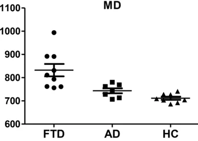
There were significant differences on MD between bvFTD and AD patients (P<0.01). MD in bvFTD patients was significantly higher than in AD patients after FWE correction. Mean MD values were calculated within the clusters wi- th significant difference. MD was 832±80 10-6 mm2/s in bvFTD patients, 743±28 10-6 mm2/s in AD patients and 711±19 10-6 mm2/s in HC as shown in Figure 3.
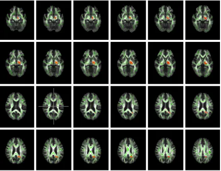
After matching with JHU White-Matter Tracto- graphy Atlas template and localization, the WM tracts that had increased MD were almost con-sistent with those with reduced FA and includ-ed right anterior thalamic radiation, bilateral cingulate gyrus, forceps minor, right inferior fr- onto-occipital fasciculus, bilateral uncinate ciculus and bilateral superior longitudinal fas-ciculus (Figure 4).
Hemisphere asymmetry of FA in bvFTD pa-tients
According to the FA distribution in both spheres of bvFTD patients, there was hemi-sphere asymmetry in FA of some regions bet- ween two hemispheres. Thus, we further calcu-lated the lateralization effect, but no significant difference was observed between bvFTD pa- tients and AD patients. After multiple compari-son FWE correction and matching with stan-dard template, significant difference was only observed in following regions: anterior thala- mic radiation, left inferior longitudinal fascicu-lus and left cingulate gyrus (Figure 5).
Discussion
Frontotemporal dementia has been a focus in recent studies. Pathological studies indicate that white matter damage is one of major
[image:5.612.93.524.69.426.2]tures of FTLD [9, 24, 25]. In addition, there are a large amount of connection fibers in the fron-tal cortex and a variety of axons which connect with other brain regions. The pathology of FTLD is characterized by the formation of inclusions containing tau, TDP-43 or FUS in the neurons and glial cells, demyelination and axonal dam-age of the white matter and excess gliosis. Currently, the knowledge on the relationship between atrophy regions which is valuable for the diagnosis of FTLD and microscopic disper-sion is still limited. As compared to morpho- logical changes, the change in diffusion tens- or may reflect earlier white matter damage in FTLD.
Impaired integrity of white matter in bvFTD patients
In the present study, TBSS with FWE (family wise error) correction was employed for analy-sis, and results showed FA in bvFTD patients
reduced significantly, and reduced FA was wi- dely distributed in the white matter including important regions such as frontal lobe, tempo-ral lobe and corpus callosum. Comparison of MD between bvFTD patients and HC indicated that MD in bvFTD patients increased markedly as compared to HC and increased MD was mainly found in the frontal lobe, temporal lobe and corpus callosum. The regions with incre- ased MD showed similar distribution to those with reduced FA. In respect of anatomy, unci-nate fascicle connects with frontal lobe and amygdale, mainly the motor speech area and orbital gyrus in the frontal lobe and orbital gyrus in the temporal lobe; superior longitudi-nal fasciculus is the longest tract of the connec-tion fibers and it originates from the prefrontal cortex and ends at the temporal lobe. It also crosses the frontal lobe, parietal lobe, occipit- al lobe and temporal lobe where it receives fibers from these lobes and projects fibers to these lobes; fasciculus frontooccipitalis
[image:6.612.92.524.71.406.2]nates from the frontal lobe, is distributed in deep superior longitudinal fasciculus and later-al caudate nucleus close to the centrlater-al laterlater-al ventricle and ends in the occipital lobe and temporal lobe in a fan form. The damage to these important connection fibers suggests the extensive white matter injury in bvFTD patients as compared to healthy controls, which was consistent with recent findings [10, 19, 20, 24].
Although it is generally accepted that bvFTD patients usually present damages to the ante-rior temporal lobe and frontal lobe, the lateral parietal and central cortex is also significantly involved at late stage of bvFTD. Arcuate fasci-cle is an important structure bridging the ante-rior and posteante-rior language area. There is evi-dence showing that arcuate fascicle is also damaged in bvFTD patients, suggesting that arcuate fascicle injury may be also related to the abnormal behaviors. The FA of anterior su- perior longitudinal fasciculus in the left hemo-sphere increased, this region connects with inferior frontal gyrus and thus may receive the projecting fibers from the arcuate fascicle. Whether this type of white matter damage is unique in bvFTD patients and the characteris-tics of white matter damage in other diseases such as AD and MCI early stage of AD are still unclear. If the white matter damage is different between AD patients and bvFTD patients, it is helpful for the differential diagnosis of bvFTD.
Difference in the white matter integrity be-tween bvFTD patients and AD patients
Our results showed the FA in bvFTD patients after FWE correction was comparable to mat- ched AD patients, but it tended to be different before correction at P<0.01. Analysis of MD showed significant difference between two gro- ups. After FWE correction, the MD in bvFTD patients was significantly higher than in AD patients at P<0.01. Following matching with template, the tracts with increased MD were similar to those with reduced FA in the distribu-tion and included left cingulate gyrus, left supe-rior longitudinal fasciculus which was in accor-dance with recent findings. Zhang et al. [20] found the FA of frontal and temporal lobes (including anterior corpus callosum, bilateral anterior and descending cingulate gyrus and uncinategyrus) reduced in bvFTD patients. St- udies on the basis of voxel-by-voxel analysis showed the FA reduced at extensive regions including frontal, temporal and parietal lobes,
but occipital white matter was not involved. However, AD patients showed FA reduction in bilateral descending cingulate gyrus, left poste-rior and anteposte-rior cingulate gyrus and left unci-nategyrus. The FA reduction in the frontal lobe of bvFTD patients is more obvious, and the reduction in FA of any brain region in AD pa- tients is still lower than that in bvFTD patients, suggesting that the white matter damage in bvFTD patients is more severe than in AD patients although the severity of dementia is similar. In addition, the white matter integrity in AD patients is near to normal. These findings indicate that, among patients with similar MMSE scores and severity of cognition impair-ment, bvFTD patients tend to present more obvious white matter injury. If the sample size is large enough, there might be significant dif-ference in FA between them. The white matter integrity is affected by multiple factors includ-ing cerebrovascular diseases and age. In the present study, cerebrovascular diseases were excluded, and included patients were relatively young. Structural imaging examinations failed to show cerebrovascular diseases which may be unlikely to bias our results.
axons. Thus, axons in these regions is suscep-tible to pathological processes including oxida-tive stress [26, 27]. Salat et al. [28] found, in the early stage of AD, some patients did not present obvious grey matter atrophy as com-pared to the hippocampal atrophy, but there was still diffusion change in the white matter around the hippocampus including the FA reduction of ventromedial prefrontal lobe and praecuneus. The structures connecting with the middle temporal lobe are closely related to the memory, indicating that the degradation of white matter around the hippocampus is likely an imaging marker of early AD. Our study did not reveal more severe damage to the white matter around the hippocampus and more obvious reduction in FA. These findings suggest that bvFTD patients may also present damages to the white matter in these regions, which is consistent with the hippocampal atrophy in some bvFTD patients [29, 30].
In present study, only white matter integrity was emphasized in DTI, and MD reduced in the grey matter of regions with brain atrophy. These also indicate the damage to the residual brain cells, and the white matter tracts connecting with these grey matters are also damaged. bvFTD patients presented MD increase in extensive grey matters, and bilateral temporal lobe, fron-tal lobe and pariefron-tal cortex were susceptible to the involvement. At late stage, the middle pari-etal cortex may be also significantly involved. In the same patient, the regions with MD increase in grey matters were highly consistent with those with brain atrophy. The close spatial rela-tionship between brain atrophy and abnormal diffusion indicates the abnormality in residual tissues. Our study also showed the MD eleva-tion in anterior hemisphere was more obvious in bvFTD patients than in AD patients, suggest-ing that regional MD increase, especially in the anterior white matters (such as frontal lobe), is helpful for the diagnosis and differential diag-nosis of bvFTD with higher sensitivity as com-pared to FA.
Laterality of white matter damage in bvFTD patients
The distribution of FA in both hemispheres showed its asymmetry. We further analyzed the lateralization effects, and results showed FA was dominant in left hemisphere in HC group, but there was no significant difference in bvFTD patients and in AD patients. The regions with
significant difference in FA between bvFTD patients and HC were matched with standard template, and results showed significant differ-ence in left thalamus radiation, left inferior lon-gitudinal fasciculus and left cingulate gyrus, suggesting that the damage is more obvious in left white matter of bvFTD patients, but the clinical significance of this asymmetry is re- quired to be further studied. Whitwell et al. [31] employed VBM to analyze the lateralization effects of dorsolateral cortex of frontal lobe, medial prefrontal lobe and orbitofrontal cortex in 80 patients with bvFTD. Results showed symmetry in 65% of patients, evident left phy in 20% of patients and obvious right atro-phy in remaining 15% of patients. Liu et al. also investigated the lateralization in AD patients with DTI, and they found the white matter around the right hippocampus displayed signifi-cant FA reduction, and FA reduction was also observed in the right front dome and left supe-rior longitudinal fasciculus in AD patients [32]. Most of other studies do not report the lateral-ization of white matter integrity in AD patients. There were still limitations in the present study. The sample size of this study was still small. Typical bvFTD usually progresses rapidly, and bvFTD patients are often unable to cooperate with imaging examinations and neuropsycho-logical assessment due to apathy and declined executive function. Thus, patient recruitment is difficult. Some studies on bvFTD with high qual-ity often employ pathological support. In the present study, the diagnosis was not confirmed by pathology, and to assure that patients with early bvFTD were recruited and the diagnosis was reliable was difficult. Thus, when the clini-cal symptoms were suspicious and imaging examinations were unable confirm the diagno-sis, patients were excluded from this study, which is a reason for small sample size.
Conclusions
Acknowledgements
This study was supported by the Shanghai Municipal Commission Health and Family Pl- anning Foundation, 2015-40314. National Science and Technology Major Project for IND (investigational new drug) 2012ZX09303- 003.
Disclosure of conflict of interest
None.
Address correspondence to: Huafang Li, Medical In- stitution Conducting Clinical Trial for Human Us- ed Drug, Shanghai Mental Health Center, Shanghai 200030, China. E-mail: lihuafangsh@sina.com; Shi- fu Xiao, Department of Geriatric, Shanghai Mental Health Center, Shanghai Jiaotong University School of Medicine, Shanghai 200030, China. E-mail: xiao-shifu@msn.com
References
[1] Knopman DS and Roberts RO. Estimating the number of persons with frontotemporal lobar degeneration in the US population. J Mol Neu-rosci 2011; 45: 330-335.
[2] Ratnavalli E, Brayne C, Dawson K and Hodges JR. The prevalence of frontotemporal demen-tia. Neurology 2002; 58: 1615-1621.
[3] Harvey RJ, Skelton-Robinson M and Rossor MN. The prevalence and causes of dementia in people under the age of 65 years. J Neurol Neurosurg Psychiatry 2003; 74: 1206-1209. [4] Borroni B, Alberici A, Grassi M, Turla M, Zanetti
O, Bianchetti A, Dalla Volta G, Rozzini R, Gil-berti N, Bellelli G and Padovani A. Is frontotem-poral lobar degeneration a rare disorder? Evi-dence from a preliminary study in Brescia county, Italy. J Alzheimers Dis 2010; 19: 111-116.
[5] Rosso SM, Donker Kaat L, Baks T, Joosse M, de Koning I, Pijnenburg Y, de Jong D, Dooijes D, Kamphorst W, Ravid R, Niermeijer MF, Verheij F, Kremer HP, Scheltens P, van Duijn CM, Heu-tink P and van Swieten JC. Frontotemporal de-mentia in The Netherlands: patient character-istics and prevalence estimates from a po- pulation-based study. Brain 2003; 126: 2016-2022.
[6] Piguet O, Hornberger M, Mioshi E and Hodges JR. Behavioural-variant frontotemporal demen-tia: diagnosis, clinical staging, and manage-ment. Lancet Neurol 2011; 10: 162-172. [7] Kipps CM, Hodges JR and Hornberger M.
Non-progressive behavioural frontotemporal de-mentia: recent developments and clinical im-plications of the ‘bvFTD phenocopy syndrome’. Curr Opin Neurol 2010; 23: 628-632.
[8] Moodley KK, Minati L, Barnes A, Dickson JC, Ell PJ and Chan D. Simultaneous PET/MRI in fron-totemporal dementia. Eur J Nucl Med Mol Im-aging 2013; 40: 468-469.
[9] Zhang Y, Tartaglia MC, Schuff N, Chiang GC, Ching C, Rosen HJ, Gorno-Tempini ML, Miller BL and Weiner MW. MRI signatures of brain macrostructural atrophy and microstructural degradation in frontotemporal lobar degenera-tion subtypes. J Alzheimers Dis 2013; 33: 431-444.
[10] Rohrer JD and Rosen HJ. Neuroimaging in fron-totemporal dementia. Int Rev Psychiatry 2013; 25: 221-229.
[11] Tosun D, Rosen H, Miller BL, Weiner MW and Schuff N. MRI patterns of atrophy and hypo-perfusion associations across brain regions in frontotemporal dementia. Neuroimage 2012; 59: 2098-2109.
[12] Tartaglia MC, Rosen HJ and Miller BL. Ne- uroimaging in dementia. Neurotherapeutics 2011; 8: 82-92.
[13] Munoz-Ruiz MA, Hartikainen P, Koikkalainen J, Wolz R, Julkunen V, Niskanen E, Herukka SK, Kivipelto M, Vanninen R, Rueckert D, Liu Y, Lot-jonen J and Soininen H. Structural MRI in fron-totemporal dementia: comparisons between hippocampal volumetry, tensor-based morph- ometry and voxel-based morphometry. PLoS One 2012; 7: e52531.
[14] Zhang Y, Schuff N, Ching C, Tosun D, Zhan W, Nezamzadeh M, Rosen HJ, Kramer JH, Gorno-Tempini ML, Miller BL and Weiner MW. Joint assessment of structural, perfusion, and diffu-sion MRI in Alzheimer’s disease and fronto-temporal dementia. Int J Alzheimers Dis 2011; 2011: 546871.
[15] Seeley WW. Anterior insula degeneration in frontotemporal dementia. Brain Struct Funct 2010; 214: 465-475.
[16] Seeley WW, Allman JM, Carlin DA, Crawford RK, Macedo MN, Greicius MD, Dearmond SJ and Miller BL. Divergent social functioning in be-havioral variant frontotemporal dementia and Alzheimer disease: reciprocal networks and neuronal evolution. Alzheimer Dis Assoc Dis-ord 2007; 21: S50-57.
[17] Seeley WW, Crawford R, Rascovsky K, Kramer JH, Weiner M, Miller BL and Gorno-Tempini ML. Frontal paralimbic network atrophy in very mild behavioral variant frontotemporal dementia. Arch Neurol 2008; 65: 249-255.
[18] Hornberger M, Geng J and Hodges JR. Conver-gent grey and white matter evidence of orbito-frontal cortex changes related to disinhibition in behavioural variant frontotemporal demen-tia. Brain 2011; 134: 2502-2512.
Josephs KA and Jack CR Jr. Gray and white matter water diffusion in the syndromic vari-ants of frontotemporal dementia. Neurology 2010; 74: 1279-1287.
[20] Zhang Y, Schuff N, Du AT, Rosen HJ, Kramer JH, Gorno-Tempini ML, Miller BL and Weiner MW. White matter damage in frontotemporal de-mentia and Alzheimer’s disease measured by diffusion MRI. Brain 2009; 132: 2579-2592. [21] Rascovsky K, Hodges JR, Knopman D, Mendez
MF, Kramer JH, Neuhaus J, van Swieten JC, Seelaar H, Dopper EG, Onyike CU, Hillis AE, Jo-sephs KA, Boeve BF, Kertesz A, Seeley WW, Rankin KP, Johnson JK, Gorno-Tempini ML, Rosen H, Prioleau-Latham CE, Lee A, Kipps CM, Lillo P, Piguet O, Rohrer JD, Rossor MN, Warren JD, Fox NC, Galasko D, Salmon DP, Black SE, Mesulam M, Weintraub S, Dickerson BC, Diehl-Schmid J, Pasquier F, Deramecourt V, Lebert F, Pijnenburg Y, Chow TW, Manes F, Grafman J, Cappa SF, Freedman M, Grossman M and Miller BL. Sensitivity of revised diagnos-tic criteria for the behavioural variant of fronto-temporal dementia. Brain 2011; 134: 2456-2477.
[22] McKhann G, Drachman D, Folstein M, Katzman R, Price D and Stadlan EM. Clinical diagnosis of Alzheimer’s disease: report of the NINCDS-ADRDA Work Group under the auspices of Department of Health and Human Services Task Force on Alzheimer’s Disease. Neurology 1984; 34: 939-944.
[23] Behrens TE, Berg HJ, Jbabdi S, Rushworth MF and Woolrich MW. Probabilistic diffusion trac-tography with multiple fibre orientations: What can we gain? Neuroimage 2007; 34: 144-155. [24] Smith SM, Jenkinson M, Johansen-Berg H, Ru-
eckert D, Nichols TE, Mackay CE, Watkins KE, Ciccarelli O, Cader MZ, Matthews PM and Beh-rens TE. Tract-based spatial statistics: voxel-wise analysis of multi-subject diffusion data. Neuroimage 2006; 31: 1487-1505.
[25] Agosta F, Scola E, Canu E, Marcone A, Magnani G, Sarro L, Copetti M, Caso F, Cerami C, Comi G, Cappa SF, Falini A and Filippi M. White mat-ter damage in frontotemporal lobar degenera-tion spectrum. Cereb Cortex 2012; 22: 2705-2714.
[26] Braak H, Del Tredici K, Schultz C and Braak E. Vulnerability of select neuronal types to Al-zheimer’s disease. Ann N Y Acad Sci 2000; 924: 53-61.
[27] Bartzokis G. Age-related myelin breakdown: a developmental model of cognitive decline and Alzheimer’s disease. Neurobiol Aging 2004; 25: 5-18; author reply 49-62.
[28] Salat DH, Tuch DS, van der Kouwe AJ, Greve DN, Pappu V, Lee SY, Hevelone ND, Zaleta AK, Growdon JH, Corkin S, Fischl B and Rosas HD. White matter pathology isolates the hippocam-pal formation in Alzheimer’s disease. Neurobi-ol Aging 2010; 31: 244-256.
[29] de Souza LC, Chupin M, Bertoux M, Lehericy S, Dubois B, Lamari F, Le Ber I, Bottlaender M, Colliot O and Sarazin M. Is hippocampal vol-ume a good marker to differentiate Alzheim-er’s disease from frontotemporal dementia? J Alzheimers Dis 2013; 36: 57-66.
[30] Onyike CU, Pletnikova O, Sloane KL, Sullivan C, Troncoso JC and Rabins PV. Hippocampal scle-rosis dementia: An amnesic variant of fronto-temporal degeneration. Dement Neuropsychol 2013; 7: 83-87.
[31] Whitwell JL, Xu J, Mandrekar J, Boeve BF, Knopman DS, Parisi JE, Senjem ML, Dickson DW, Petersen RC, Rademakers R, Jack CR Jr and Josephs KA. Frontal asymmetry in behav-ioral variant frontotemporal dementia: clinico-imaging and pathogenetic correlates. Neuro-biol Aging 2013; 34: 636-639.