Original Article
Correlation of LF-PRL-R expression with ER/PR and
HER-2 expression in breast cancer
Ronghui Zheng1,2, Xunxing Guan3, Xiaojun Tan2, Jie Zhou2, Zhiwei Liao3, Yawei Yuan1
1Department of Radiation Oncology, Nanfang Hospital, Southern Medical University, Guangzhou 510515,
Guang-dong, P. R. China; 2Cancer Center of Guangzhou Medical University, Guangzhou 510095, Guangdong, P. R. China; 3Department of Radiotherapy, Sun Yat-sen University Cancer Center, Guangzhou 510060, Guangdong, P. R. China
Received September 6, 2015; Accepted February 22, 2016; Epub March 15, 2016; Published March 30, 2016
Abstract: Background and objective: The activation of prolactin receptor (PRL-R) may contribute to the development and progression of breast cancer, which is mainly mediated by the long form of PRL-R (LF-PRL-R). Therefore, we ana-lyzed the correlation of LF-PRL-R with ER, PR, and HER-2 expression in breast cancer. Methods: One hundred and thirty female patients with breast cancer (median age, 46 years; age range 26-77 years) undergone surgery without new adjuvant therapy at Sun Yat-sen University Cancer Center between Jan 2000 and Jun 2001 were included. The expression of LF-PRL-R, ER, PR, and HER-2 in the primary lesion from each patient was detected by immunohisto-chemistry. The correlation of LF-PRL-R expression with ER, PR, and HER-2 in breast cancer was assessed by Chi-square test. Results: Among 130 patients, 89 showed positive LF-PRL-R expression. Stratification of the statistical analysis showed that in the HER-2-positive sub-layer, LF-PRL-R expression was positively correlated with ER and PR expression (P < 0.05), while no correlation was noted in the HER-2-negative sub-layer (P > 0.05). In the ER (or PR) positive sub-layer, LF-PRL-R expression was positively correlated with HER-2 expression (P < 0.05), while no such correlation was noted in the ER (or PR)-negative sub-layer (P > 0.05). Conclusion: The positive correlation of LF-PRL-R expression with ELF-PRL-R/PLF-PRL-R in breast cancer relies on the positive expression of HELF-PRL-R-2, while the positive correlation with HER-2 expression relies on the positive expression of ER/PR, which suggesting combined anticancer therapy based on the individual target site may benefit patients with breast cancer.
Keywords: Breast cancer, long form of prolactin receptor (LR-PRL-R), ER, PR, HER-2
Introduction
Approximately 70% of human breast cancer tis-sues express prolactin receptor (PRL-R), which may contribute to the development and pro-gression of breast cancer. In fact, the biological effect of PRL-R is mainly induced through the long form of PRL-R (LF-PRL-R) [1-6]. The expres-sion of ER/PR and HER-2 is an important prog-nostic indicator of breast cancer. Anticancer therapies based on these two targets have been considered as two important therapeutic methods for breast cancer. Laboratory studies have indicated that positive regulation exists between PRL-R and ER/PR, as well as HER-2, which suggests that there may be some com-plex interrelationships among PRL-R, ER/PR and HER-2 [7-10]. Studies on these
interrela-tionships among them may help to find out
some clues for optimizing anti-PRL-R, anti-ER/ PR, and anti-HER-2 combination therapy. Thus,
we attempted to elucidate the complex interre-lationship between LF-PLR-R expression and the expression of ER/PR and HER-2 based on clinical pathological evaluation.
Materials and methods
Patients
One hundred and thirty female breast cancer patients undergone surgery without new adju-vant chemotherapy from Jan 2000 to Jun 2001 at Sun Yat-sen University Cancer Center were
retrospectively collected. Paraffin-embedded
primary cancer tissues were well-preserved. The patients were 26-77 years of age (median age: 46 years old). There were 80 pre- and 50 post-menopausal patients. The primary lesions were in the inner/central and outer quadrants in 48 and 82 patients, respectively. According
by WHO, 119 patients had invasive ductal car-cinoma, 5 had early invasive ductal carcar-cinoma, and 6 had other types of carcinoma.
Immunohistochemistry
The expression of LF-PRL-R, ER, PR and HER-2 in post-operative samples was detected by
immunohistochemistry with LSAB kit according
to the manufacturers’ instructions. Briefly,
6-µm slices were obtained from the biopsy
specimen embedded in paraffin, heated in the
[image:2.629.331.531.78.228.2]thermostat at 60°C for 2 h and cooled in liquid at 37°C. Then, the tissue slices were placed into a fresh xylene tank twice (5 min each time), dipped into 95% ethanol twice (3 min each time), 70% ethanol twice (3 min each time), dis-tilled water for at least 30 s, and pre-heated 0.01 M citric acid buffer solution (100°C, pH 8.0) for 20 min, and stood still at room temper-ature for 20 min. After dipped in distilled water for 3 min, the tissue slices were delineated from 2 mm away with an anti-seepage pen. Then, the slices were incubated with solution A (3% H2O2) and solution B (normal serum) for 10 min at room temperature, respectively. Then the tissue slices were incubated with mouse primary monoclonal antibodies (1:50) (ZYMED Laboratories, U.S.A) overnight at 4°C. The pri-mary antibody was discarded and the tissue slices were thrice-dipped (5 min each time) in
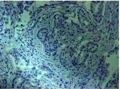
[image:2.629.99.297.79.226.2]Figure 1. PRL-R negative expression (×200).
[image:2.629.332.532.268.413.2]Figure 3. ER negative expression (×200).
Figure 5. PR negative expression (×200).
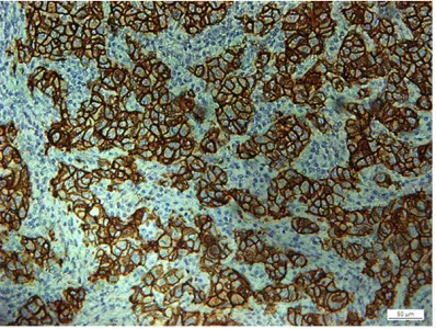
[image:2.629.99.297.269.414.2]Figure 2. PRL-R positive expression (×200).
[image:2.629.99.298.456.604.2]bated with solution C (biotin-labeled secondary antibody) and solution D (horseradish peroxi-dase-labeled streptavidin) at 37°C for 15 min respectively and developed with fresh DAB developer solution for 10 min. After rinsed with running water and stained in hematoxylin
solu-tion for approximately 30 s, the slices were dried and sealed in neutral balsam for observation.
The entire tissue slice was observed under an optical microscope by pathologists with exten-sive experience who were blinded to the clinical data. When the percentage of positive cells were < 10% of the total number of cells, the tis-sue was categorized as “negative expression” and when the percentage was > 10%, the tis-sue was categorized as “positive expression.” Statistical analysis
SPSS 13.0 for Windows was used for statistical analysis. The correlation of LF-PRL-R expres-sion with ER, PR, and HER-2 expresexpres-sion in breast cancer was determined by Chi-square
test. P < 0.05 was defined as statistically significant.
Results
Overall results
Among the 130 patients, 89 showed positive LF-PRL-R expression (positive rate: 68.5%), 70 showed positive ER expression (positive rate: 53.8%), 88 showed positive PR expression (positive rate: 67.7%) and 97 showed positive HER-2 expression (positive rate: 74.6%), as shown in Figures 1 to 8. The positive rate of LF-PRL-R expression in ER-positive patients was greater than ER-negative patients (P < 0.05), suggesting LF-PRL-R expression is tively correlated with ER expression. The posi-tive rates of LF-PRL-R expression in PR-negaposi-tive and -positive patents, and HER-2-negative and -positive patients were not statistically differ-ent (P > 0.05) (Table 1).
Results of stratification analysis
[image:3.629.98.299.80.236.2]The positive rate of LF-PRL-R expression in ER-positive patients was greater than ER-negative patients (LF-PRL-R expression was positively correlated with ER expression). This correlation was only limited in the HER-2-positive sub-layer (P < 0.05), while no such cor-relation was noted in the HER-2-negative sub-layer (P > 0.05). Similarly, the positive rate of LF-PRL-R expression in PR-positive patients was greater than PR-negative patients (LF-PRL-R expression was positively correlated with PR expression), which was only shown in
[image:3.629.99.297.275.426.2]Figure 6. PR positive expression (×200).
Figure 7. HER-2 negative expression (×200).
[image:3.629.99.298.467.617.2]the HER-2-positive sub-layer (P = 0.034 < 0.05), but not in the HER-2-negative sub-layer (P > 0.05) (Table 2).
The positive rate of LF-PRL-R expression in HER-2 positive patients was higher than HER-2-negative patients (LF-PRL-R expression was positively correlated with HER-2 expression), which was only shown in the ER (or PR)-positive sub-layer (P = 0.006 and 0.027 < 0.05, respec-tively), but not in the ER (or PR)-negative sub-layer (P > 0.05) (Table 3).
potential factors. The studies mentioned above were not subjected to a thorough analysis [14].
LF-PRL-R was specifically detected in this study.
The overall results showed that LF-PRL-R expression was positively correlated with ER
[image:4.629.98.370.107.209.2]expression. However, according to the stratifi -cation of HER-2 expression, the aforemen-tioned difference was only limited in the HER-2-positive sub-layer (not noted in the HER-2- negative sub-layer). Thus, HER-2 expression may be a strong impact factor for the correla-tion between LF-PRL-R and ER expression, and Table 1. Correlation of LF-PRL-R expression with ER, PR, and
HER-2 expression in all patients Clinical
factor casesTotal PRL-R (+) cases Positive rate (%) X2 value P value
ER - 60 35 58.1 5.294 0.021
+ 70 54 77.1
PR - 42 25 59.5 2.295 > 0.05
+ 88 64 72.7
HER-2 - 33 19 57.6 2.472 > 0.05
[image:4.629.99.370.258.386.2]+ 97 70 72.2
Table 2. Correlation of LF-PRL-R expression with ER and PR
expression after stratificated by HER-2 expression
HER-2 sub-layer Cases LF-PRL-R (+) cases Positive rate (%) X2 value P value
HER-2 (-) ER - 16 10 62.5 0.308 > 0.05
+ 17 9 52.9
PR - 11 7 63.6 0.248 > 0.05
+ 22 12 54.5
HER-2 (+) ER - 44 25 56.8 9.442 0.002
+ 53 45 84.9
PR - 31 18 58.1 4.510 0.034
+ 66 52 78.8
Table 3. Correlation of LF-PRL-R expression with HER-2
expres-sion after stratificated by ER/PR expresexpres-sion
Stratification
factor Sub-layer HER-2 Cases (+) casesLF-PRL-R Positive rate (%) X2 value P value
ER - - 16 10 62.5 0.156 > 0.05
+ 44 25 56.8
+ - 17 9 52.9 7.458 0.006
+ 53 45 84.9
PR - - 11 7 63.6 0.105 > 0.05
+ 31 18 58.1
+ - 22 12 54.5 4.889 0.027
+ 66 52 78.8
Discussion
[image:4.629.101.368.435.565.2]the positive correlation relies on the positive expression of HER-2.
According to previous study, the correlation between PRL-R and PR is lower than the corre-lation between PRL-R and ER [11]. Most stud-ies reported negative results [11, 14, 15]. No correlation between LF-PRL-R expression and PR expression was noted in all patients in our
study. However, according to the stratification
of HER-2, in the HER-2-positive sub-layer, LF-PRL-R expression was positively correlated with PR expression (no such correlation was noted in the HER-2-negative sub-layer). Thus, HER-2 expression may be also a strong impact factor for the correlation between PRLR expres-sion and PR expresexpres-sion. Only when HER-2 was positive did a positive correlation exist between PRLR expression and PR expression.
It has beenshown that the biological effect mediated by PRL-R may enhance the biological activity of HER-2 [8, 9]. However, up to now we have only found one study about the correla-tion between PRL-R expression and HER-2 expression,which didn’t demonstrated any pos-itive correlations [13]. No correlation was dem-onstrated between LF-PRL-R expression and HER-2 expression for all patients in our study.
However, according to the stratification of ER or
PR expression, LF-PRL-R expression was posi-tively correlated with HER-2 expression in the ER- or PR-positive sub-layer, but not in the ER- or PR-negative sub-layer. The results suggest that the positive correlation between LF-PRL-R expression and HER-2 expression relies on the positive expression of ER or PR. ER/PR are thus the impact factors for the correlation between LF-PRL-P and HER-2.
This study investigated the expression of PRL-R subtype (LF-PRL-R) in breast cancer tissues, which was closest to the occurrence and devel-opment of breast cancer. We also analyzed the complicated correlation between LF-PRL-R and ER, PR, and HER-2. Based on clinical pathology,
this study confirmed the complex regulation
and dependence which exists between LF-PRL-R expression and ER/PR and HER-2 expression, which provides potential clues for combined therapy based on PRL-R, ER/PR, and HER-2 target sites and also put forward a new thinking for the anti-PRL-R treatment on breast cancer.
Disclosure of conflict of interest
None.
Address correspondence to: Yawei Yuan, Depart- ment of Radiation Oncology, Nanfang Hospital, Southern Medical University, Guangzhou 510515, Guangdong, P. R. China. E-mail: yuanyw66@hotmail. com
References
[1] Tan D, Tang P, Huang J, Zhang J, Zhou W and Walker AM. Expression of a constitutively ac-tive prolactin receptor causes histone trimeth-ylation of the p53 gene in breast cancer. Chin Med J (Engl) 2014; 127: 1077-1083.
[2] Bostanci Z, Alam S, Soybel DI and Kelleher SL. Prolactin receptor attenuation induces zinc pool redistribution through ZnT2 and decreas-es invasion in MDA-MB-453 breast cancer cells. Exp Cell Res 2014; 321: 190-200. [3] McHale K, Tomaszewski JE, Puthiyaveettil R,
Livolsi VA and Clevenger CV. Altered expression of prolactin receptor-associated signaling pro-teins in human breast carcinoma. Mod Pathol 2008; 21: 565-571.
[4] Xu J, Sun D, Jiang J, Deng L, Zhang Y, Yu H, Bahl D, Langenheim JF, Chen WY, Fuchs SY and Frank SJ. The role of prolactin receptor in GH signaling in breast cancer cells. Mol Endo-crinol 2013; 27: 266-279.
[5] Howell SJ, Anderson E, Hunter T, Farnie G and Clarke RB. Prolactin receptor antagonism re-duces the clonogenic capacity of breast can-cer cells and potentiates doxorubicin and pa-clitaxel cytotoxicity. Breast Cancer Res 2008; 10: R68.
[6] Hu ZZ, Zhuang L, Meng J, Tsai-Morris CH and Dufau ML. Complex 5’ genomic structure of the human prolactin receptor: multiple alterna-tive exons 1 and promoter utilization. Endocri-nology 2002; 143: 2139-2142.
[7] Gutzman JH, Miller KK and Schuler LA. Endog-enous human prolactin and not exogEndog-enous hu-man prolactin induces estrogen receptor alpha and prolactin receptor expression and increas-es increas-estrogen rincreas-esponsivenincreas-ess in breast cancer cells. J Steroid Biochem Mol Biol 2004; 88: 69-77.
[9] Xu C, Langenheim JF and Chen WY. Stromal-epithelial interactions modulate cross-talk be-tween prolactin receptor and HER2/Neu in breast cancer. Breast Cancer Res Treat 2012; 134: 157-169.
[10] Fiorillo AA, Medler TR, Feeney YB, Wetz SM, Tommerdahl KL and Clevenger CV. The prolac-tin receptor transactivation domain is associ-ated with steroid hormone receptor expression and malignant progression of breast cancer. Am J Pathol 2013; 182: 217-233.
[11] Gill S, Peston D, Vonderhaar BK and Shousha S. Expression of prolactin receptors in normal, benign, and malignant breast tissue: an immu-nohistological study. J Clin Pathol 2001; 54: 956-960.
[12] Touraine P, Martini JF, Zafrani B, Durand JC, Labaille F, Malet C, Nicolas A, Trivin C, Postel-Vinay MC, Kuttenn F and Kelly PA. Increased expression of prolactin receptor gene as-sessed by quantitative polymerase chain reac-tion in human breast tumors versus normal breast tissues. J Clin Endocrinol Metab 1998; 83: 667-674.
[13] Faupel-Badger JM, Duggan MA, Sherman ME, Garcia-Closas M, Yang XR, Lissowska J, Brinton LA, Peplonska B, Vonderhaar BK and Figueroa JD. Prolactin receptor expression and breast cancer: relationships with tumor characteris-tics among pre- and post-menopausal women in a population-based case-control study from Poland. Horm Cancer 2014; 5: 42-50.
[14] De Placido S, Gallo C, Perrone F, Marinelli A, Pagliarulol C, Carlomagno C, Petrella G, D’Istria M, Delrio G and Bianco AR. Prolactin receptor does not correlate with oestrogen and proges-terone receptors in primary breast cancer and lacks prognostic significance. Ten year results of the Naples adjuvant (GUN) study. Br J Can-cer 1990; 62: 643-646.