Original Article
Application of self assembling peptide
as drug carrier for extending the GLP-1 stability
Ying Li1, Xuemin Zheng2, Fancui Meng2, Min Gong2,3,4
1Tianjin Neurological Institute, Tianjin Medical University General Hospital, Tianjin, China; 2Tianjin Institute of Pharmaceutical Research, Tianjin, China; 3Department of Pharmacy, Tianjin Medical University, Tianjin, China; 4Department of Oncology, University of Oxford, Oxford, UK
Received November 20, 2015; Accepted February 3, 2016; Epub May 15, 2016; Published May 30, 2016
Abstract: The multiple physiological characterizations of glucagon-like peptide-1 (GLP-1) make it a promising drug candidate for the therapy of type 2 diabetes. However, the extremely poor stability of GLP-1, proteolysis by dipeptidyl peptidase-IV (DPP IV), limits its development in the clinical utility significantly. In order to avoid the pocket docking of GLP-1 against DPP IV, self assembling peptide was employed in this study. Self assembling peptide (pp1) is capable of forming supramolecule contain a cavity which load GLP-1 molecules inside. In this study, the formation procedure of GLP-1/pp1 complex was computationally simulated, in order to further investigate the interaction between GLP-1 and pp1. In this study, the amino acid residues in GLP-1 and pp1 those involved in forming hydrogen bonds were ascertained which benefits the further mutation and improvement on pp1. In addition, the glucose tolerance test in rodent animals was employed to ascertain the prolonged stability of GLP-1 followed the complex formation with pp1. Our results clarified the mechanism of the formation of GLP-1 and pp1 stable complex, which is significant for its development into clinical trial.
Keywords: Self-assembled peptide, molecular dynamics, simulations, GLP-1, sustained complex
Introduction
Glucagon-like peptide 1 (GLP-1) is a gut hor-mone released from intestinal L cells after oral glucose administration [1]. It serves as an incretin factor stimulating the secretion of insu-lin, and reduces the levels of blood glucose in normal subjects and patients with type 2 diabe-tes mellitus [2]. GLP-1 stimuladiabe-tes the secretion of insulin and suppresses glucagon secretion in glucose-dependent manner, which minimizes
the risk of hypoglycemia [3, 4].It was also found that GLP-1 restores the glucose sensitivity of pancreatic β-cells, which possibly depend on the increasing level of GLUT2 and glucokinase expression [5]. Furthermore, GLP-1 promotes β-cell regeneration, proposing that it is effec-tive for the treatment of type 1 diabetes [6, 7]. Report proved that GLP-1 inhibits apoptosis (programmed cell death) in cells, consequently improves their survival [8]. Evidence has dem-onstrated that GLP-1 was identified to play a crucial role in the regulation of glucose metabo-lism, and the infusion of GLP-1 induces the
decreasing level of blood glucose [9, 10]. The deficiency of GLP-1 secretion in type 2 diabetes suggested that GLP-1 acts as a promising potential therapy for type 2 diabetes [11]. The most serious challenge is that GLP-1 has an exceptionally short half-life of less than 2 mins in vivo [12], due to the rapid degradation of the enzyme dipeptidyl peptidase IV (DPP-IV) [13]. This caused that the therapeutic administration of GLP-1 seemed impractical, thus many efforts have focused on amending the pharmacokinet-ic properties of GLP-1 in a series of derivatives and analogues [14].
residues of this amphiphilic α-helix all interact with the ECD, whereas Lys26 is the only one of the hydrophilic residues is possible to interact directly with the ECD. Previous Ala scanning assay has demonstrated the importance of Phe28, Ile29, and Leu32 in GLP-1 binding [20]. The mutation of the residues on the hydropho-bic side of GLP-1 causes loss of ECD binding affinity, while that on the hydrophilic face does not [21, 22]. Moreover, the N-terminal trunca-tion of GLP-1 are unable to bind and activate GLP-1R [23], this segment is an obstacle for long-acting GLP-1 analogs as the main site of DPP-IV degradation. Two GLP-1 analogous, Exenatide and Liraglutide, were approved by FDA for the treatment of type 2 diabetes in 2005 and 2010, respectively [24, 25]. Com- pared with Exendin-4 and two GLP-1 analogs, the utility of hGLP-1 is quite probably to avoid any possible immunotoxicity of n on-mammali-an product on-mammali-and improve the biotoleron-mammali-ance in turn.
In our previous study, the self-assemble pep-tide (pp1) was employed to form a stable com-plex with native hGLP-1 [26]. The serum stabili-zation assay of GLP-1/pp1 complex illuminated that the complex formation leads to an improve-ment of GLP-1 stability in vitro and in vivo [26]. It was presumed that pp1 formed a complex which provided with a ‘protecting surface’ pro-tects the GLP-1 molecule inside against the enzymatic degradation [26]. In order to clarify the formation of stable complex of GLP-1 and pp1 deeply, the molecular dynamics simula-tions were performed and the interior hydrogen bonds were analyzed via molecular dynamics simulations. Upon the simulations, hydrogen bonds and torsion angle were analyzed; the amino acid residues of either pp1 or GLP-1 involved in the formation of hydrogen bonds were ascertained as well. The simulations data helped us to investigate the mechanism of the complex formation and perform the further mutation for improved assembling activity. Combined with simulation data, the glucose tol-erance test was performed in order to ascer-tain the prolonged stability of complex in exper-imental animals.
Self-assembled peptides display the advanta-geous properties of stability, injectability, bio-degradability and biocompatibility. These pep-tides, by self-assembling, can be widely applied in such fields as drug delivery (small molecules and large molecules), regenerative medicine
and nanobiotechnology. The various physical, chemical and biological aspects of self assem-bling peptide family lead to different applica-tion field. Clarificaapplica-tion the mechanism of self assembling peptide makes these novel bio-medical materials to be applied much more available.
Materials and methods
Animals
All studies were carried out with permits from the Animal Experiments Inspectorate, China. Male SD rats were obtained from SLAC La- boratory Animal, China Academy of Sciences (Shanghai, China).
Simulation procedures
In the current work, molecular dynamics (MD) simulations were carried out for two different systems containing different amount of pp1 peptides and one GLP-1 peptide. The two sys-tems represent GLP-1 in pp1 solutions with dif-ferent concentrations. AMBER force field and GBSA model, which count solvent effect implic-itly, were chosen for the simulations. Simula- tions were performed by AMBER 99 software package. In the MD run, Langvin thermostat method was used to fix the temperature at 300 K. Large box size, which equivalents to no peri-odic boundary condition, was used in the runs. The initial array of system I was that the GLP-1 was immersed in ten and twenty randomly arrayed pp1 peptides. For each system, MD simulations were carried out for 1 ms. The time-step size for the integration was 1 ms, and trajectories at each of 500 time-steps were col-lected for analysis.
Peptide synthesis
GLP-1 (sequence as: HAEGTFTSDVSSYLEGQ- AAKEFIAWLVKGRG) and pp1 (sequence as: GLWWKAWWKAWWKSLWWRKRKR) were pur-chased from Sangon Biotech (Shanghai, China) Co., Ltd (HPLC-purified; purity >90%, identified by MS). The freeze-dried peptides were weighed and dissolved in pure water to make 10 mg/ml stock solutions for further analysis.
Glucose tolerance test upon single dose ad-ministration
injected subcutaneously into fasting SD rats (n=10 per group, male) 30 min prior to glucose administration. GLP-1 was injected into the control animals. Rats were given 2 g glucose/ kg body weight via intraperitoneal injection. Blood was drawn from the tail vein, and glucose levels were measured using a glucometer 30 min after glucose administration. Chronic glu-cose (2 g/kg body weight) was administrated
30 min prior to each blood glucose measure-ment time point during the 100-h experimeasure-mental period.
Statistical analyses
[image:3.612.90.525.69.499.2]Student’s t-test was employed for analyses of the data. Unless otherwise stated, the results were reported as the means ± standard error. P
values less than 0.05 were considered significant.
Results and discussions
Binding mode of GLP-1 and pp1 peptide
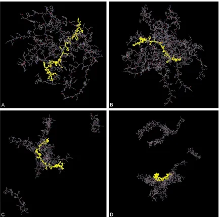
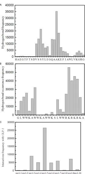
[image:4.612.93.372.62.628.2]Our previous findings indicat-ed the formation of a supra-molecular complex of GLP-1 and pp1 by HPLC assays. The incubating mixture of pp1 and GLP-1 was loaded onto C18 column and the spectra fea-ture was compared to those from GLP-1 and pp1 alone, respectively. Data demonst- rated the existence of supra-molecule and it was clarified as complex of GLP-1 and pp1 upon the ratio of 1:6 using MALDI-TOF. In current study, Figure 1C and 1D showed the snapshots of systems I (initial assay as Figure 1A) and II (ini-tial assay as Figure 1B) at the ends of the MD runs. We found that GLP-1 was wrapped by five pp1 peptides in system I and seven pp1 peptides in system II. From Figure 1C and Figure 2. Hydrogen bonds analysis of pp1 and GLP-1 complex in system I. A.
Hydrogen bonds formation frequency of GLP-1 residues with pp1 peptide. B. Hydrogen bonds formation frequency of pp1 residues with GLP-1. C. The numbered pp1 peptides formed complex with GLP-1 by hydrogen bonds. Conditions: The hydrogen bonds were determined by checking the distance between polarized hydrogen (H-N, H-O) and electronegative atoms O and N. Legend: The hydrogen bonds between residues of GLP-1 and residues of
neighboring pp1 peptides in systems I was analyzed. It was observed that
1D, it was observed that GLP-1 could form a complex with pp1 in system I and sys-tem II. Like showed in Figure 1, the GLP-1 molecule was wrapped by pp1 peptides to form a stable complex whi- ch might provide the inside GLP-1 molecule against the degradation of DPP IV.
The frequency of occurrence of hydrogen bonds between GLP-1 and pp1
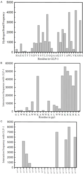
It was identified that the pp1 peptides bound GLP-1 via hy- drogen bonds, by investigat-ing the distance between po- larized hydrogen (H-N, H-O) and electronegative atoms O and N (Figures 2 and 3). Bar charts showed the frequency of occurrence of the hydrogen bonds between residues of GLP-1 and residues of neigh-boring pp1 peptides in sys-Figure 3. Hydrogen bonds analysis of pp1 and GLP-1 complex in system II.
A. Hydrogen bonds formation frequency of GLP-1 residues with pp1 peptide. B. Hydrogen bonds formation frequency of pp1 residues with GLP-1. C. The numbered pp1 peptides formed complex with GLP-1 by hydrogen bonds. Conditions: The hydrogen bonds were determined by checking the distance
[image:5.612.90.373.71.669.2]tems I and II. It was observed that hydrogen bonds were formed mainly in the middle region and C-terminus of GLP-1 as showed in Figures 2A and 3A. Interestingly, the N-terminus of GLP-1 exhibited very low probability of forming hydrogen bond with closed pp1 peptides. The simulation data provided irrefutable evidence to the previous finding: the N-terminal of GLP-1 plays an important role in their physiological functions by interacting with ECD of GLP-1 receptor. Thus it was considered that the for-mation of hydrogen bonds in N-terminal seg-ments of GLP-1 would impair the docking of GLP-1 to receptor inevitably, but not in C-terminal. These presumed that pp1 provided a protection role for the GLP-1 inside and remained their activities successfully. Figures 2C and 3C indicated that 7 (peptide numbered 3, 4, 5, 6, 7, 9 and 10 in system I, Figure 2C) or 8 (peptide numbered 3, 4, 5, 14, 17, 18, 19 and 20 in system II, Figure 3C) interacted with GLP-1 upon high hydrogen bonds frequency, which is coincided with our previous results that GLP-1 could form stable complexes at the molar ratio of 1:6 by reverse phase HPLC and S-200 gel filtration column [26].
In addition, results presented that both N- and C-terminus of pp1 showed high hydrogen bond occurrence frequency, suggesting that they are vital for the formation of hydrogen bonds with GLP-1 (Figures 2B and 3B). Structural analysis of pp1 illuminated that pp1 possesses ‘L shape’
Data proved that these three residues interact-ed with GLP-1 only, and importantly suggestinteract-ed the mutations at these positions should be carefully in order to avoid the self-aggregation of pp1 or loss of binding affinity with GLP-1. Torsion angle analysis of GLP-1/pp1
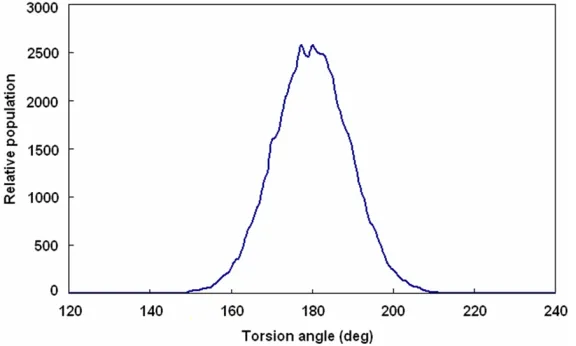
Torsion angle analysis was carried out to inves-tigate whether pp1 binding changed the confor-mation of GLP-1. Figure 5 showed that the main frame torsion angles of GLP-1 and closed pp1 peptides in system I are all populated arou- nd 180°, indicating that GLP-1 maintained its active form even upon the wrapping of pp1 pep-tides and the existence of pp1 is unable to influ-ence the conformational of GLP-1 and its phy- siological activities, such as insulin secretion, cAMP and blood glucose regulation.
Anti-diabetic activity of complex of GLP-1 and pp1 in long-acting manner
[image:6.612.91.379.73.246.2]To ascertain the long-lasting effects on glucose tolerance of complex of GLP-1 and pp1, glucose tolerance tests (GTTs) were performed 120 h after single dose administration into Sprague Dawley rats (n=6 per group, male). As shown in Figure 6, the rats treated with wild-type GLP-1 had high blood glucose levels at 11 mmol/L approximately in the whole experimental period of 100 h since the GLP-1 had been degraded rapidly. However, as predicted, the rats injected Figure 4. Analysis of torsion angles in GLP-1 and pp1 complex. Legend: It
showed that these angles are all populated around 180° indicating that GLP-1 maintaining its active form even upon the wrapping of pp1 peptides. Results illuminated that the existence of pp1 unable to induce the conforma-tional change of GLP-1.
helix containing hydrophobic and hydrophilic tail end, fur-thermore, the amphipathic te- rminuses are key domains interacting with GLP-1. Our result also revealed that the residues W, K, and R of pp1 have high probability of the formation of hydrogen bonds with GLP-1.
with GLP-1 complex showed improved glucose tolerance in this experiment. The blood glucose levels were maintained at 8 mmol/L after 60 h, P<0.01.
Conclusion
The discovery of self-assemble peptides pro-vides a platform for a drug sustained releasing system. Self-assemble peptides possess a self-assembly property which depends on amphip-athic domains. GLP-1 is considered to be potent tool for treatment of type 2 diabetes since
dis-then prolonged the stability of GLP-1. We also performed further analysis to clarify the re- sidue(s) involved in this interaction in both pp1 and GLP-1 peptides. Elucidation the crucial res-idues for this interaction will be helpful to fur-ther mutate of pp1 peptide in order to achieve an improved the assembling property.
Acknowledgements
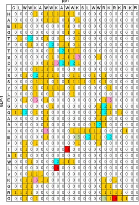
[image:7.612.93.376.76.495.2]This study was supported by the National Science and Technology Major Project of the Ministry of Science and Technology of China Figure 5. Occurrences of hydrogen bonds between GLP-1 and pp1 peptides
(the numbers in the matrix are the counting numbers divided by 1000). Leg-end: The occurrences of hydrogen bonds formed between GLP-1 and pp1 molecule was obtained by MD simulations. Data illuminated the residues in either GLP-1 or pp1 with higher frequency for hydrogen bonds formation.
(2014ZX09507005-003), the National Natural Science Funding (81400932), Major Project of Science and Technology of Tianjin (13RCGF- SY19700) and the Natural Science Foundation of Tianjin, China (12JCYBJC31500).
Disclosure of conflict of interest
None.
Address correspondence to: Min Gong, Tianjin Institute of Pharmaceutical Research, Tianjin, Chi- na. Tel: +862223003529; Fax: +862223006862; E-mail: [email protected]
References
[1] Weir GC, Mojsov S, Hendrick GK, Habener JF. Glucagonlike peptide I (7-37) actions on endo-crine pancreas. Diabetes 1989; 38: 338-42. [2] Lugari R, Dell’Anna C, Ugolotti D, Dei Cas A,
Barilli AL, Zandomeneghi R, Marani B, Iotti M, Orlandini A, Gnudi A. Effect of nutrient inges-tion on glucagon-like peptide 1 (7-36 amide) secretion in human type 1 and type 2 diabetes. Horm Metab Res 2000; 32: 424-8.
[3] Yamato E, Noma Y, Tahara Y, Ikegami H, Yamamoto Y, Cha T, Yoneda H, Ogihara T, Ohboshi C, Hirota M, et al. Suppression of
syn-proliferation, differentiation, and apoptosis. Mol Endocrinol 2003; 17: 161-71.
[8] Vilsboll T. The effects of glucagon-like pep-tide-1 on the beta cell. Diabetes Obes Metab 2009; 11 Suppl 3: 11-8.
[9] Vella A, Shah P, Basu R, Basu A, Holst JJ, Rizza RA. Effect of glucagon-like peptide 1 (7-36) amide on glucose effectiveness and insulin ac-tion in people with type 2 diabetes. Diabetes 2000; 49: 611-7.
[10] Straub SG, Sharp GW. Glucose-stimulated sig-naling pathways in biphasic insulin secretion. Diabetes Metab Res Rev 2002; 18: 451-63. [11] Nauck MA, Holst JJ, Willms B, Schmiegel W.
Glucagon-like peptide 1 (GLP-1) as a new ther-apeutic approach for type 2-diabetes. Exp Clin Endocrinol Diabetes 1997; 105: 187-95. [12] Scheen AJ. [Glucagon-like peptide-1 (GLP-1),
new target for the treatment of type 2 diabe-tes]. Rev Med Liege 2007; 62: 217-21. [13] Davidson JA. Advances in therapy for type 2
diabetes: GLP-1 receptor agonists and DPP-4 inhibitors. Cleve Clin J Med 2009; 76 Suppl 5: S28-38.
[14] Scheen AJ. New therapeutic approaches in type 2 diabetes. Acta Clin Belg 2008; 63: 402-7.
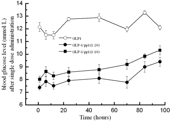
[image:8.612.91.376.73.278.2][15] Ohneda A, Ohneda K, Ohneda M, Koizumi F, Ohashi S, Kawai K, Suzuki S. The structure-function relationship of GLP-1 related peptides Figure 6.GLP-1/peptide complexes improve glucose regulation in ZDF rats.
Conditions: The intraperitoneal injections of GLP-1/peptide 1 complex (mo-lecular ratio at 1:10 ●; and ratio at 1:20 ■) (300 µg GLP-1/kg body weight) were administrated once in the whole experimental period of 100 hours and then glucose levels were measured by a glucometer at indicated times. GLP-1 (▲) (300 mg/kg body weight) was administrated daily and Exenatide (♦) (100 mg/kg body weight) was injected intraperitoneally twice daily. Saline (□) was injected in controls. Legend: Results indicated that the GLP-1/peptide 1 treated rats maintained relatively constant glucose levels within 70 hours.
thesis and release of glu-cagon by gluglu-cagon-like pe- ptide-1 (7-36 amide) with-out affect on mRNA level in isolated rat islets. Bio- chem Biophys Res Com- mun 1990; 167: 431-7. [4] Parhofer KG.
[Glucagon-like peptide 1 (GLP-1)]. MMW Fortschr Med 2007; 149: 41-3.
[5] Villanueva-Peñacarrillo ML, Puente J, Redondo A, Cle- mente F, Valverde I. Effect of GLP-1 treatment on GLUT2 and GLUT4 expres-sion in type 1 and type 2 rat diabetic models. Endocrine 2001; 15: 241-8.
[6] Brubaker PL, Drucker DJ. Minireview: Glucagon-like peptides regulate cell pro-liferation and apoptosis in the pancreas, gut, and central nervous system. Endocrinology 2004; 145: 2653-9.
in the endocrine function of the canine pan-creas. Tohoku J Exp Med 1991; 165: 209-21. [16] Hui H, Zhao X, Perfetti R. Structure and
func-tion studies of glucagon-like peptide-1 (GLP-1): the designing of a novel pharmacological agent for the treatment of diabetes. Diabetes Metab Res Rev 2005; 21: 313-31.
[17] Underwood CR, Garibay P, Knudsen LB, Hastrup S, Peters GH, Rudolph R, Reedtz-Runge S. Crystal structure of glucagon-like peptide-1 in complex with the extracellular do-main of the glucagon-like peptide-1 receptor. J Biol Chem 2010; 285: 723-30.
[18] Graziano MP, Hey PJ, Strader CD. The amino terminal domain of the glucagon-like peptide-1 receptor is a critical determinant of subtype specificity. Receptors Channels 1996; 4: 9-17. [19] Runge S, Wulff BS, Madsen K,
Bräuner-Osborne H, Knudsen LB. Different domains of the glucagon and glucagon-like peptide-1 re-ceptors provide the critical determinants of li-gand selectivity. Br J Pharmacol 2003; 138: 787-94.
[20] Adelhorst K, Hedegaard BB, Knudsen LB, Kirk O. Structure-activity studies of glucagon-like peptide-1. J Biol Chem 1994; 269: 6275-8.
[21] Al-Sabah S, Donnelly D. A model for receptor-peptide binding at the glucagon-like receptor-peptide-1 (GLP-1) receptor through the analysis of trun-cated ligands and receptors. Br J Pharmacol 2003; 140: 339-46.
[22] Gallwitz B, Witt M, Paetzold G, Morys-Wortmann C, Zimmermann B, Eckart K, Fölsch UR, Schmidt WE. Structure/activity charac- terization of glucagon-like peptide-1. Eur J Biochem 1994; 225: 1151-6.
[23] Persson B, Argos P. Prediction of membrane protein topology utilizing multiple sequence alignments. J Protein Chem 1997; 16: 453-7. [24] Briones M, Bajaj M. Exenatide: a GLP-1
recep-tor agonist as novel therapy for Type 2 diabetes mellitus. Expert Opin Pharmacother 2006; 7: 1055-64.
[25] Drucker DJ, Dritselis A, Kirkpatrick P. Lira- glutide. Nat Rev Drug Discov 2010; 9: 267-8. [26] Zheng X, Li Y, Li X, Tang L, Xu W, Gong M.